- 48 -
서 론
혈관근종(angiomyoma)은 혈관 중간막(tunica media)에 서 기원하/는 양성 평활근 종양으로,1) 그 빈도가 드문 것으로 알려져 있다. 이 종양은 신체 어느 부위에서든 발생할 수 있으 나, 주로 하지에 많이 발생하며, 피하조직에 호발한다. 두경부
에 발생하는 혈관근종은 그 중에서도 빈도가 더욱 드문 종양 으로 주로 아형(subtype)인 정맥형(venous type)의 형태로 발 견되며, 후두(larynx) 및 비갑개(turbinate) 등에서 주로 발생하 고 그 빈도는 9% 정도로 보고되고 있다.1)
혈관근종(angiomyoma)은 혈관 평활근종(angioleiomyoma, vascular leiomyoma)으로도 불린다. 혈관근종은 평활근 세포 및 혈관으로 이루어져 있으며, 대부분의 증례에서 단일성의 피 하조직 내 종양으로 발견된다. 본 증례 보고에서는 안면부 비익 (alae)의 피부에 발생한 혈관근종을 소개하고자 한다.
증 례
63세 남자 환자가 내원 1년 전에 발생하여 서서히 자라는, Received : May 29, 2013 / Revised : July 3, 2013
Accepted : July 5, 2013
이 논문은 2008년도 정부(교육과학기술부)의 재원으로 한국과학 재단의 지원을 받아 수행된 연구임(R11-2005-065).
교신저자 : 장 학, 110-744 서울 종로구 대학로 101 서울대학교 의과대학 서울대학교병원 성형외과학교실 전화 : (02) 2072-3086 ・ 전송 : (02) 3675-7792 E-mail : [email protected]
대한 두경부 종양 학회지 제 29 권 제 2 호 2013
안면부에 발생한 혈관근종의 치험례 : 증례보고
서울대학교 의과대학 서울대학교병원 성형외과학교실
최 준 호·장 학
=
Abstract
=Angiomyoma on Face : A Case Report
Junho Choi, MD, Hak Chang, MD, PhD
Department of Plastic and Reconstructive Surgery, Seoul National University Hospital, College of Medicine, Seoul, Korea
Angiomyoma is a rare, benign smooth muscle cell tumor. These tumors may be found anywhere in the body. They frequently occur in the lower extremities except venous type. Angiomyoma in the head and neck area is very rare, and only a few cases have been reported. A 63 year-old male patient visited to outpatient clinic due to size-grow- ing nodule-like lesion in the Lt. alar area. Excisional biopsy was done for diagnosis. The lesion was totally excised with 2 mm safety margin. Frozen biopsy of the lesion was requested, and all resection margins were proved neg- ative. To cover the raw surface, full thickness skin grafting was performed. The graft was harvested from Rt. pos- terior auricular area. Tie over dressing was applyed on Lt. alar area. The graft was well taken and healed without any complication in both short term and long term follow up periods of 2 weeks, 1 month, 2 months, and 6 months.
Donor site completed healed without any complications. The leiomyoma is benign tumor originated from smooth muscle, and it can be classified into solid leiomyoma, angiomyoma, and epithelioid leiomyoma. Especially, the an- giomyoma consists of smooth muscle cell and blood vessel, and it is originated from the tunica media of blood vessel. Angiomyoma alone frequently occurs in the lower extremities as solitary painless subcutaneous tumor. Ve- nous type of angiomyoma in the oral cavity was reported in other references, but on the facial surface it may be the first case reported as paper. So this report can be very meaningful.
KEY WORDS
:AngiomyomaㆍLeiomyomaㆍSmooth muscle tumorㆍVenous typeㆍTumor on facial skin.online©MLComm
- 49 -
좌측 비익부의 1.0×0.8 cm 크기의 타원형의 융기된 종괴를 주소로 2011년 5월 25일 성형외과 외래에 내원하였다(Fig. 1).환자는 이전에 외상을 입었던 병력이나 기타 치료력에 대해서 는 부인하였으며, 종괴의 정확한 진단 및 완전 제거를 목표로 절제 생검술(excisional biopsy)의 시행에 동의하였다. 수술 전 에 시행한 혈액 검사에서 특이 소견은 발견되지 않았으며, 2011 년 5월 31일 국소 마취 하에 수술이 진행되었다. 2 mm의 안전 경계(safety margin)를 두고 종괴가 절제되었고, 곧바로 시행 된 동결절편 검사(frozen section biopsy)에서는 절제연에서 모두 음성의 결과가 나왔다. 절제 후의 피부 결손 부위에는 우 측 이개후부(Rt. posterior auricular area)에서 전층 피부(full thickness skin graft)를 채취하여 이식하였고, 거즈와 실을 이용하여 봉합 고정 드레싱(tie over dressing)을 시행하였다.
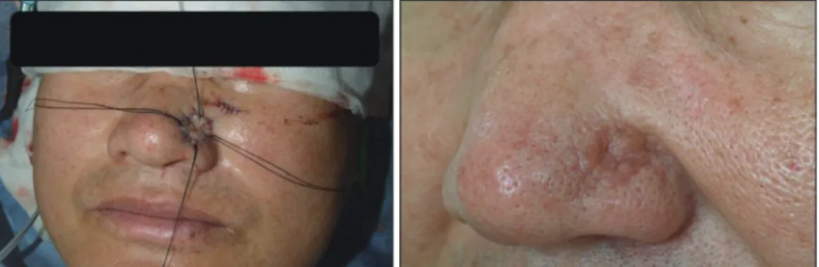
수술 시행 후 4일째에 봉합 고정 드레싱을 제거하였고, 8일 째에 수술 부위 봉합사를 제거하였다. 수술 후 각각 2주, 1개 월, 2개월, 그리고 6개월까지의 외래 추적 관찰 결과 절제 부 위의 합병증 발생 및 재발 소견은 없었으며, 공여부 또한 합병 증 발생 없이 치유되었다(Fig. 2).
고 찰
평활근종(leiomyoma)은 평활근(smooth muscle)에서 기원 하는 양성 종양으로, 조직학적으로는 고형 평활근종(solid leio- myoma), 혈관근종(angiomyoma), 및 상피양 평활근종(epithe- lioid leiomyoma)으로 분류된다.1) 그 중 혈관근종(angiomy- oma)은 평활근 세포와 혈관으로 구성된 종양으로, 혈관 중간 막(tunica media)에서 기원하는 것으로 알려져 있다. 혈관근종 은 조직학적인 특성에 따라 모세혈관형(capillary type), 해면형 (cavernous type), 그리고 정맥형(venous type)으로 분류되며,2) 두경부에 발생하는 혈관근종은 주로 정맥형으로 알려져 있 다. 혈관근종은 40대에서 50대의 남성에 호발하며, 하지에 주 로 발생하는 것으로 알려져 있으며, 단일성의 통증을 수반하 지 않는 피하조직 층의 종양으로 발견되는 경우가 많다.3) 두경 부에 발생하는 혈관근종의 예로는 후두(larynx) 및 비갑개(tur- binate)에 발생한 경우가 많았으며, 그외 구강내(oral cavity),4) 턱밑(submandibular area)5,6) 등에 발생한 예가 있었다. 하지만
Fig. 1. Preoperative feature of Angiomyoma on Lt. alar area.
Fig. 2. Postoperative features. Left : Immediate postoperative feature, Right : 6 months after the operation.
Fig. 3. Microscopic findings of the excised lesion. Proliferation of vascular smooth muscle cell can be examined.
- 50 -
본 증례에서와 같이, 안면부 피부에 발생한 혈관근종은 이제 껏 보고된 적이 없는 첫 번째 증례이다(Fig. 3).임상적으로는 혈관근종(angiomyoma)을 악성종양인 혈관 육종(angiosarcoma)과 혈관 평활근육종(vascular leiomyosar- coma) 등과 감별진단하는 것이 중요하다. 또한 혈관종(hem- angioma), 혈관섬유종(angiofibroma), 그리고 섬유근종(fibro- myoma) 등의 양성 종양과도 감별되어야 하겠다.7)
대부분의 다른 증례에서 혈관근종은 피막(capsule)을 형성 하고 주변부과 잘 구분되어 있어 주변 조직으로부터 쉽게 떼 낼 수 있었고, 단순절제술로써 재발없이 완치되었다.8) 본 증 례보고에서는 안면부 피부의 혈관근종(angiomyoma)을 단순 절제술로 제거하고 전층피부이식술(full thickness skin graft) 로써 재건한 증례에 대해 보고하였다. 본 혈관근종의 임상소 견은 안면부 피부에 발생하는 여러 양성 종양뿐만 아니라, 악 성 종양인 기저 세포암(basal cell carcinoma), 편평 상피 세포 암(squamous cell carcinoma) 등의 초기 육안 소견과 비슷할 수 있어, 이에 대한 감별진단이 중요할 것으로 생각된다. 이에 의미있는 증례로서 보고하는 바이다.
중심 단어 : 혈관근종 ・평활근종 ・정맥형 ・안면부종양 ・양성 종양.
References
1) Enzinger FM, Weiss SW. Benign tumors of smooth muscle. In:
Soft Tissue Tumors. 4th ed. St Louis: Mosby; 2001. p.699-700.
2) Morimoto N. Angiomyoma: A clinicopathological study. Med J Kagoshima Univ. 1973;24:663-683.
3) Brook JK, Nikitakis NG, Goodman NJ. Clinicopathologic char- acterization of oral angioleiomyomas. Oral Surg Oral Med Oral Pathol Radiol Endod. 2002;94:221-227.
4) Toida M, Koizumi H, Shimokawa K. Painful angiomyoma of the oral cavity: Report of a case and review of the literature. J Oral Maxillofac Surg. 2000;58:450-453.
5) Wong SK, Ahuja A, Chow J. Angioleiomyoma in the subman- dibular region: An unusual tumor in an unusual site. Otolaryn- gol Head Neck Surg. 2000;122:144-145.
6) Ide F, Mishima K, Saito I. Angiomyoma in the submandibular gland: A rare location for a ubiquitous tumor. J Laryngol Otol.
2003;117:1001-1002.
7) Singh R, Hazarika P, Balakrishnan R. Leiomyoma of the nasal septum. Indian J Cancer. 2008;45:173-175.
8) Wang CP, Chang YL, Sheen TS. Vascular leiomyoma of the head and neck. Laryngoscope. 2004;114:661-665.