Cellular Localization and Translocation of Duplication and Alternative Splicing Variants of Olive Flounder Phospholipase C-δ1
Na Young Kim1, Moo-Sang Kim2, Sung Hee Jung1, Myoung Sug Kim1, Mi Young Cho1, Joon Ki Chung2 and Sang Jung Ahn3*
1Pathology Research Division, National Institute of Fisheries Science (NIFS), 216 GijangHaean-Ro, Gijang-up, Gijang-Gun, Busan 46083, Korea
2Department of Aquatic Life Medicine, Pukyong National University, Busan 48513, Korea
3Fisheries R&D Management Center, Korea Institute of Marine Science & Technology Promotion (KIMST), Seoul 06775, Korea
Received September 15, 2017 /Revised September 28, 2017 /Accepted October 26, 2017
The purpose of this study was to investigate the cellular characterization of phospholipase C-δ1 in olive flounders (Paralichthys olivaceus). In general, phospholipase C signaling pathways are distributed in nuclei at plasma membranes and in cytoplasms, although the pathways' nuclear localization mecha- nisms are unclear. P. olivaceus duplicates type-A PoPLC-δ1 (PoPLC-δ1A), which has a high similarity to the human isoform PLC-δ; type-B PoPLC-δ1 (PoPLC-δ1B [Sf]), which has a low similarity to the human isoform PLC-δ and the alternative splice variant PoPLC-δ1B (Lf), which has a nuclear local- ization signal (NLS) and a nuclear export signal (NES) for nuclear imports and exports, respectively.
This study confirmed the effects of the cellular localization and translocation of GFP-tagged PoPLC-δ 1A, PoPLC-δ1B (Sf) and PoPLC-δ1B (Lf). It administered treatments of Ca2+ ionophore ionomycin and endoplasmic reticulum (ER)–Ca2+ pump inhibitor thapsigargin to hirame natural-embryo (HINAE) cells. A laser-scanning confocal microscope was used. GFP-tagged PoPLC-δ1A was distributed to the cellular organelles, rather than to the cytoplasms and cytomembranes, when PoPLC-δ1B (Lf) and PoPLC-δ1B (Sf) were localized at the plasma membranes. The treatments of ionomycin and thapsi- gargin showed the accumulation of PoPLC-δ1A in the nuclei when PoPLC-δ1B (Lf) nucleocytoplasmic shuttling and PoPLC-δ1B (Sf) nucleocytoplasmic shuttling were not observed. The results were the first evidence that PoPLC-δ1A, which contains functional, intact NES sequences, has a main role in nucleocytoplasmic shuttling and translocation in fish.
Key words : Cell localization, olive flounder, Paralichthys olivaceus, phospholipase C (PLC), translocation
*Corresponding author
*Tel : +82-2-3460-4057, Fax : +82-2-3461-4090
*E-mail : [email protected]
This is an Open-Access article distributed under the terms of the Creative Commons Attribution Non-Commercial License (http://creativecommons.org/licenses/by-nc/3.0) which permits unrestricted non-commercial use, distribution, and reproduction in any medium, provided the original work is properly cited.
Journal of Life Science 2017 Vol. 27. No. 11. 1369~1375 DOI : https://doi.org/10.5352/JLS.2017.27.11.1369
Introduction
Among the six phospholipase C (PLC) family enzymes (PLC-β1-4, PLC-γ1-2, PLC-δ1, 3, 4, PLC-ε, PLC-ζ and PLC-η 1, 2), PLC δ types are evolutionarily conserved from lower to higher eukaryotes, and these isozymes are thought to be the primary forms expressed in mammals [6, 13, 17]. There are three PLC δ isozymes, PLC-δ1, -δ3, and -δ4 [5]. It has been suggested that PLC-δ1 is involved in Alzheimer’s dis- ease [14] and essential hypertension [7]. Analysis of PLC-δ1 knockout (KO) mice and found that PLC-δ1 has an im- portant role in skin homeostasis [4], and other studies showed that PLC-δ1 and PLC-δ3 play important roles in nor-
mal development of the placenta [9, 10]. PLC-δ1 is generally found in the cytoplasm of quiescent cells and it has both nuclear export and import sequences that contribute to its shuttling between the cytoplasm and nucleus [15, 16]. In ad- dition, PLC-δ1 is involved in osmotic response and pathway from hypo-osmotic activation of GAP43 (a membrane-anch- ored neuronal protein implicated in axonal growth and syn- aptic plasticity) to Ca2+ increase [1].
However, the exact physiological function and activation mechanism of the PLC-δ isoform family has not been com- pletely resolved. Recently, Kim et al. [8] reported that PLC-δ 1 in olive flounder P. olivaceus had three variants: two dupli- cated gene (PoPLC-δ1A and δ1B(Sf)) and the N-terminal splice variants of PoPLC-δ1B(Lf) , which were shared from exon 3 (including PH domain) to exon 16, but differ at the exon 1 (Short form: Sf) and novel exon 2 (Long form: Lf) of the transcripts. Although they have different tissue-specif- ic expression patterns and enzymatic characterization, cel- lular mechanisms of PLC-δ1 isozymes in fish remain to be studied.
- Note -
Table 1. Oligonucleotide primers used for pEGFP cloning of olive flounder phospholipase C isoforms
Primer name 5’-3’ sequence Information
CMV-ProF EGFP-NR
AAATGG GCGGTAGGCGTG
CGTCGCCGTCCAGCTC Sequencing primers
PoPLC DIA-F-GFP PoPLC DIA-R-GFP PoPLC Lf-F-GFP PoPLC Sf-F-GFP PoPLC DIB-R-GFP
CGGCTCGAGATGGAGGCAAATGGCACAGCTGGA GTGCATCTCATGCTCATGGACGCATCCCGGGGGA CGGCTCGAGATGAGCTGCCTGCAGAGACAAGCCAA CGGCTCGAGATGGAAATGAATGGGGTCGAAAATACA GTACACGTCATGGTCGTCGATGTTAACTCCCGGGGGA
PoPLC-δ1A PoPLC-δ1B(Lf) PoPLC-δ1B(Sf) PoPLC-δ1Bs In this study, we investigated the cellular localization and
translocation of duplicated gene variants and N-terminal splice variant phospholipase C-δ1 in fish using laser scan- ning confocal microscope and GFP-tagged PoPLC-δ1 genes with Ca2+ ionophore ionomycin and ER Ca2+-pump inhibitor thapsigargin treatments.
Materials and Methods
Animal cell culture
HINAE cells were maintained in Leibovitz L-15 medium with 10%(v/v) heat-inactivated fetal bovine serum (FBS;
Gibco BRL) and 1%(v/v) penicillin-streptomycin (PS; Gibco BRL) at 20°C.
Construction of recombinant PoPLC-δ1s/GFP-fused plasmids
Amplification of the ORFs of PoPLC-δ1s was carried out using Ex Taq® DNA polymerase (Takara BIO, Inc., Japan) with each gene specific primers based on their nucleotide sequences. To create GFP-fused PoPLC-δ1s‘ expression con- structs, primers were designed containing SmaI and XhoI re- striction sites at their 5‘ and 3’ ends, respectively. The primer sequences are shown in Table 1. The amplified PoPLC-δ1 cDNA fragments were cloned into pEGFP-N1 vector (Clon- tech Laboratories, Inc., USA), respectively. The sequences of the constructs were confirmed by sequencing.
Transfection of GFP-tagged PoPLC-δ1s into HINAE cells
HINAE cells were seeded on glass coverslips in 12-well plate and transiently transfected with 1 μg of GFP-tagged PoPLC-δ1A, PoPLC-δ1B (Lf) and PoPLC-δ1B (Sf), or empty vector (GFP only) using FuGENE® 6 transfection reagent (Roche Diagnostics, UK). The day before transfection, con- fluent HINAE were trypsinized, counted, and plated into 12-well dishes to reach 50-60% confluency on the day of
transfection. One microliter (1 ug) of vector and 1.5 ul of FuGENE® 6 transfection reagent were diluted with 47 ul of serum-free DMEM. After 45 min of incubation at room tem- perature, 50 ul DNA-medium mixtures were added into 2 ml of cell culture medium. The cells were cultured for 2 days until confluency. At 48 hr after transfection, the cells were washed twice with a PBS and fixed with 3.7% formaldehyde for 10 min at room temperature, mounted using VECTASHIELD® with DAPI (VectorLabs, USA), and viewed using an LSM700 laser scanning confocal microscope (CarlZeiss Micro Imaging). The localization of GFP-tagged PoPLC-δ1A, PoPLC-δ1B (Lf), PoPLC-δ1B(Sf) and GFP alone in the cells was demonstrated by direct fluorescence; stacks of optical sections were acquired by sequential acquisition and analyzed using ZEN 2009 software.
Translocation in PoPLC-δ1s
To determine the intracellular distribution of PoPLC-δ1s, GFP-tagged PoPLC-δ1A, PoPLC-δ1B(Lf), PoPLC-δ1B(Sf) and GFP transiently transfected cells were treated with the in- dicated concentrations of ionomycin (0.1-5 uM) or thapsi- gargin (0.1-5 uM) for 30 min, after which the culture medium was replaced and the cells grown for 24 hr. Cells were fixed with 3.7% formaldehyde in PBS (pH 7.4) for 10 min at room temperature, and cells were washed twice with PBS, and fixed with 3.7% formaldehyde for 10 min at room temper- atures. After fixation, cells mounted using VECTASHIELD®
with DAPI (VectorLabs, USA), and we acquired stacks of optical sections using the same methods as for transfected cells.
Results and Discussion
Fish PLC-δ1s have NLS and NES sequences for nuclear import and export
Phosphoinositides (PIs) involved in the PI signaling path- way are localized in the nucleus as well as at the plasma
A
B
Fig. 1. The putative NES and NLS sequences of PoPLC-δ1s. (A) Corresponding sequences in the EF-hand domains of olive flounder PLC-δ1A, -δ1B (Lf), -δ1B (Sf), mouse PLC-δ1A, -δ1B (Lf), -δ1B (Sf) and human PLC-δ1A, -δ3, -δ4 are aligned for comparison with typical leucine-rich NES sequences in EF hand domain. A consensus sequence ‘LLLxLLxxLxLx (LxxxLxxLxL)’, in which a white “x” denotes any amino acid, is shown at the bottom. (B) Sequence alignment of the C-terminus of the X domain and XY-linker in various PI-PLC-δs. Both the lysine residues (K432 and K434) in X domain, which are important for the nuclear import of PLC, was is shown at the bottom. Conserved lysine residues of the X domain and XY-linker in in various PLC-δ1s are indicated by asterisks (*).
membrane and in the cytoplasm, although their nuclear lo- calization mechanisms have not been clarified in detail. In general, PI pathway enzymes that shuttle between the cyto- plasm and nucleus contain nuclear localization signal (NLS) and nuclear export signal (NES) sequences for nuclear im- port and export, respectively [2, 3]. It is important to know when and how these PI pathway enzymes are regulated and localized. Approximately 200 amino acid residues at the N-terminus of PLC-δ1 are necessary for nuclear export and the rest is sufficient for nuclear import. The sequenced of residues 164-177 in PLC-δ1 that functions as an NES [19].
NES sequence is that the first proposed of the Leu-rich NES consensus is ‘LLLxLLxxLxLx (LxxxLxxLxL)’ where X is any amino acid and some PLC isoforms have canonical leu- cine-rich NES sequences in their Ca2+-binding helix-loop-he- lix domain (EF-hand domain) [18]. There is no apparent clas- sical NLS sequence in PLC-δ1. Strategic deletion studies re- vealed that a basic amino acid-rich region covering the C-ter- minus X domain and the XY-linker is necessary for the nu- clear import of PLC-δ1. Two lysine residues (K432 and K434) in the region are important for nuclear import, because a
deletion mutant lacking the region or a site-directed mutant of the lysine residues does not accumulate in the nucleus, even in the presence of leptomycin B (LMB), a drug that binds to and inhibits CRM1 (Chromosomal Maintenance 1, also known as Exportin 1) is the major mammalian export protein across the nuclear membrane to the cytoplasm. Thus the NLS-like region was identified in PLC-δ1 [11].
As shown in Fig. 1, PoPLC-δ1A, PoPLC-δ1B (Lf) and PoPLC- δ1B (Sf) have NES sequences and NLS like sequences.
Results from amino acid alignment show that only PoPLC-δ 1A has the leucine residue (LxxxLxxLxL) like human PLC-δ1 (Fig. 1A). However, three isoforms of olive flounder PLC-δ1 did not exist the leucine residue (LxxxLxxLxL) in EF hand domain although mammalian PLC-δ1s have the leucine resi- due (Fig. 1A). While most of PLC-δ1 proteins were the lysine residues (K432 and K434) in X domain, comparing NLS se- quence, both human and mouse were first residue of com- mon lysine residues ‘KxKxxKxK’ in XY-linker. In olive flounder, only the PoPLC-δ1A has lysine residues (Fig. 1B).
Consequently, we expected that PoPLC-δ1A plays a main role in nucleocytoplasmic shuttling in olive flounder.
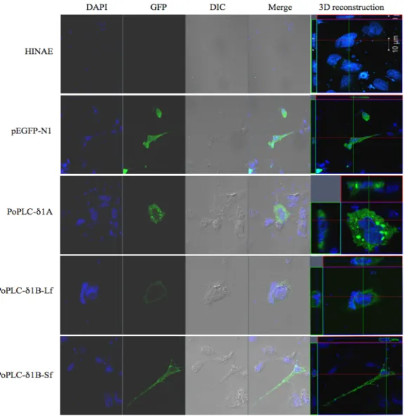
Fig. 2. Cellular characterization of PoPLC-δ1A, PoPLC-δ1B (Lf) and PoPLC-δ1B (Sf). Transfection with GFP-tagged PoPLC-δ1A, PoPLC-δ1B (Lf) and PoPLC-δ1B (Sf) isoforms in HIANE cell and viewed using an LSM700 laser scanning confocal microscope (Carl Zeiss Micro Imaging).
PoPLC-δ1A is main role of cell localization and translocation in fish
To understand the cellular localization and translocation of fish PLC-δ1 isozymes, GFP fused PoPLC-δ1s expression vectors were constructed to study the cellular localization.
As shown in Fig. 2, PoPLC-δ1A was observed as scattered patches and localized in cell organelles when PoPLC-δ1B (Lf) and PoPLC-δ1B (Sf) were localized at the plasma membrane.
To study changes in the cellular localization and trans- location of fish PLC-δ1s in the occurrence of calcium signals, we used treatment with ionomycin and thapsigargin into GFP fused PoPLC-δ1s, which transfected into HINAE cell lines (Fig. 3). After ionomycin treatment, we were able to
see the accumulation of expressed PoPLC-δ1A after treat- ment of serum-starved cells with drug (1 uM-5 uM).
However, PoPLC-δ1B (Lf) and PoPLC-δ1B (Sf) were not ac- cumulated in the nucleus. Treatment of thapsigargin, which is useful in examining the effects of increasing cytosolic cal- cium concentrations, also facilitates the nuclear import of PoPLC-δ1A. These results suggest that raising the extracel- lular Ca2+ concentration would facilitate the nuclear import PLC-δ1 in fish. In fact, a portion (10%-20%) of serum-starved MDCK cells accumulated PLC-δ1 within 2 min after the treatment of cells with ATP (100 mM), suggesting that the activation of purinergic receptors causes the nuclear import of PLC-δ1. Recent studies have indicated that the Ca2+ in- crease activates PLC isoforms and causes a decrease in
A
B
Fig. 3. Effect of ionomycin and thapsigargin after transfection with GFP-fused PoPLC-δ1A, PoPLC-δ1B (Lf) and PoPLC-δ1B (Sf) isoforms in HIANE cell. (A) PoPLC-δ1A transfected in HINAE cell and treatment two drugs for 20 min. (B) PoPLC-δ1B (Lf). (C) PoPLC-δ1B (Sf).
C
Fig. 3. Continued.
Phosphatidylinositol 4,5-bisphosphate [PtdIns(4,5)P2] levels at the plasma membrane, releasing mammalian PLC-δ1 from the plasma membrane to increase the levels in the cytoplasm. The efficacy of mammalian PLC-δ1 binding to nuclear import machineries then increases and more nuclear import complexes are formed and transported into the nu- cleus [12, 15].
Taken together, our results suggest that elevated intra- cellular calcium levels would facilitate the nuclear import of PLC-δ1. Among the three isoforms of olive flounder PLC- δ1s, only GFP-tagged PoPLC-δ1A which contains the intact functional NES and the NLS sequences in the EF-hand do- main and the C-terminus of the X domain and the XY-linker, was observed nucleocytoplasmic shuttling. That means PoPLC-δ1A is main role of cell localization and translocation in fish.
Acknowledgment
This work was supported by a grant from the National Institute of Fisheries Science (R2017064).
References
1. Caprini, M., Gomis, A., Cabedo, H., Planells-Cases, R., Belmonte, C., Viana, F. and Ferrer-Montiel, A. 2003. GAP43 stimulates inositol triphosphate-mediated calcium release in
response to hypotonicity. EMBO J. 22, 3004-3014.
2. Dingwall, C. and Laskey, R.A. 1991. Nuclear targeting se- quences-A consensus? Trends. Biochem. Sci. 16, 478-481.
3. Fukuda, M., Asano, S., Nakamura, T., Adachi, M., Yoshida, M., Yanagida, M. and Nishida, E. 1997. CRM1 is responsible for intracellular transport mediated by the nuclear export signal. Nature 390, 308-311.
4. Ichinohe, M., Nakamura, Y., Sai, K., Nakahara, M., Yamaguchi, H. and Fukami, K. 2007. Lack of phospholipase C delta1 induces skin inflammation. Biochem. Biophys. Res.
Commun. 356, 912-918.
5. Irino, Y., Cho, H., Nakamura, Y., Nakahara, M., Furutani, M., Suh, P. G., Takenawa, T. and Fukami, K. 2004.
Phospholipase C delta-type consists of three isozymes: bo- vine PLCdelta2 is a homologue of human/mouse PLCdelta4. Biochem. Biophys. Res. Commun. 320, 537-543.
6. Katan, M. 1998. Families of phosphoinositide-specific phos- pholipase C: structure and function. Biochim. Biophys. Acta.
1436, 5-17.
7. Kato, H., Fukami, K., Shibasaki, F., Homma, Y. and Takenawa, T. 1992. Enhancement of phospholipase C delta 1 activity in the aortas of spontaneously hypertensive rats.
J. Biol. Chem. 267, 6483-6487.
8. Kim, N. Y., Kim, M. S., Ahn, S. J., Seo, J. S., Bak, H. J., Kim, B. S., Jo, H. I., Jang, H. Y., Jo, H. S., Lee, H. H. and Chung, J. K. 2013. Functional analysis of duplicated genes and N- terminal splice variant of phospholipase C-δ1 in Paralichthys olivaceus. Comp. Biochem. Physiol. B. 165, 201-210.
9. Nakamura, Y., Fukami, K., Yu, H., Takenaka, K., Kataoka, Y., Shirakata, Y., Nishikawa, S., Hashimoto, K., Yoshida, N.
and Takenawa, T. 2003. Phospholipase C δ1 is required for skin stem cell lineage commitment. EMBO J. 22, 2981-2991.
초록:넙치 3가지 타입 인지질가수분해효소(PLC-δ1)의 세포 내 위치 및 이동
김나영1․김무상2․정승희1․김명석1․조미영1․정준기2․안상중3*
(1국립수산과학원, 2부경대학교 수산생명의학과, 3해양수산과학기술진흥원)
본 연구의 목적은 넙치 인지질가수분해효소(PLC- δ1) 3가지 타입의 세포내 특성을 규명하고자 하였다. 일반적 으로 인지질가수분해효소(PLC)의 신호전달경로는 핵, 세포막, 세포질에 분포한다고 알려져 있으나, 핵내 위치 메 커니즘은 여전히 불분명하다. PoPLC-δ1A, PoPLC-δ1B (Sf)과 PoPLC-δ1B (Lf)의 3타입의 유전자들은 각각 핵위치 신호(NLS)와 핵방출서열(NES)을 포함하고 있다. 본 연구에서는, 넙치 3가지 타입 인지질가수분해효소(PLC-δ1)의 세포내 위치이동 메커니즘 분석을 위해 GFP 벡터에 유전자를 삽입하여 ionomycin과 thasogargin처리 후 세포 위치와 이동양상을 공초점 레이저 주사현미경으로 관찰하였다. PoPLC-δ1A는 PoPLC-δ1B (Lf)와 PoPLC-δ1B (Sf) 가 원형질막에 국한되어 분포할때 세포질과 세포막보다 세포 소기관에 분포되어 있었다. PoPLC-δ1B (Lf) 및 PoPLC-δ1 (Sf)이 핵 세포질내 이동양상을 보이지 않을 때, PoPLC-δ1A은 ionomycin과 thapsigargin 처리에 의해 핵 내에 축적되는 양상을 나타냈다. 이런 결과는 손상되지 않은 기능적 NES 서열을 포함하는 PoPLC-δ1A가 어류 에서 핵 세포질 내 왕복 및 이동의 주된 역할을 한다는 것을 보여주고 있다.
10. Nakamura, Y., Hamada, Y., Fujiwara, T., Enomoto, H., Hiroe, T., Tanaka, S., Nose, M., Nakahara, M., Yoshida, N., Takenawa, T. and Fukami, K. 2005. Phospholipase C-delta1 and -delta3 are essential in the trophoblast for placental development. Mol. Cell. Biol. 25, 10979-10988.
11. Okada, M., Fujii, M., Yamaga, M., Sugimoto, H., Sadano, H., Osumi, T., Kamata, H., Hirata, H. and Yagisawa, H.
2002. Carboxyl-terminal basic amino acids in the X domain are essential for the nuclear import of phospholipase C δ1.
Genes Cells 7, 985-996.
12. Okada, M., Ishimoto, T., Naito, Y., Hirata, H. and Yagisawa, H. 2005. Phospholipase Cδ1 associates with importin β1 and translocates into the nucleus in a Ca2+-dependent manner.
FEBS Lett. 579, 4949-4954.
13. Rhee, S. G. 2001. Regulation of phosphoinositide-specific phospholipase C. Annu. Rev. Biochem. 70, 281-312.
14. Shimohama, S., Homma, Y., Suenaga, T., Fujimoto, S., Taniguchi, T., Araki, W., Yamaoka, Y., Takenawa, T. and Kimura, J. 1991. Aberrant accumulation of phospholipase C-δ in Alzheimer brains. Am. J. Pathol. 139, 737-742.
15. Stallings, J. D., Tall, E. G., Pentyala, S. and Rebecchi, M.
J. 2005. Nuclear translocation of phospholipase C-delta1 is linked to the cell cycle and nuclear phosphatidylinositol 4,5-bisphosphate. J. Biol. Chem. 280, 22060-22069.
16. Stallings, J. D., Zeng, Y. X., Narvaez, F. and Rebecchi, M.
J. 2008. Phospholipase C-delta1 is linked to proliferation, DNA synthesis and cyclin E levels. J. Biol. Chem. 283, 13992- 14001.
17. Stewart, A. J., Mukherjee, J., Roberts, S. J., Lester, D. and Farquharson, C. 2005. Identification of a novel class of mam- malian phosphoinositol-specific phospholipase C enzymes.
Int. J. Mol. Med. 15, 117-121.
18. Yagisawa, H., Okada, M., Naito, Y., Sasaki, K., Yamaga, M.
and Fujii, M. 2006. Coordinated intracellular translocation of phosphoinositide-specific phospholipase C-δ with the cell cycle. Biochim. Biophys. Acta. 1761, 522-534.
19. Yamaga, M., Fujii, M., Kamata, H., Hirata, H. and Yagisawa, H. 1999. Phospholipase C- δ 1 contains a functional nuclear export signal sequence. J. Biol. Chem. 274, 28537-28541.