Effects of Ethanol Extract of Sargassum horneri on Adipocyte Differentiation and Adipogenesis in 3T3-L1 Preadipocytes
Da Hye Kwon1, Yung Hyun Choi1,2,3, Byung Woo Kim3,4 and Hye Jin Hwang3,5*
1Anti-aging Research Center, Dong-eui University, Busan 614-714, Korea
2Department of Oriental Medicine, Dong-eui University, Busan 614-052, Korea
3Blue Bio Industry RIC., Dong-eui University, Busan 614-714, Korea
4Division of Applied Bioengineering, Dong-eui University, Busan 614-714, Korea
5Department of Food and Nutrition, Dong-eui University, Busan 614-714, Korea
Received October 11, 2018 /Revised November 9, 2018 /Accepted November 14, 2018
Sargassum horneri (Turner) C. Agardh is a marine brown algae widely distributed in the North Pacific Ocean. It is known for its anti-inflammatory and anti-atopic effects. In this study, we determined the effects of ethanol extract of Sargassum horneri (Turner) C. Agardh (EESH) on anti-obesity activities in 3T3-L1 preadipocytes. Our results indicated that treatment with EESH decreased the differentiation of 3T3-L1 preadipocytes in a dose-dependent manner, as confirmed by a decrease in lipid droplet content observed by oil red O staining. The concentrations of cellular triglycerides were also reduced in 3T3-L1 cells after treatment with EESH. Triglyceride content was inhibited by 13%, 16%, and 23% after treatment with 250, 500, and 1,000 μg/ml of EESH in 3T3-L1 cells, respectively. Western blotting anal- ysis showed that EESH suppressed adipogenic transcription factor expression in a dose dependent manner. Specifically, it suppressed cytidine-cytidine-adinosine-adenosine-thymidine (CCAAT) /enhancer binding proteins (C/EBP)α, C/EBPβ and peroxisome proliferator-activated receptor (PPAR)γ. This in- dicated that EESH could control the expression of adipogenic transcription factors and inhibit the dif- ferentiation of adipocytes. Taken together, these findings demonstrated that EESH showed anti-obesity effects and could have potential uses in the field of nutraceuticals.
Key words : Adipogenic transcription factor, obesity, Sargassum horneri, triglyceride, 3T3-L1
*Corresponding author
*Tel : +82-51-890-1594, Fax : +82-51-890-2646
*E-mail : [email protected]
This is an Open-Access article distributed under the terms of the Creative Commons Attribution Non-Commercial License (http://creativecommons.org/licenses/by-nc/3.0) which permits unrestricted non-commercial use, distribution, and reproduction in any medium, provided the original work is properly cited.
Journal of Life Science 2019 Vol. 29. No. 2. 209~214 DOI : https://doi.org/10.5352/JLS.2019.29.2.209
서 론
비만은 에너지 섭취와 소비의 불균형으로 체내에 지방이 과다하게 축적되어 생긴 대사성 질환으로, 이는 고혈압, 고지 혈증, 동맥경화 등 각종 대사성 질환들을 유발시킬 수 있기 때문에 그 위험성이 많이 강조되고 있다[7, 14]. 유전적, 환경적 요인도 비만의 원인으로 작용되지만, 식습관, 생활습관 등이 가장 큰 원인으로써 작용된다[18, 19]. 이를 치료하기 위한 방 법으로 adipogenesis의 발현과 지방전구세포의 분화를 억제 하여 triglyceride (TG)의 축적을 억제하는 것이 잘 알려져 있 다[1].
지방전구세포의 분화는 confluence, hormonal induction, mitotic clonal expanssion, growth arrest, terminal differ- entiation의 5단계로 이루어진다. 이때 adipogenic transcrip-
tion factor가 관여하게 되어 분화단계를 조절하게 된다[6]. 세 포분열 물질과 insulin, dexamethasone, 3-isobutyl-1-methyl- xanthine (IBMX)와 같은 hormonal cocktail 등의 자극에 의해 분화가 시작되면 다양한 인자들에 의해 CCAAT/enhancer binding proteins (C/EBP) β와 C/EBPδ의 발현이 증가되게 된다[2, 22]. 이들에 의해 일시적이고 빠르게 세포분열과 함께 세포수를 증가시키게 되고, C/EBPα와 peroxisomeprolifer- ator-activated receptor γ (PPARγ)의 발현을 조절하게 된다.
Adipogenesis의 master regulator라고도 불리는 C/EBPα와 PPARγ는 서로 상호작용으로 발현이 증가되어 분화를 촉진하 고, adiponectin, glucose transporter type 4 (GLUT4), fatty acid synthase 등에 의해 terminal marker의 발현을 증가시켜 둥근 지방세포의 모양으로 분화를 완성시키게 된다[15, 16].
본 연구에서 사용된 물질은 괭생이모자반(Sargassum hor- neri (Turner) C. Agardh)으로 생물학적으로 식물계 갈조식물문 갈조강 모자반목 모자반과로 분류되고, 북태평양 서안에 주로 분포하고 있으며 조간대 하부에서 자란다. 현재 아토피 억제 효과[13], 항염증효과[10], 중금속 흡착 효과[17]에 대해 보고가 되어 있으며, 스크리닝 실험을 통해 항산화[8, 11], 지방세포 생성 억제 효과[11], 암세포 증식 억제 효과[5], 항 혈액 응고 효과[12] 등이 보고되어 있다. 그러나 그 기전에 대한 연구는
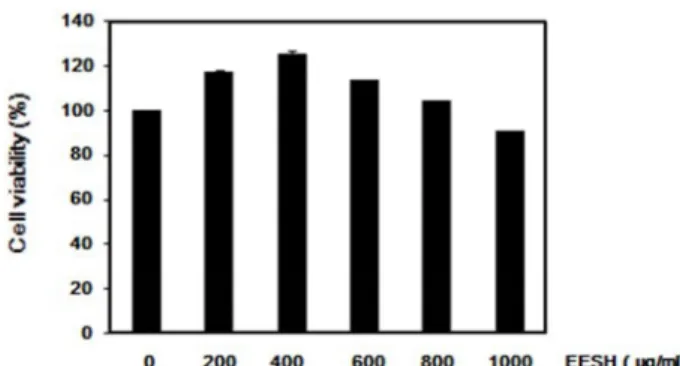
Fig. 1. Effects of EESH on the cell viability in 3T3-L1 preadipo- cytes. Cultured 3T3-L1 preadipocytes were treated with concentrations of EESH for 72 hr, and cell viability was determined by MTT assay. Data were expressed as per- centage of control. Each value indicates the mean ± S.D.
and is representative of results obtained from three in- dependent experiments.
Oil red O staining, triglyceride content와 adipogenesis의 발 현에 대해 측정함으로써 괭생이모자반 에탄올 추출물의 항비 만 효과에 대해 알아보고, 기능성 소재로서의 활용 가능성에 대해 검증해보고자 하였다.
재료 및 방법
시료
본 연구에 사용된 70% 괭생이모자반 에탄올 추출물(Etha- nol Extract of Sargassum horneri, EESH)은 제주유용생물자원 추출물은행(Jeju Bio-Resource Extract Bank, Jeju, Korea)에서 구입하여 사용하였다.
실험재료
3T3-L1 preadipocyte를 배양하기 위해 사용된 Dulbecco's Modified Eagle's Medium (DMEM), Bovine Calf serum (BCS), Fetal Bovine serum (FBS)은 Welgene Inc. (Gyeongsan, Korea)에서 구입하였으며, Penicillin-Streptomycin solution 은 Hyclone Inc. (Utah, USA)에서 구입하여 사용하였다. 분화 를 유도하기 위해 사용된 3-isobutyl-1-methylxanthine (IBMX), dexamethasone, insulin과 지방을 염색시키기 위해 사용된 Oil Red O 시약은 Sigma-Aldrich Chemical Co. (St.
Luis, MO, USA)에서 구입하여 사용하였다. 또한 western blot assay를 위해 1차 항체로 사용된 C/ EBPα, C/EBPβ, PPARγ 및 Actin과 2차 항체로 사용된 Rabbit, Goat는 Santa Cruz Biotechnology Inc. (Santa Cruz, CA, USA) 및 Cell signaling Technology (Beverly, MA, USA)에서 구입하여 사용하였다.
세포배양
실험에 사용된 3T3-L1 preadipocyte (Mouse embryonic fi- broblast cell)는 한국 세포주은행에서 분양받았으며, 90%
Dulbecco's Modified Eagle's Medium (DMEM)에 10% Bovine Calf Serum (BCS)과 1% Penicillin-Streptomycin solution을 혼합한 배지를 사용하여 37℃, 5% CO2 조건에서 48시간 배양 하였다.
MTT assay
세포 배양용 6 well plate에 3T3-L1 preadipocyte를 con- fluent 상태까지 배양하였으며, EESH를 농도별로 처리하였다.
72시간 후에 배지를 제거하고, 0.5 mg/ml 농도의 thiazolyl blue tetrazolium bromide (MTT, AMRESCO) 시약을 well당 2 ml 씩 분주하여 37℃, 5% CO2 incubator에서 2시간 더 배양 하였다. 모든 시약을 제거하고 DMSO (AMRESCO)를 넣어 well에 생성된 formazan을 모두 녹였으며, 96 well plate에 200 μl 씩 옮겨 540 nm에서 흡광도를 측정하였다. 각 세포에 대한
분화유도
3T3-L1 preadipocyte를 분화유도 하기 위해 10% BCS가 함 유된 DMEM 배지로 세포 배양용 plate에 배양한 후, confluent 상태가 되면 10% FBS가 함유된 배지로 바꿔 배양해주었다.
48시간 뒤에 0.5 mM 3-isobutyl-1-methylxanthine (IBMX), 1 μM dexamethasone과 5 μg/ml insulin이 함유된 배지로 교환 하여 2일간 지방전구세포의 분화를 유도하였으며, 이때 EESH도 같이 처리 하였다 (day 0). 2일마다 5 μg/ml insulin이 함유된 배지로 교환하여 3T3-L1 preadipocyte의 분화를 촉진하였다 (day 8).
Oil Red O staining & Triglyceride 측정
3T3-L1 preadipocyte에서 EESH가 지방세포 분화 억제에 효과가 있는지 확인하기 위해 Oil Red O staining을 실시하였 으며, 이를 정량하여 triglyceride content (%)로 나타내었다.
분화가 완료되면 배지를 제거하고, 1XPBS로 세척 한 다음 3.7% formalin 용액을 넣고 5~10분간 고정해 주었다. 이 후 60% isopropyl alcohol 용액을 넣고 세척한 다음 20분간 Oil Red O 용액을 넣고 염색해주었다. 염색이 완료되면 Oil Red O 시약을 모두 제거하고, 1XPBS와 60% isopropyl alcohol로 세척한 다음, 염색된 세포를 위상차 현미경으로 관찰하였다.
Triglyceride content는 100% isopropyl alcohol로 지방을 모두 용해시킨 후 96 well plate에 200 µl씩 넣고 500 nm에서 흡광도 를 측정하였으며, 대조군 흡광도 값에 대한 백분율로 나타내 었다.
Western blot assay
EESH가 adipogenic transcription factor의 발현에 어떠한
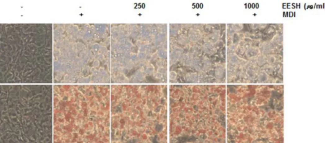
Fig. 2. Effects of EESH on the phase contrast microscopic morphological changes of differentiated 3T3-L1 preadipocyte. 3T3-L1 preadipocyte's differentiation was initiated with MDI (0.5 mM IBMX, 1 μM dexamethasone and 5 μg/ml insulin) and FBS medium in the absence or presence of EESH (day 2). And then every 2 days, medium was changed with 5 μg/ml insulin and FBS medium for maintain differentiation. After 8 day, differentiated 3T3-L1 adipocyte were visualized by phase contrast microscope (Magnification, X200).
Fig. 3. Effects of EESH on the lipid droplet accumulation in- hibition of differentiated 3T3-L1 preadipocyte. 3T3-L1 preadipocyte's differentiation was initiated with MDI (0.5 mM IBMX, 1 μM dexamethasone and 5 μg/ml in- sulin) and FBS medium in the absence or presence of EESH (day 2). Every 2 days, medium was changed with 5 μg/ml insulin and FBS medium for maintain differ- entiation. After 8 day, differentiated 3T3-L1 adipocyte were gauge triglyceride contents for ELISA reader. Data were expressed as percentage of positive control. Each value indicates the mean ± S.D. and is representative of results obtained from three independent experiments.
영향을 미치는지 알아보기 위해 western blot assay를 실시하 였다. 분화가 완료된 세포들을 harvest 한 후, lysis buffer를 첨가하여 4℃에서 1시간 반응 시켰으며, 14,000 rpm에서 30 분 원심 분리하여 상층액에 있는 단백질을 분리하였다. Bio- Rad 단백질 정량 시약을 통해 단백질의 농도를 정량하였고, 그 값에 따라 sample, lysis buffer (Cell signaling), 2X Laemmli sample buffer (Bio-rad)와 2-mercaptoethanol (Sigma)을 첨가 하였다. 동량의 sample을 sodium dodecyl sulphate (SDS)- polyacrylamide gel을 이용하여 전기영동으로 분리하였고, PVDF membrane (Bio-rad)에 전이시켰다. 5% skim milk (BD) 를 처리하여 비특이적인 단백질 반응을 막았으며, 1차 항체를 4℃에서 over-night 시킨 후 그에 맞는 2차 항체를 처리하여 상온에서 1시간 반응 시켰다. 특정 단백질의 발현 양상은 Enhanced Chemiluminoesence (ECL) solution (Thermo sci- entific)을 사용하여 Alpha innotech 기기를 통해 분석하였다.
통계학적인 분석
모든 실험 결과는 Statistical Package for the Social Sciences (SPSS) 통계 프로그램을 이용하여 평균(mean)±표준편차(S.D) 로 나타내었다. 각 실험 결과에 대한 유의성은 Student's t-test 와 Duncan's multiple range test를 이용하여 p<0.05 수준에서 검증하였다.
결과 및 고찰
EESH가 3T3-L1 preadipocyte 세포 생존율에 미치는 영향 3T3-L1 지방전구세포에서 EESH의 세포독성 유무를 확인 하고, 실험에 사용할 농도를 결정하기 위해 MTT assay를 실시 하였다. 200~1,000 μg/ml 농도까지 EESH를 처리한 후 72시간
뒤에 세포 독성을 분석한 결과 800 μg/ml까지의 농도에서는 control보다 더 높은 세포 생존율을 보였다(Fig. 1). Choi [3]의 연구에서도 3T3-L1 preadipocyte에서 갈조류인 톳의 분획물 의 세포생존율을 비교한 결과, 부탄올, 에탄올, 디클로메탄 분 획물의 세포생존율이 증가하는 결과를 보고한 바 있다.
Fig. 1과 같이 최고농도인 1,000 μg/ml 농도에서는 세포생 존율이 감소하여 90%의 세포 생존율을 확인 할 수 있었다.
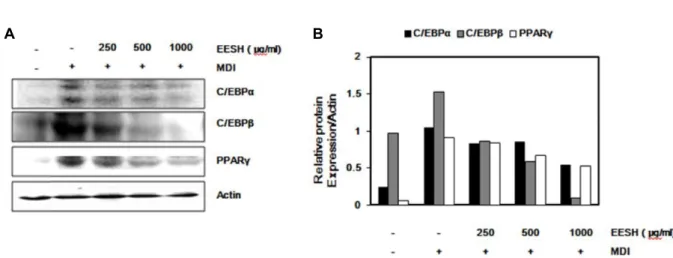
Fig. 4. Effects of EESH on the levels of adipogenic transcription factors protein expression by Western blot analysis in differentiated 3T3-L1 preadipocyte. (A) 3T3-L1 preadipocyte's differentiation was initiated with MDI (0.5 mM IBMX, 1 μM dexamethasone and 5 μg/ml insulin) and FBS medium in the absence or presence of EESH (day 2). And then every 2 days, medium was changed with 5 μg/ml insulin and FBS medium for maintain differentiation. And then harvest cell lysates, cellular proteins were separated by SDS-polyacrylamide gels and transferred onto PVDF membranes. The membranes were attach with the indicated antibodies. Proteins were visualized using an ECL detection system. (B) adipogenic transcription factor's expression was quantified with Image J software and appeared radio. Actin was used as control.
따라서 본 실험에서는 세포 독성을 보이지 않은 1,000 μg/ml 농도까지 실험조건으로 설정하여 비만 실험을 진행하였다.
EESH가 지방세포 분화 억제 및 triglyceride 생성 억제에 미치는 영향
3T3-L1 지방전구세포에서 EESH가 지방세포 분화 억제에 미치는 영향을 확인하기 위해 Oil Red O 시약으로 분화 시 나타나는 lipid droplet을 염색하였으며, 위상차 현미경으로 관찰하여 나타내었다. Fig. 2에 나타낸 바와 같이 분화유도제 를 처리하지 않은 군에 비해 분화유도제 처리가 지방구를 활 발하게 형성하였으며, EESH를 처리하였을 때는 lipid droplet 이 농도 의존적으로 감소됨을 관찰 할 수 있었다. 또한 이를 정량하여 triglyceride content (%)로 나타낸 결과(Fig. 3), EESH 처리가 분화유도제만 처리한 대조군에 비해 250, 500, 1,000 μg/ml 농도에서 13%, 16%, 23%의 triglyceride 생성 억 제 효과를 확인 할 수 있었다. 이는 Kim [9]의 연구에서 바위수 염 메탄올 추출물이 100, 300, 500 μg/ml 농도에서 triglyceride contents가 35%, 42%, 76% 억제하는 효과에 비해 낮은 수준으 로 관찰되었다. 그러나 Kim [11]의 모자반 3종류 에탄올 추출 물을 이용하여 지방생성 억제효과를 비교 검토하는 연구에서 괭생이모자반 99.9% 에탄올 추출물 1 mg/ml가 MDI 처리한 군에 비해 12% 감소한 보고에 비해 높은 수준으로 관찰되었 다. 따라서 EESH는 Oil Red O staining과 triglyceride content 를 통해 3T3-L1 지방전구세포에서 지방세포로 분화되는 과정 을 억제하는데 효과가 있음을 알 수 있었다.
EESH가 adipogenic transcription factor의 발현에 미 치는 영향
EESH의 지방세포 분화억제에 대한 효과가 adipogenic transcription factor의 발현에 어떠한 영향을 미치는지 알아보 기 위하여 Western blot assay를 실시하였으며(Fig. 4A), 이를 정량하여 비율로 나타내었다(Fig. 4B). MDI를 처리하여 분화 를 유도하였을 때, adipogenic transcription factor인 C/EBPα, C/EBPβ, PPARγ의 발현이 증가하였으나, EESH를 250, 500, 1,000 μg/ml 농도로 처리하였을 때는 농도 의존적으로 전사인 자들의 발현이 감소됨을 확인 할 수 있었다. 3T3-L1 전지방세 포가 지방 세포로 분화되는 과정에서 초기분화는 C/EBPβ와 C/EBPδ를 중심으로 조절되고, 후기분화는 PPARγ와 C/EBP α를 중심으로 조절된다. 세포분열 유도물질(mitogen)의 자극 으로 초기 분화가 시작되면 여러 인자들에 의해 C/EBPβ와 C/EBPδ의 발현은 상승되고[2, 21] 이는 이어서 PPARγ와 C/EBPα의 발현을 조절한다[19-21]. Kim [9]과 Choi [4]의 연 구에서도 MDI로 유도된 3T3-L1 cell에서 갈조류인 바위수염 과 잎파래 메탄올 추출물의 처리가 C/EBPα, C/EBPβ, PPARγ 의 발현을 농도 의존적으로 감소시키는 결과를 보고하였다.
이러한 결과들을 통해 괭생이모자반 에탄올 추출물은 adi- pogenic transcription factor의 발현을 억제하여 지방세포의 분화를 억제하였으며, 지방구 형성과 triglycerde의 생성을 감 소하여 항비만 효과를 나타낸다는 것을 알 수 있었다.
References
1. Chen, H. C. and Farese, R. V. 2005. Inhibition of triglyceride synthesis as a treatment strategy for obesity : lessons from DGAT1-deficient mice. Arterioscle. Thromb. Vasc. Biol. 25, 482-486.
2. Chen, Z., Torrens, J. I., Anand, A., Spiegelman, B. M. and
Friedman, J. M. 2005. Krox20 stimulates adipogenesis via C/EBPbeta-dependent and -independent mechanisms. Cell Metab. 1, 93-106.
3. Choi, E. O., Kim, H. S., Han, M. H., Choi, Y. H., Kim, B.
W., Hwang, J. A. and Hwang, H. J. 2012. Effects of Hizikia fusiforme extracts on adipocyte differentiation and adipo- genesis in 3T3-L1 preadpocytes. J. Life Sci. 22, 1399-1406.
4. Choi, E. O., Kim, H. S., Han, M. H., Park, C., Choi, Y. H.
and Hwang, H. J. 2012. Anti-obesity activities of methyl al- cohol extract of Enteromorpha linza in 3T3-L1 preadipocytes.
Cancer Prev. Res. 18, 80-87.
5. Choi, H. J., Kil, J. H., Bak, S. S., Kong, C. S., Park, K. Y., Sep, Y. W. and Lim, S. Y. 2006. Inhibitory effects of solvent extracts from seven brown algae on mutagenicity and growth of human cancer cells. J. Life Sci. 16, 1080-1086.
6. Fajas, L., Fruchart, J. C. and Auwerx, J. 1998. Transcriptional control of adipogenesis. Curr. Opinion. Cell Biol. 10, 165-173.
7. Haslam, D. W. and James, W. P. 2005. Obesity. Lancet 366, 1197-1209.
8. Kim, B. M., Jun, J. Y., Park, Y. B. and Jeong, I. H. 2006.
Antioxidative activity of methanolic extracts from seaweeds.
J. Kor. Soc. Food. Sci. Nutr. 35, 1097-1101.
9. Kim, H. S., Kwon, D. H., Cheon, J. M., Choi, E. O., Kim, J. H., Han, M. H., Choi, Y. H., Kim, B. W. and Hwang, H.
J. 2015. Effects of Myelophycus Simplex Papenfuss methanol extract on adipocyte differentiation and adipogenesis in 3T3-L1 preadipocytes. J. Life Sci. 25, 62-67.
10. Kim, M. E., Jung, Y. C., Jung, I. A., Lee, H. W., Youn, H.
Y. and Lee, J. S. 2014. Anti-inflammatory effects of ethanolic extract from Sargassum horneri (Turner) C. Agardh on lip- opolysaccharide-stimulated macrophage activation via NF- kB pathway regulation. Immunol. Invest. 44, 137-183.
11. Yu, K. H., Ahn, B. N., Lee, S. H., Kim, M. H., Mun, O.
J., Karadeniz, F., Kong, C. S., Kim, Y. Y. and Kwon, M.
S. 2014. Sargassum sp. attenuates oxidative stress and sup- presses lipid accumulation in vitro. J. Life Sci. 24, 273-283.
12. Kim, Y. M., Kim, D. S. and Choi, T. S. 2004. Anticoagulant activities of brown seaweed extracts in korea. Kor. J. Food
Sci. Technol. 36, 1008-1013.
13. Lee, C. J. 2010. Inhibitory Effects of Sargassum horneri extracts on atopic disease. M.D. dissertation, Pukyong National University. Busan, Korea.
14. Leung, W. Y., Thomas, G. N., Chan, J. C. and Tomlinson, B. 2003. Weight management and current options in phar- macotherapy : orlistat and sibutramine. Clin. Ther. 25, 58-80.
15. Morrison, R. F. and Farmer, S. R. 2000. Hormonal signaling and transcriptional control of adipocyte differentiation.
Nutrition 130, 3116S-3121S.
16. Rosen, E. D. and MacDougald, O. A. 2006. Adipocyte differ- entiation from the inside out. Nat. Rev. Mol. Cell. Biol. 7, 885-896.
17. Suh, K. H., Ahn, K. H. and Cho, M. C. 1999. Biosorption of Pb1+ and Cr3+ by using Sargassum horneri. J. Kor. Environ.
Sci. Soc. 8, 387-391.
18. Wellman, N. S. and Friedberg, B. 2002. Causes and con- sequences of adult obesity : health, social and economic im- pacts in the United States. Asia Pac. J. Clin. Nutr. 11, S705- 709.
19. Wu, Z., Bucher, N. L. and Farmer, S. R. 1996. Induction of peroxisome proliferator-activated receptor gamma during the conversion of 3T3 fibroblasts into adipocytes is mediated by C/EBPbeta, C/EBPdelta, and glucocorticoids. Mol. Cell Biol. 16, 4128-4136.
20. Wu, Z., Xie, Y., Bucher, N. L. and Farmer, S. R. 1995.
Conditional ectopic expression of C/EBP beta in NIH-3T3 cells induces PPAR gamma and stimulates adipogenesis.
Genes Dev. 9, 2350-2363.
21. Yeh, W. C., Cao, Z., Classon, M. and Mcknight, S. L. 1995.
Cascade regulation of terminal adipocyte differentiation by three members of the C/EBP family of leucine zipper proteins. Genes Dev. 9, 168-181.
22. Zhang, J. W., Klemm, D. J., Vinson, C. and Lane, M. D. 2004.
Role of CREB in transcriptional regulation of CCAAT/en- hancer-binding protein beta gene during adipogenesis. J.
Biol. Chem. 279, 4471-4478.
초록:괭생이모자반 에탄올 추출물이 3T3-L1 지방전구세포의 분화 및 adipogenesis에 미치는 영향
권다혜1․최영현1,2,3․김병우3,4․황혜진3,5*
(1동의대학교 항노화 연구소, 2동의대학교 한의학과, 3블루바이오 소재개발 및 실용화지원센터, 4동의대학교 바이오
응용공학부, 5동의대학교 식품영양학과)
본 연구에서는 괭생이모자반 에탄올 추출물의 항비만 효과에 대해 알아보고자 3T3-L1 preadipocyte에서 Oil Red O staining과 triglyceride content, adipogenesis의 발현에 대해 조사하였다. MTT assay를 통해 괭생이모자반 에탄올 추출물의 세포 독성을 확인 해 본 결과 200~1,000 μg/ml 농도에서 90% 이상의 세포 생존율을 확인 할 수 있었다. 또한 insulin, dexamethasone과 IBMX 복합물을 처리하여 분화를 유도하였을 때는 분화유도제를 처리 하지 않은 군에 비해 활발하게 지방구를 형성하였으나, 괭생이모자반 에탄올 추출물을 250, 500, 1,000 μg/ml 농 도로 처리하였을 때는 농도 의존적으로 지방구 형성을 억제하였다. 이를 정량하여 triglyceride content (%)로 나타 낸 결과 농도별로 13%, 16%, 23% 억제 효과를 나타내었다. 또한 Western blot assay를 통해 adipogenic tran- scription factor의 발현양상을 비교해 본 결과, 분화유도제 처리에 의해 C/EBPα, C/EBPβ와 PPARγ의 발현이 증 가하였으나, 괭생이모자반 에탄올 추출물을 처리하였을 때는 이러한 전사인자들의 발현이 농도 의존적으로 감소 됨을 확인 할 수 있었다. 이상의 결과를 통해 괭생이모자반 에탄올 추출물은 adipogenic transcription factor의 발현을 조절하여 지방세포의 분화를 억제하였으며, 지방구와 triglyceride 생성을 감소시켜 최종적으로 비만억제 효과를 나타낸다는 것을 알 수 있었다.