PREVENTION RESEARCH □ ORIGINAL ARTICLE □
371 책임저자:황혜진, 614-714, 부산시 부산진구 엄광로 995번지
동의대학교 생활과학대학 식품영양학과 Tel: 051-890-1594, Fax: 051-890-2646 E-mail: [email protected]
접수일:2011년 11월 22일, 1차 수정일:2011년 11월 29일, 2차 수정일:2011년 12월 5일, 게재승인일:2011년 12월 9일
Correspondence to:Hye-Jin Hwang
Department of Food and Nutrition, College of Human Ecology, Dongeui University, 995, Eomgwang-no, Busanjin-gu, Busan 614-714, Korea Tel: +82-51-890-1594, Fax: +82-51-890-2646
E-mail: [email protected]
3T3-L1 지방전구세포의 분화 및 Adipogenesis에 미치는 하수오 추출물의 영향
동의대학교 1생활과학대학 식품영양학과, 2한방식품연구소, 3한의학과,
4블루바이오소재개발센터, 5자연과학대학 생명응용학과, 6숭의여자대학 식품영양학과
최은옥1,2ㆍ한민호3ㆍ김향숙2ㆍ최성희1,4ㆍ최영현3,4ㆍ김병우4,5ㆍ김수연6ㆍ황혜진1,2,4
Effects of Cynanchi wilfordii Radix Extracts on Adipocyte Differentiation and Adipogenesis in 3T3-L1 Preadipocytes
Eun-Ok Choi1,2, Min-Ho Han3, Hyang Suk Kim2, Sung Hee Choi1,4, Yung-Hyun Choi3,.4, Byung-Woo Kim4,5, Soo-Yeon Kim6 and Hye-Jin Hwang1,2,4
1Department of Food and Nutrition, College of Human Ecology, 2Dongeui Food Research Institute, 3Departments of Oriental Medicine, 4Blue-Bio Industry Regional Innovation Center, 5Department of Life Science and Biotechnology,
College of Natural Science, Dongeui University, Busan 614-714, 6Department of Food and Nutrition, Soongeui Womens College, Seoul 100-751, Korea
The present study aimed to evaluate the effects of various extracts of Cynanchi wilfordii Radix on the effects of adipocyte differentiation and adipogenesis using 3T3-L1 preadipocyte cell line. For this study, the extracts of methanol of C. wilfordii (MECWR) was isolated, and then the extracts were fractionated into dichloromethane (CFCWR), ethyl acetate (EAFCWR), butanol (BFCWR) and water (WFCWR) partition layers. The extracts of C. wilfordii showed significant inhibitory activity on adipocyte differentiation in the 3T3-L1 preadipocytes without affecting cell viability as assessed by measuring fat accumulation using Oil Red O staining, and this effect was higher in EAFCWR than in other extracts.
In addition, the inhibitory effects of extracts of C. wilfordii were associated with modulation of adipogenic transcription factors such as peroxisome proliferator-activated receptorγ (PPARγ), CCAAT/enhancer binding proteinα (C/EBPα) and C/EBPβ in both translational and transcriptional levels. These results demonstrate that C. wilfordii extracts may be an ideal candidate for obesity relief to disturb adipocyte differentiation and that further studies will be needed for identification of active compounds. (Cancer Prev Res 16, 371-378, 2011)
Key Words: Cynanchi wilfordii radix, Adipocyte differentiation, Adipogenesis, PPARγ, C/EBP
서 론
지방세포(adipocyte)는 에너지 항상성 및 대사에서 매 우 중요한 역할을 한다.1) 지방세포 내에 과도한 지방의
축적은 당뇨병, 고혈압, 심혈관 질환 및 암을 포함한 만 성질병의 유발 원인으로 알려져 있다.2,3) 따라서 세포내 지방의 축적을 예방하거나 축적된 지방을 분해하도록 자극하는 방안에 대한 연구가 최근 많은 관심을 끌고 있 으며 항비만 기능성 신소재 발굴에 관한 연구가 전 세계
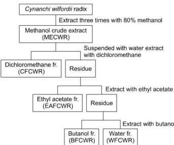
Fig. 1. Fractionation procedure of C. wilfordii Radix. Frac- tionations were separated by separatory funnel.
Table 1. Sequence of primers used for RT-PCR
Gene name Sequence
GAPDH Sense 5'-CGG-AGT-CAA-CGG-ATT-TGG-TCG-TAT-3'
Antisense 5'-AGC-CTT-CTC-CAT-GGT-GGT-GAA-GAC-3'
PPARγ Sense 5'-CGC-TGA-TGC-ACT-GCC-TAT-GA-3'
Antisense 5'-TGC-GAG-TGG-TCT-TCC-ATC-AC-3'
C/EBPα Sense 5'-GTG-TGC-ACG-TCT-ATG-CTA-AAC-CA-3'
Antisense 5'-GCC-GTT-AGT-GAA-GAG-TCT-CAG-TTT-G-3'
C/EBPβ Sense 5'-GTT-TCG-GGA-GTT-GAT-GCA-ATC-3'
Antisense 5'-AAC-AAC-CCC-GCA-GGA-ACA-T-3'
적으로 활발히 진행되고 있다.
3T3-L1 전지방세포(preadipocyte)가 지방세포로 분화되 는 과정은 CCAAT/enhancer binding protein에 속한 C/EBP β와 C/EBPδ를 중심으로 조절되는 초기 분화와 perox- isome proliferator activated receptor γ (PPARγ)와 C/EBPα 를 중심으로 조절되는 후기 분화로 나누어진다. 세포분 열 유도물질(mitogen)과 호르몬의 자극으로 초기 분화가 시작되면 다양한 인자들에 의해 C/EBPβ와 C/EBPδ의 발현은 상승된다.4,5) C/EBPβ와 C/EBPδ는 서로 협동적 으로 혹은 단독으로 PPARγ와 C/EBPα의 발현을 조절 한다.6~9) PPARγ와 C/EBPα는 adipogenesis를 조절하는 핵심 전사인자로서 분화의 후기에 높게 발현되어 adipo- nectin (ADIPOQ)과 glucose transporter (GLUT) 4 등을 포함 한 adipogenesis의 terminal marker의 발현을 유도한다.10~13) 전통 동약의학에서 자양, 강장 및 보혈 효능과 함께 면역증강 작용이 뛰어나 수명 연장에 도움을 줄 수 있는
것으로 알려진 하수오는14) 적하수오(Polygoni multiflori Radix)와 백하수오(Cynanchi wilfordii Radix)로 크게 분류된 다. 백하수오는 박주과리과(Asclepiadaceae)의 덩이뿌리로 한국이 원산지이며, 적하수오는 마디풀과(Polygonaceae) 의 덩이뿌리로써 중국이 원산지이다.15,16) 전통 약재로서 중국, 대만 및 일본에서는 주로 적하수오를 사용하고 있 지만, 국내에서는 백하수오가 쇠약, 빈혈, 조기백발, 신 경쇠약 등의 치료에 많이 사용되고 있다. 최근 하수오 추출물의 다양한 생리활성 효능에 대한 연구가 부분적 으로 시도된 바 있는데, 현재까지 보고된 하수오의 약리 작용으로는 면역 증강과 연계된 함염증 작용이 대표적
이며,17~20) 최근 암세포의 증식억제 효능도 있음이 보고
된 바 있다.21) 그러나 하수오 추출물의 생리활성에 관한 체계적인 연구는 매우 미흡한 실정이며, 특히 항비만 효 능에 대한 구체적인 자료는 거의 발표된 바가 없다.
본 연구에서는 3T3-L1 지방전구세포를 이용하여 다양 한 하수오 분획물에 의한 항비만 효과 및 그에 따른 분 자적 기전을 규명하기 위하여, insulin, dexamethasone 및 3-isobutyl-1-methylxanthine (IBMX) 등의 비만유도인자에 의하여 인위적으로 유발된 adipogenesis 과정에 이들 추출 물이 어떠한 영향을 미치는지를 조사하였고, 특히 지방 세포의 분화에 관여하는 PPARγ, C/EBPα, C/EBPβ 등 과 같은 adipogenic transcription factor 들의 발현에 미치는 다양한 하수오 추출물의 영향을 조사하였다.
재료 및 방법 1. 하수오 분획물의 제조
다양한 하수오 분획물을 얻기 위하여 건조된 백하수 오 1.5 kg을 80% 메탄올에 24시간 동안 침지시킨 후 환 류 냉각시키면서 80oC 수욕상에서 3시간씩 3회 반복 추 출한 후 이 추출액을 여지로 여과하고 감압 추출 농축장 치(EYELA, Japan)로 메탄올 추출물(extracts of methanol of
C. wilfordii, MECWR)을 얻은 후, 이를 다시 Fig. 1과 같이 분획하였다. 즉 메탄올 추출물을 증류수에 현탁하고, 분 액깔때기에서 dichloromethane (CFCWR), ethyl acetate (EAFCWR), butanol (BFCWR) 및 물(WFCWR)을 순차적으 로 계층 분획한 뒤 여과 후 회전 진공 농축기로 45oC에서 감압 농축하여 각각의 분획물을 얻었다.
2. 실험재료
mRNA 발현양 분석을 위하여 Bioneer (Taejeon, Korea)에 서 구입한 primer 염기서열은 Table 1에 나타내었으며, 단 백질 벌현 변화 분석을 위하여 사용된 PPARγ, C/EBPα, C/EBPβ 및 actin 항체는 Santa Cruz Biotechnology Inc.
(Santa Cruz, CA, USA) 및 Cell Signaling Technology (Beverly, MA, USA)에서 구입하였다. Immunoblotting을 위해 2차 항체로 사용된 peroxidase-labeled donkey anti-rabbit 및 per- oxidase- labeled sheep anti-mouse immunoglobulin은 Amer- sham Life Science Co. (Arlington Heights, IL, USA)에서 구입 하였다. 또한 3T3-L1 지방전구세포의 분화를 위하여 사 용된 insulin, dexamethasone 및 IBMX와 지방세포 내 trigly- ceride 생성을 확인하기 위하여 사용된 Oil Red O는 Sigma- Aldrich (St. Luis, MO, USA)에서 구입하였다.
3. 3T3-L1 지방전구세포의 배양
실험에 사용된 3T3-L1 지방전구세포는 American Type Culture Collection (Manassas, VA, USA)에서 구입하였으며, 10% bovine calf serum (BCS) 및 1% penicillin 및 streptomycin (Gibco BRL, Grand Island, NY, USA)이 함유된 Dulbecco’s Modified EaDCRT Media (DMEM, Gibco BRL)를 사용하여 37oC, 5% CO2 조건하에서 배양하였다. 세포수의 증식에 따른 과밀도 현상을 해소하기 위하여 성장배지의 교환 을 매 48시간마다 실시하여 적정수의 세포를 유지하였 다.
4. 3T3-L1 지방전구세포의 분화 유도
3T3-L1 지방전구세포를 세포 배양용 6 well plate에서 confluent 상태까지 배양한 후 10μg/ml insulin, 0.1 mM dexamethasone 및 0.5 mM 3-isobutyl-1-methylxanthin (IBMX) 가 포함된 분화 배지로 교환하여 2일간 배양하였으며, 그 후 매 2일마다 10μg/ml insulin이 포함된 배지로 교환 하였다. Adipogenesis의 진행과정에서 하수오 추출물의 영향을 확인하기 위해, 배지를 교환할 때마다 하수오 추 출물을 함께 처리하였다.
5. MTT assay에 의한 3T3-L1 지방전구세포의 증식 억제 조사
세포 배양용 6 well plate에 3T3-L1 지방전구세포를 분 주하여 confluent 상태까지 배양한 후 하수오 추출물을 배지에 희석하여 각 well 당 적정농도로 처리하였다. 72 시간 후 희석된 0.5 mg/ml 농도의 tetrazolium bromide salt (MTT, Amresco, Solon, OH, USA)를 200μl씩 분주하고 2시 간 동안 CO2 incubator에서 배양시킨 다음 배지와 MTT 시약을 깨끗하게 제거하고 DMSO를 1ml 씩 분주하여 well에 생성된 formazin을 모두 녹인 후 96 well plate에 200 μl씩 옮겨서 ELISA reader (Molecular Devices, Sunnyvale, CA, USA)로 540 nm에서 흡광도를 측정하였다. 각 세포 에 대한 독성은 대조군의 평균 흡광도 값을 구하여 평균 흡광도 값에 대한 백분율로 나타내었다.
6. Trypan blue assay에 의한 3T3-L1 지방전구세포 의 생존율의 측정
동일 조건에서 배양된 3T3-L1 지방전구세포를 2,000 rpm에서 5분간 원심분리하여 배지를 제거하고 phos- phate-buffered saline (PBS)를 각 well 당 1 ml을 첨가하여 세포를 부유시킨 다음 0.5% trypan blue solution (Gibco BRL)을 동량으로 첨가하여 2분 간 처리하였다. 처리된 세포을 hemocytometer에 적용한 후 도립 현미경을 이용 하여 살아있는 세포를 계수하였으며, 이에 따른 결과를 Microsoft EXCEL program을 사용하여 분석하였다.
7. 3T3-L1 지방전구세포의 형태의 관찰
하수오 추출물 처리에 의한 3T3-L1 지방전구세포의 분화 및 lipid droplet 생성 정도를 확인하기 위하여 세포 배양용 6 well plate에 3T3-L1 지방전구세포를 분주하여 confluent 상태까지 배양한 후 하수오 추출물을 적정농도 로 희석 처리하면서 분화를 유도하였다. 분화가 끝난 후 하수오 추출물 처리농도에 따른 분화 및 lipid droplet 생 성 정도를 도립 현미경을 이용하여 200배의 배율로 관찰 하였다.
8. Oil Red O 염색 및 정량
세포 내 lipid droplet 생성을 확인하기 위하여 Oil Red O 염색을 실시하기 위하여 준비된 3T3-L1 세포를 PBS로 세척한 후 3.7% formalin으로 10분간 고정하고 60% iso- propanol을 이용하여 세척한 다음 Oil Red O solution을 처 리하여 실온에서 20분 간 염색하였다. 염색 후 Oil Red O solution을 제거하고 증류수로 4회 세척한 다음 염색된
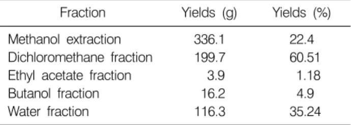
Table 2. Yields of methanol extracts and various solvent fractions of C. wilfordii Radix
Fraction Yields (g) Yields (%)
Methanol extraction 336.1 22.4
Dichloromethane fraction 199.7 60.51 Ethyl acetate fraction 3.9 1.18
Butanol fraction 16.2 4.9
Water fraction 116.3 35.24
세포를 위상차 현미경을 이용하여 관찰하였다. 또한 정 량적 분석을 위하여 100% isopropanol을 이용하여 지방을 추출한 후 96 well plate에 200μl씩 옮겨서 ELISA reader로 500 nm에서 흡광도를 측정하였고 대조군의 흡광도 값에 대한 백분율로 나타내었다.
9. Reverse transcriptase polymerase chain re- action (RT-PCR)에 의한 mRNA의 분석
준비된 3T3-L1 세포에 TRIzol reagent (Invitrogen Co., Carlsbad, CA, USA) 1 ml을 첨가하여 4oC에서 1시간 동안 반응시킨 후 200μl의 chloroform을 첨가하여 4oC에서 30 분 동안 반응시켰다. 14,000 rpm, 4oC에서 15분간 원심분 리시켜 400μl의 상층액을 분리하여 동량의 isopropanol을 첨가하여 4oC에서 30분 동안 반응시킨 후 14,000 rpm, 4oC에서 30분간 원심분리시켜 total RNA를 분리하였다.
분리된 RNA를 DEPC water를 이용하여 녹이고 정량한 후, SuperScriptⓇ III kit (Invitrogen Co.)를 사용하여 1차 cDNA를 제작하였였으며, 1μg의 cDNA를 Mastercycler gradient (EPPendorf, Hamburg, Germany)를 이용하여 역전 사 및 증폭하였다. 각 PCR 산물들의 양적 차이를 확인하 기 위하여 1×TAE buffer로 1% agarose gel을 만들고 well 당 각각의 primer에 해당하는 PCR 산물에 DNA gel load- ing solution을 섞어서 loading한 후 100V에서 전기영동을 하였다. 전기영동으로 DNA 분리가 끝난 gel을 ethidium bromide (EtBr)을 이용하여 염색한 후 UV하에서 확인하 였으며, house keeping 유전자인 glyceraldegyde-3-phosphate dehydrogenase (GAPDH)를 internal control로 사용하였다.
10. Western blot analysis에 의한 단백질 발현의 분석
동일한 조건에서 배양된 3T3-L1 세포에 적당량의 lysis buffer [25 mM Tris-Cl (pH 7.5), 250 mM NaCl, 5 mM EDTA, 1% NP-40, 1 mM phenymethylsulfonyl fluoride (PM- SF), 5 mM dithiothreitol (DTT)]를 첨가하여 4oC에서 1시간 동안 반응시킨 후, 14,000 rpm으로 30분간 원심분리하여 상층액에 있는 총 단백질을 분리하였다. 상층액의 단백 질 농도는 Bio-Rad 단백질 정량 시약(Bio-Rad, Hercules, CA, USA)과 그 사용방법에 따라 정량 한 다음 동량의 Laemmli sample buffer (Bio-Rad)를 섞어서 sample을 만들었 다. 동량의 sample을 sodium dodecyl sulphate (SDS)- poly- acrylamide gel을 이용하여 전기영동으로 분리한 후, nitro- cellulose membrane (Schleicher and Schuell, Keene, NG, USA) 으로 electroblotting에 의해 전이시켰다. 분리된 단백질이 전이된 nitrocellulose membrane을 5% skim milk를 처리하 여 비특이적인 단백질들에 대한 blocking을 실시하고 1차
항체를 처리하여 상온에서 2시간 이상 또는 4oC에서 over night 시킨 다음 PBS-T로 세척하고 처리된 1차 항체에 맞 는 2차 항체를 사용하여 상온에서 1시간 정도 반응시켰 다. 반응이 끝난 후 암실에서 Enhanced Chemiluminoe- sence (ECL) solution (Amersham Life Science Co.)을 적용시킨 다음 X-ray film에 감광시켜 특정 단백질의 발현 양을 분 석하였다.
11. Statistical analysis
모든 실험 결과는 Statistical Package for the Social Sciences (SPSS) 통계 프로그램을 이용하여 평균(mean)±표 준편차(S.D.)로 나타냈다. 각 실험군의 분석 항복별 통계 의 유의성은 Duncan's multiple range test를 이용하여 p<
0.05 수준에서 검증하였다.
결과 및 고찰 1. 하수오 분획물의 제조
하수오 분말로부터 methanol로 추출한 후 분획하여 분 획물을 제조한 결과를 Table 2에 나타냈다. 하수오 meth- anol 추출물(MECWR)의 경우, 1.5 kg으로부터 추출물 336.1 g을 획득하여 22.4%의 수율을 나타냈다. 하수오 methanol 추출물 330 g으로부터 분획물을 제조한 결과, ethyl acetate 분획물(EAFCWR)은 3.9 g을 획득하여 1.18%
의 수율로 가장 낮은 수율을 나타내었다. Dichloromethan 분획물(CFCWR)은 199.7 g을 획득하여 60.51%의 수율을 나타내었고, butanol 분획물(BFCWR)은 16.2 g을 획득하 여 4.9%의 수율을 나타냈다. 또한 물 분획물은 116.3 g을 획득하여 35.24%의 수율을 나타냈다. 따라서 분획물의 수율만 비교할 경우 dichloromethan 분획물이 가장 높은 수율을 나타냈다. 이로 인해 하수오의 성분은 수용성 성 분보다는 지용성 성분을 더 함유하고 있음을 확인할 수 있었다.
Fig. 2. Effects of C. wilfordii Radix extracts on the cell viability in 3T3-L1 mouse preadipocytes. Cells were treated with the indicated concentrations of various C. wilfordii Radix extracts for 72 h. Cell numbers were determined by hemocytometer counts of trypan blue excluding cells. Results are expressed as average of two separate experiments (MECWR: methanol extracts of C. wilfordii, CFCWR: dichloromethane fraction of C.
wilfordii, EAFCWR: ethyl acetate fraction of C. wilfordii, BFCWR:
butanol fraction of C. wilfordii, WFCWR: water fraction of C.
wilfordii).
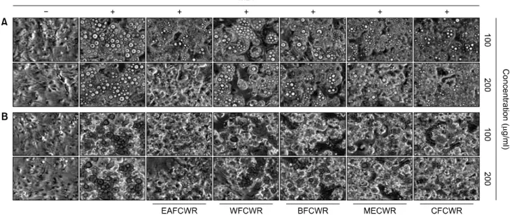
Fig. 3. Effects of C. wilfordii Radix extracts on lipid droplet accumulation of differentiated 3T3-L1 mouse preadipocytes. (A) Differentiation of confluent 3T3-L1 preadipocytes was initiated with MDI (0.5 M 3-isobutyl-1-methylxanthine, 1 mM dexamethasone and 10 mg/ml insulin) and maintained DMEM-5% FBS medium in the absence or presence of C. wilfordii Radix extracts. After day 8, differentiating 3T3-L1 cells were visualized by a light microscope. Magnification, ×200. (B) Cells grown under the same conditions as (A) were fixed and stained with Oil Red O to visualize lipid droplets by a light microscope. Magnification, ×200.
2. 3T3-L1 세포의 증식 및 생존에 미치는 하수오 추출 물의 영향
3T3-L1 세포의 분화 및 adipogenesis 실험의 조건 설정 을 위한 하수오 추출물의 세포독성 여부를 조사하기 위 해서 MTT assay를 실시하였다. MTT assay 결과에 의하면 100μg/ml 농도에서 다섯 가지 분획물 모두 90% 이상의 생존율을 보인 반면, 200μg/ml의 농도에서는 EAFCWR 분획물 처리군이 80% 이하의 생존율을 보였다. 하지만 200μg/ml의 농도에서 대조군에 비해 80% 이하의 생존 률을 보인 EAFCWR 분획물 처리군을 위상차 현미경으 로 관찰한 결과 세포의 형태적 손상이 관찰되지 않았다 (data not shown). 따라서 3T3-L1 지방전구세포의 생존율 을 다른 방법으로 알아보기 위하여 trypan blue assay를 실 시하였다. 이를 위하여 3T3-L1 지방전구세포에 다섯 가 지 분획물을 적정 농도로 72시간 처리한 후, 살아있는 세포의 수를 계수한 결과를 Fig. 2에 나타내었다. 결과에 서 알 수 있듯이 EAFCWR 분획물 처리군 외의 네 가지 분획물은 400μg/ml 농도까지 생존율의 감소가 완만하게 이루어지는 반면 EAFCWR 분획물 처리군은 300μg/ml의 농도에서부터 다른 분획물에 비해 생존율이 크게 감소하 여 400μg/ml 에서는 생존율이 매우 낮음을 확인할 수 있 었다. 이러한 결과로 하수오의 다섯 가지 분획물 처리군
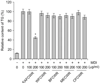
Fig. 4. Effects of C. wilfordii Radix extracts on triglyceride accumulation of differentiated 3T3-L1 mouse preadipocytes.
Triglyceride contents were determined by Oil Red O staining after treatment with C. wilfordii Radix extracts as described in Fig. 3. The rates of triglyceride contents were measured at λ
=500 nm wavelength by the ELISA reader. Data are expressed as the means±S.D. *p<0.05.
Fig. 5. Effects of C. wilfordii Radix extracts on the levels of adipogenic transcription factors mRNA and protein expression in differentiated 3T3-L1 mouse preadipocytes. Differentiation of confluent 3T3-L1 mouse preadipocytes was incubated with the absence or presence of C. wilfordii Radix extracts for 8 day after initiated with MDI. (A) Total RNAs were isolated and reverse-transcribed. The resulting cDNAs were subjected to PCR with the indicated primers and the reaction products were subjected to electrophoresis in 1% agarose gel and visualized by EtBr staining. GAPDH was used as an internal control. (B) Cells were lysed and cellular proteins were separated by SDS-polyacrylamide gels and transferred onto nitrocellulose membranes.
The membranes were probed with the indicated antibodies. Proteins were visualized using an ECL detection system. Actin was used as an internal control.
에서 생존율에 큰 영향을 미치지 않았던 100μg/ml 및
200μg/ml 처리군을 실험 조건으로 설정하였다. 3. 3T3-L1 지방전구세포의 lipid droplet 생성에 미치 는 하수오 추출물의 영향
다음은 3T3-L1 지방전구세포가 지방세포로의 분화과 정에 나타나는 lipid droplet 생성이 하수오 추출물 처리에 의하여 억제되는지의 여부를 조사하였다. 이를 위하여 Oil Red O 염색 전후로 구분하여 lipid droplet 생성 정도를 위상차 현미경으로 관찰하였다. Fig. 3에서 볼 수 있듯이 하수오 추출물을 처리하지 않고 분화를 유도하였을 경 우에 세포질 내 lipid droplet이 과다하게 축적되었음을 관 찰할 수 있었다. 그러나 하수오 추출물을 처리하였을 경 우, 특히 EAFCWR 처리군에서 lipid droplet 형성이 매우 억제되었다. 이를 바탕으로 다양한 하수오 추출물 처리 에 의한 세포내 triglyceride 축적의 정도를 정량적으로 비 교 분석한 결과(Fig. 4), 100μg/ml의 EAFCWR이 처리된 경우 대조군과 유의적인 차이는 없었으나, 200μg/ml 처 리군에서는 55% 이상의 triglyceride 축적 감소 효과를 보 였다. 그리고 나머지 네 가지 추출물 처리군의 경우 lipid droplet의 크기는 조금 줄어드는 것으로 보였으나 trigly- ceride 축적 억제에는 유의적인 효과를 보여주지 못하였 다. 따라서 다섯 종류의 추출물 중 EAFCWR 처리군이 3T3-L1 지방전구세포에서 지방세포로의 분화 억제에 가 장 효과가 높았음을 알 수 있었다.
4. Adipogenic transcription factor들의 발현에 미 치는 하수오 추출물의 영향
지방조직 및 지방세포의 주요 기능인 adipogenesis에는 다양한 조절 인자들이 작용하는데, 그 중 C/EBPβ는 C/EBPα와 PPARγ를 조절하는 대표적인 전사 인자이
며,22~24) PPARγ와 C/EBPα는 adipogenesis를 조절하는 핵
심 전사인자로서 분화의 후기에 높게 발현되어 adipo- nectin과 GLUT4 등을 포함한 adipogenesis의 terminal mark- er의 발현을 유도한다.11,25) 이렇게 분화된 세포는 lipid droplet 생성 및 세포의 크기 증가 등과 같은 형태적 특징 과 더불어 특이적인 유전자의 발현을 유발함으로서 지 방세포로서의 특징을 지니게 된다. 따라서 본 연구에서 는 하수오 추출물이 adipogenic transcription factor들의 발 현에 어떠한 영향을 미치는지를 RT-PCR 및 Western blot- ting 방법을 통하여 전사 및 번역 수준에서 조사하였다.
Fig. 5에 나타난 바와 같이 분화유도인자가 단독으로 처 리된 3T3-L1 세포의 경우 PPARγ, C/EBPα 및 C/EBPβ 의 발현이 현저하게 증가되었으나, 분화유도 과정에서 하수오 추출물을 처리하였을 경우, EAFCWR 처리군에서 이들 유전자들의 발현이 mRNA 및 단백질 수준에서 현 저하게 감소되었다. CFCWR 분획물 처리군에서도 Oil Red O 결과와는 달리 mRNA 수준에서는 C/EBPα 및 C/EBPβ의 발현이, 단백질 수준에서는 PPARγ의 발현 이 감소한 것으로 나타났는데 mRNA 수준에서 PPARγ 의 발현이 감소하지 않았고 단백질 수준에서 C/EBPα 및 C/EBPβ의 발현이 감소하지는 않았다. 이러한 추출물 에 따른 이들 유전자 발현 변화에 대한 구체적인 추가실 험이 요구되지만, EAFCWR 처리군에서의 결과는 세포내 lipid droplet 형성 및 triglyceride 축적 억제와 매우 일치되 는 결과였다. 이는 또한 MTT assay 결과와 비교했을 때, 세포의 형태학적 손상이 없었음에도 불구하고 미토콘드 리아 활성이 떨어진 것과 관련이 있을 것으로 추정이 되 며(data not shown), 세포독성 또한 EAFCWR 처리군이 다 른 추출물 처리군에 비하여 높았기 때문에 지방세포의 apoptosis 유도 가능성도 있을 것으로 추정된다.
이상의 결과를 살펴볼 때 하수오 추출물 중 EAFCWR 처리군이 3T3-L1 지방전구세포에서 adipogenic transcrip- tion factor인 PPARγ, C/EBPα 및 C/EBPβ의 발현을 억제 함으로서 lipid droplet 생성을 감소시켜 지방세포로의 분 화를 억제시킬 수 있을 것으로 추정된다.
결 론
본 연구에서는 다양한 하수오 추출물의 항비만 효과 및 그에 따른 생화학적 기전의 해석을 위하여 하수오 분 획물이 비만유도인자에 의하여 인위적으로 유발된 adi- pogenesis 과정에 있어서 어떠한 영향을 미치는 지를 조 사하였고, 이때 PPARγ, C/EBPα, C/EBPβ 등과 같은 adipogenic transcription factor들의 발현에 어떠한 변화가 유발되었는지를 조사하였다. 하수오 추출물 처리가 3T3-L1 지방전구세포의 분화 및 droplet 생성에 미치는 영향을 확인한 결과, EAFCWR 처리군이 lipid droplet의 형성 및 triglyceride 축적 억제에 효과가 가장 높게 나타 났으며, 이는 PPARγ, C/EBPα 및 C/EBPβ의 발현 감소 와 연관성이 있었음을 확인하였다. 이상의 결과를 살펴 볼 때 하수오 추출물 중 특히 EAFCWR이 3T3-L1 지방전 구세포에서 adipogenic transcription factor인 PPARγ, C/EBPα 및 C/EBPβ의 발현을 억제함으로서 lipid droplet 생성을 감소시켜 지방세포로의 분화를 억제시킬 수 있 는 항비만 효능이 높다고 생각된다.
감사의 글
이 연구는 지식경제부ㆍ부산광역시 지원 지역혁신센 터사업(RIC08-06-07) 동의대학교 블루바이오 소재 개발 및 실용화 지원 센터의 지원으로 이루어졌습니다.
참 고 문 헌
1) Spiegelman BM, Flier JS. Obesity and the regulation of energy balance. Cell 104, 531-543, 2001.
2) Kopelman PG. Obesity as a medical problem. Nature 404, 635-643, 2000.
3) Visscher TL. The public health impact of obesity. Annu Rev Publ Health 22, 355-375, 2001.
4) Chen Z, Torrens JI, Anand A, Spiegelman BM, Friendman JM. Krox20 stimulates adipogenesis via C/EBPbeta-dependent and -independent mechanisms. Cell Metab 1, 93-106, 2005.
5) Zhang JW, Klemm DJ, Vinson C, Lane MD. Role of CREB in transcriptional regulation of CCAAT/enhancer-binding protein beta gene during adipogenesis. J Biol Chem 279, 4471-4478, 2004.
6) Cao Z, Umek, RM, McKnight SL. Regulated expression of three C/EBP isoforms during adipose conversion of 3T3-L1 cells. Genes Dev 5, 1538-1552, 1991.
7) Wu Z, Bucher NL, Farmer SR. Induction of peroxisome pro- liferator-activated receptor gamma during the conversion of
3T3 fibroblasts into adipocytes is mediated by C/EBPbeta, C/EBPdelta, and glucocorticoids. Mol Cell Biol 16, 4128- 4136, 1996.
8) Wu Z, Xie Y, Bucher NL, Farmer SR. Conditional ectopic expression of C/EBP beta in NIH-3T3 cells induces PPAR gamma and stimulates adipogenesis. Genes Dev 9, 2350-2363, 1995.
9) Yeh WC, Cao Z, Classon M, McKnight SL. Cascade re- gulation of terminal adipocyte differentiation by three mem- bers of the C/EBP family of leucine zipper proteins. Genes Dev 9, 168-181, 1995.
10) Morrison RF, Farmer SR. Hormonal signaling and trans- criptional control of adipocyte differentiation. J Nutr 130, 3116S-3121S, 2000.
11) Rosen ED, Macdougald OA. Adipocyte differentiation from the inside out. Nat Rev Mol Cell Biol 7, 885-896, 2006.
12) Freytag SO, Paielli DL, Gilbert JD. Ectopic expression of the CCAAT/enhancer-binding protein alpha promotes the adi- pogenic program in a variety of mouse fibroblastic cells. Genes Dev 8, 1654-1663, 1994.
13) Tontonoz P, Hu E, Spiegelman BM. stimulation of adipo- genesis in fibroblasts by PPAR gamma 2, a lipid-activated transcription factor. Cell 79, 1147-1156, 1994.
14) Xiao PG, Xing ST, Wang LW. Immunological aspects of Chinese medicinal plants as antiageing drugs. J Ethnophar- macol 38, 167-175, 1993.
15) Kim CM, Shin MG. Ahn DH, Lee GS. Chinese herbal me- dicine. Seoul, Korea, Jungdam, pp 2152-2153, 5947-5955, 1998.
16) Korea Food & Drug Administration. The Korean Pharma- copoeia & Korea herbal pharmacopoeia. Seoul, Korea, Doung Won Cultural history, pp 169, 397, 2002.
17) Xue B, Liang A, Yang Q, Fu M, Wang J. Antifebrile and
anti-inflammatory effects of radix Cynanchi atrati. Zhongguo Zhong Yao Za Zhi 20, 751-752, 1995.
18) Liang A, Xue B, Yang Q, Fu M, Wang J. Antitussive, expectorant and anti-inflammatory effects of rhizoma Cynanchi stauntonii. Zhongguo Zhong Yao Za Zhi 21, 173-175, 1996.
19) Gao LJ, Wang JH, Cui JH, Wang HZ. Studies on immu- noregulation of polysaccharides-la from Radix Cynanchi Bun- gei. Zhongguo Zhong Yao Za Zhi 30, 1352-1355, 2005.
20) Choi JH, Jung BH, Kang OH, Choi HJ, Park PS, Cho SH, Kim YC, Sohn DH, Park H, Lee JH, Kwon DY. The anti-inflammatory and anti-nociceptive effects of ethyl acetate fraction of Cynanchi paniculati radix. Biol Pharm Bull 29, 971-975, 2006.
21) Jeon J, Park KA, Lee H, Shin S, Zhang T, Won M, Yoon HK, Choi MK, Kim HG, Son CG, Hong JH, Hur GM.
Water extract of Cynanchi atrati Radix regulates inflammation and apoptotic cell death through suppression of IKK-mediated NF-κB signaling. J Ethnopharmacol 137, 626-634, 2011.
22) Burton GR, Nagarajan R, Peterson CA, McGehee Jr RE.
Microarray analysis of differentiation specific gene expression during 3T3-L1 adipogenesis. Gene 329, 167-185, 2004.
23) Christy RJ, Kaestner KH, Geiman DE, Lane MD. CCAAT/
enhancer binding protein gene promoter: binding of nuclear factors during differentiation of 3T3-L1 preadipocytes. Proc Natl Acad Sci USA 88, 2593-2597, 1991.
24) Hamm JK, Park BH, Farmer SR. A role for C/EBPbeta in regulating peroxisome proliferator activated receptor gamma activity during adipogenesis in 3T3-L1 preadipocytes. J Biol Chem 276, 18464-18471, 2001.
25) Morrison RF, Farmer SR. Hormonal signaling and transcrip- tional control of adipocyte differentiation. J Nutr 130, 3116S- 3121S, 2000.