Histone H3K4 Methyltransferase SET1A Stimulates the Adipogenesis of 3T3-L1 Preadipocytes
Seon Hoo Kim and Myeong Ho Jung*
Division of Longevity and Bifunctional Medicine, School of Korean Medicine, Pusan National University, Yangsan 50612, Korea Received August 24, 2017 /Revised October 17, 2017 /Accepted October 20, 2017
SET1A is a histone H3K4 methyltransferase that catalyzes di- and trimethylation of histone H3 at ly- sine 4 (H3K4). Mono-, di-, and trimethylations on H3K4 (H3K4me1, H3K4me2, and H3K4me3, re- spectively) are generally correlated with gene activation. Although H3K4 methylation is associated with the stimulation of adipogenesis of 3T3-L1 preadipocytes, it remains unknown whether SET1A plays a role in the regulation of adipogenesis of 3T3-L1 preadipocytes. Here, we investigated whether SET1A regulates 3T3-L1 preadipocytes’ adipogenesis and characterized the mechanism involved in this regulation. SET1A expression increased during 3T3-L1 preadipocytes’ adipogenesis. Consistent with the increased SET1A expression, the global H3K4me3 level had also increased on day 2 after the induction of adipogenesis in 3T3-L1 adipocytes. SET1A knockdown using siRNA in 3T3-L1 preadipocytes in- hibited 3T3-L1 preadipocytes’ adipogenesis, as assessed by Oil Red O staining and the expression of adipogenic genes, indicating that SET1A stimulates the adipogenesis of 3T3-L1 preadipocytes. SET1A knock- down inhibited the cell proliferation of 3T3-L1 cells during mitotic clonal expansion (MCE) via down- regulation of the cell cycle gene cyclin E1, as well as the DNA synthesis gene, dihydrofolate reductase.
Furthermore, SET1A knockdown repressed peroxisome proliferator-activated receptor gamma (PPARγ) expression during the late stage of adipogenesis. These results indicate that SET1A stimulates MCE and PPARγ expression, which leads to the promotion of 3T3-L1 preadipocytes’ adipogenesis.
Key words : Adipogenesis, histone methylation, mitotic clonal expansion, peroxisome proliferator- activated receptor γ, SET1A
*Corresponding author
*Tel : +82-51-510-8468, Fax : +82-51-510-8437
*E-mail : [email protected]
This is an Open-Access article distributed under the terms of the Creative Commons Attribution Non-Commercial License (http://creativecommons.org/licenses/by-nc/3.0) which permits unrestricted non-commercial use, distribution, and reproduction in any medium, provided the original work is properly cited.
Journal of Life Science 2017 Vol. 27. No. 10. 1104~1110 DOI : https://doi.org/10.5352/JLS.2017.27.10.1104
Introduction
The differentiation of preadipocytes into adipocytes (adi- pogenesis) is modulated by diverse transcription factors that coordinate the expression of genes responsible for determin- ing the mature fat-cell feature [4, 7]. At the early stage of adipogenesis, the transcription factors, including the CCAAT/
enhancer-binding protein (C/EBP) β/δ, glucocorticoid re- ceptor (GR), Krüppel-like factor 5 (KLF5), cAMP response element-binding protein (CREB), early growth response pro- tein 2 (EGR2 or Krox20), and sterol regulatory element-bind- ing protein 1c (SREBP-1c) are induced, thereby stimulating the expression of the peroxisome proliferator-activated re- ceptor γ (PPARγ) and C/EBPα. The key adipogenic factors, PPARγ and C/EBPα, in turn stimulate the expression of the
genes for the mature adipocyte phenotype, including adipo- cyte fatty acid-binding protein (aP2), CD36, and adiponectin.
In addition, PPARγ and C/EBPα reciprocally stimulate each other to mediate the transition of preadipocyte to the adipo- cyte phenotype. In contrast, the Wnt/β-catenin pathway serves as negative regulator of adipocyte differentiation.
The methylation of lysine residues in histones is a main epigenetic modification in the regulation of eukaryotic gene expression. Mono-, di- and tri-methylations on histone H3 at the lysine 4 (H3K4) (H3K4me1, H3K4me2 and H3K4me3, respectively) are generally correlated with gene activation.
Genome-wide analyses show that H3K4me1 and H3K4me2 are associated with open chromatin and are often enriched on cis-regulatory regions [8]. H3K4me1, along with H3K27 acetylation, is often enriched on enhancers [5]. H3K4me3 is enriched around transcription start sites and correlates well with gene expression level [1]. H3K4 methylation is cata- lyzed by histone methyltransferases that share a conserved SET domain. Several specific H3K4 methyltransferases have been identified including SET1A, SET1B, and 4 mixed-line- age leukemia (MLL) family HMTs (MLL1-4) [9]. PTIP and PA1 are both unique components of the MLL3/MLL4-con-
Table 1. List of primers for q- PCR
Gene Forward primer Reverse primer
SET1A PPARγ aP2 CyclinA2 CyclinE1 DHFR C/EBPα C/EBPβ
GTCATGGGCAACATCATT GTGCCAGTTTCGATCCGTAGA ACACCGAGATTTCCTTCAAAC ACGCGTCGACGCCTGCTCTCG TCC AGA CCC ACA CCA ACAGC CGCTCAGGAACGAGTTCAAGT CAAGAACAGCAACGAGTACCG AAGCTGAGCGACGAGTACAAG
TGAGGAGTGTAAGAGCCAT GGCCAGCATCGTGTAGATGA CCATCTAGGGTTATGATGCTCTTCA CGGGATCCCACTTAGTGTCTCTGG TGT CAG GAC CAC ACT CGGAG TGCCAATTCCGGTTGTTCAATAA GTCACTGGTCAACTCCAGCAC GTCAGCTCCAGCACCTTGTG taining histone H3K4 methyltransferase complexes [3]. PTIP
and associated MLL3/MLL4 complexes regulate adipo- genesis through increasing H3K4me3 levels on PPARγ and C/EBPα promoters [2, 6]. SET1A is a major H3K4 tri-methyl- transferase in mammals [9]. However, the role of SET1A in the regulation of adipogenesis remains unknown. In this study, we investigated whether SET1A regulates adipo- genesis in 3T3-L1 preadipocytes.
Materials and Methods
Cell culture and differentiation of 3T3-L1
The 3T3-L1 preadipocytes used in this study were ob- tained from the American Type Culture Collection (Manassas, VA, USA) and were cultured in Dulbecco’s modified Eagle’s medium (DMEM) containing 10% fetal bovine serum (FBS), and 1% penicillin/streptomycin. To differentiate the 3T3-L1 preadipocytes, 90% confluent preadipocytes at day 0 were incubated in a differentiation medium containing 20 nM in- sulin, 1 nM T3, 125 μM indomethacin, 500 μM isobutylme- thylxanthine (IBMX), and 0.5 μM dexamethasone for 2 days, and then treated with the differentiation medium supple- mented with 20 nM insulin and 1 nM T3. Insulin, T3, in- domethacin, IBMX, and dexamethasone were purchased from Sigma Aldrich (St. Louis, MO, USA). DMEM, FBS, and penicillin/streptomycin were purchased from Life Technolo- gies (Grand Island, NY, USA).
Oil Red O staining
The 3T3-L1 adipocytes were fixed overnight with 4% par- aformaldehyde, washed with 60% isopropyl alcohol, and stained with Oil Red O solution (0.21% Oil Red O in 60%
isopropyl alcohol) for 1 hr at room temperature. Cells were washed with water and photographed.
Transfection of 3T3-L1 cells with SET1A siRNA To deplete SET1A, siRNA targeting SET1A was purchased
from Sigma Aldrich. The 3T3-L1 preadipocytes were trans- fected with the siRNA using the lipofectamine RNAiMAX reagent kit (Invitrogen, Carlsbad, CA, USA). The transfected cells were differentiated in the differentiation medium for 6 days after transfection.
Total RNA preparation and quantitative real-time PCR (qPCR)
Total RNA was extracted using TRIZOL® (Invitrogen) ac- cording to the manufacturer's instructions. The cDNA was generated from 1 μg of total RNA using the GoScript™
Reverse Transcription System (Promega, Madison, WI, USA) according to the manufacturer's protocol. PCR amplification was performed using gene specific primers. The primers used in this study are listed in Table 1.
Western blot
Equal amounts of protein (40 μg/lane) from the 3T3-L1 cell lysates were resolved by 8% SDS-polyacrylamide gel electrophoresis (SDS-PAGE), transferred to polyvinylidene difluoride membranes (Millipore, Billerica, MA, USA), and immunoblotted with antibodies against SET1A, H3K4me3, H3K9me3, and histone H3. Antibodies for SET1A, H3K4me3, H3K9me3 and H3 were purchased from Millipore. The pro- teins were detected using an enhanced chemiluminescence western blot detection kit (Amersham, Uppsala, Sweden).
Statistical analysis
Data are expressed as the mean ± SEM. Statistically sig- nificant differences were determined by the two-tailed Student's t-test. For all statistical analyses, p values below 0.05 were considered significant.
Results
SET1A expression is induced during adipogenesis of 3T3-L1 preadipocytes
A
B
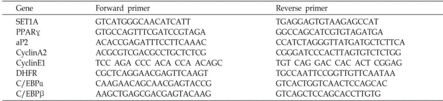
Fig. 1. SET1A expression is induced during differ- entiation of 3T3-L1 preadipocytes. (A) The 3T3- L1 preadipocytes were differentiated in the dif- ferentiation medium. At the indicated time, SET1A expression was measured by qPCR.
Data are presented as means±SEM from three independent experiments. *p<0.05 vs. 3T3-L1 preadipocyte (D0). (B) H3K4 me3 level was measured by western blot.
A B
C D
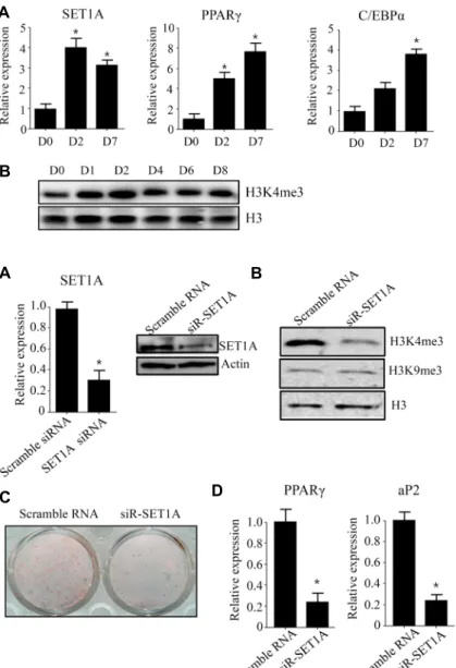
Fig. 2. Knockdown of SET1A decreased differ- entiation of 3T3-L1 preadipocytes. The 3T3- L1 preadipocytes were transfected with SET1A siRNA and differentiated in the dif- ferentiation medium for six days. (A) SET1A expression was measured at days 0 by qPCR.
(B) The levels of H3K4me3, and H3K9me3 were measured by western blot. (C) The 3T3-L1 adipocyte cells were stained with Oil Red O. (D) The expression of adipogenic genes, PPARγ and aP2, was measured by qPCR. Data are presented as means±SEM from three independent experiments. *p<0.05 vs. scramble RNA.
To determine whether SET1A expression is correlated with adipogenesis of 3T3-L1 cells, we examined SET1A ex- pression and epigenetic mark of H3K4 me3 level at day 2 (D2) and day 7 (D7) after induction of adipogenesis. As shown in Fig. 1A, SET1A expression increased at D2 and D7. Consistent with increased SET1A expression, global H3K4 me3 level also increased at D2 (Fig. 1B). These results suggest that SET1A may play a role in regulation of adipo- genesis in 3T3-L1 preadipocytes.
SET1A stimulates adipogenesis in 3T3-L1 preadi- pocytes
To see the role of SET1A in adipogenesis, we examined adipogenesis in SET1A- knockdowned 3T3-L1 preadipocytes using siRNA. The 3T3-L1 preadipocytes were treated with SET1A siRNA or scramble RNA and then were stimulated
to differentiate in a differentiation medium for 6 days. To verify the knockdown of SET1A, we determined the SET1A expression in SET1A siRNA-transfected 3T3-L1 preadi- pocytes. The qPCR and western blot analysis showed that transfection with SET1A siRNA efficiently reduced SET1A expression in 3T3-L1 preadipocyte (Fig. 2A). Consistent with decreased SET1A expression, SET1A knockdown sig- nificantly decreased global H3K4me3 level compared with scramble RNA-transfected 3T3-L1 preadipocyte cells (Fig.
2B). However, H3K9me3level was unchanged. Then, we as- sessed adipogenesis of SET1A siRNA-transfected 3T3-L1 preadipocytes. Oil Red O (ORO) staining showed that SET1A knockdown inhibited adipogenesis of 3T3-L1 pre- adipocyte (Fig. 2C), which was also detected by expression of adipogenic genes such as, PPARγ and aP2 (Fig. 2D).
A B
C
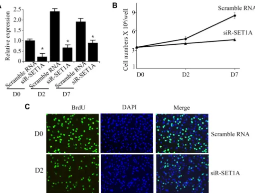
Fig. 3. Knockdown of SET1A inhibits cell proliferation of 3T3-L1 cells during mitotic cell expansion. (A) The 3T3-L1 preadipocytes were transfected with SET1A siRNA and differentiated in the differentiation medium. SET1A expression was measured at the indicated times by qPCR. (B) The 3T3-L1 preadipocytes were transfected with SET1A siRNA and after induction of differentiation, cell numbers were determined at the indicated times. (C) The 3T3-L1 preadipocytes were transfected with SET1A siRNA and differentiated in the presence of BrdU for 24 hr. Cells that incorporated BrdU were observed under a fluorescence microscope. Data are presented as means±SEM from three independent experiments. *p<0.05 vs. scramble RNA.
SET1A stimulates mitotic clonal expansion during early stage of adipogenesis
In the early adipogenic stage, the number of adipocytes greatly increases due to mitotic clonal expansion (MCE) [7].
Because SET1A expression increased at day 2, we deter- mined whether SET1A affects MCE. To this end, we exam- ined the effects of SET1A on proliferation of SET1A-knock- downed 3T3-L1 cells after induction of differentiation. We observed that SET1A siRNA efficiently reduced SET1A ex- pression at D0, D2 and D7 (Fig. 3A). SET1A-knockdown sig- nificantly decreased the cell numbers of 3T3-L1 adipocytes during day 2, as compared to the cell numbers of scramble RNA-transfected adipocyte cells (Fig. 3B). Consistent with these results, transfection of 3T3-L1 preadipocytes with SET1A siRNA resulted in reduced levels of bromodeoxyur- idine (BrdU) incorporation into cells compared to those of scramble RNA-transfected adipocyte cells (Fig. 3C), indicat- ing that SET1A increases proliferation of 3T3-L1 cells in the MCE process.
Cell cycle progression is regulated by cyclin-dependent kinases (CDKs) and their associated corresponding regu-
latory cyclins [10]. To examine whether these cell cycle regu- lators are affected by SET1A, we measured expression of cell cycle regulators in SET1A knockdown-3T3-L1 cells by qPCR.
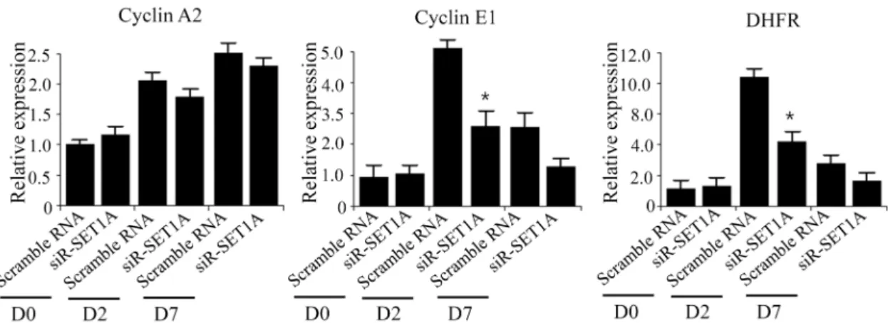
As shown in Fig. 4, SET1A knockdown decreased mRNA levels of cyclins E1 and dihydrofolate reductase (DHFR) at D2. These results suggest that SET1A stimulates MCE through upregulation of cell cycle regulator and DNA syn- thesis genes such as cyclins E1 and DHFR, which leads to stimulate adipogenesis of 3T3-L1 preadipocytes.
SET1A stimulates PPARγ expression during late stage of adipogenesis
Furthermore, we examined the expression of adipogenic regulators in SET1A knockdowned 3T3-L1 adipocyte cells.
The 3T3-L1 preadipocytes were transfected with either siRNA or scramble RNA, and stimulated to differentiate for 6 days. The expression of adipogenic regulators including PPARγ, C/EBPα, and C/EBPβ was determined at D0, D2, D4, and D7. The qPCR analysis showed that SET1A ex- pression was significantly decreased in the 3T3-L1 adipocyte cells by SET1A siRNA. Concomitant with the decreased
Fig. 4. Knockdown of SET1A downregulates the expression of cyclin E1 and DHFR. The 3T3-L1 preadipocytes were transfected with SET1A siRNA and after induction of differentiation, expression of cell cycle regulators were determined at the indicated times by qPCR. Data are presented as means±SEM from three independent experiments. *p<0.05 vs. scramble RNA.
Fig. 5. Knockdown of SET1A downregulates PPARγ expression. The 3T3-L1 preadipocytes were transfected with SET1A siRNA and differentiated in the differentiation medium. The expression of SET1A, PPARγ, C/EBPα, and C/EBPβ was measured at the indicated times by qPCR. Data are presented as means±SEM from three independent experiments. *p<0.05 vs. scramble RNA.
SET1A expression, PPARγ expression was significantly de- creased in SET1A siRNA-transfected 3T3-L1 cells at D0, D2, D4, and D7 compared with scramble RNA-transfected 3T3-L1 cells (Fig. 5). However, C/EBPβ expression was not affected by treatment with SET1A siRNA. These results sug- gest that SET1A stimulates PPARγ expression during adipo- genesis in 3T3-L1 preadipocytes.
Discussion
In this study, we demonstrated that SET1A stimulates adi- pogenesis of 3T3-L1 cells. ORO staining and the expression of adipogenic genes such as PPARγ and aP2 showed that knockdown of SET1A using siRNA inhibits adipogenesis of 3T3-L1 cells. Adipocyte differentiation occurs via three stages, the growth arrest of confluent preadipocytes, MCE,
and terminal differentiation [7]. After induction of differ- entiation with MDI, the growth-arrested preadipocytes syn- chronously re-enter the cell cycle for two additional rounds of division. The cells progress from the G0/G1 to S and G2/M phases at 24 h, and cell proliferation increases, which is known as the MCE process during the early stage of adi- pocyte differentiation. Our current data revealed that knock- down of SET1A significantly decreased the cell numbers of MDI-treated 3T3-L1 adipocytes during day 2, as compared to the cell numbers of scramble siRNA-treated adipocyte cells. Consistent with these results, knockdown of SET1A resulted in reduced levels of BrdU incorporation into cells compared to those of scramble siRNA-treated adipocyte cells, indicating that SET1A increase proliferation of 3T3-L1 cells in the MCE process and stimulates the adipogenesis of 3T3-L1 cells. CDKs and their associated regulatory cyclins play a key role in the regulation of cell cycle processes [10].
Our results revealed that knockdown of SET1A resulted in a decrease in expression of cyclin E1 and DHFR. Thus, SET1A increases the expression of cyclin E1 and DHFR, and stimulates MCE, which may contribute to stimulation of adi- pogenesis of 3T3-L1 preadipocytes.
PPARγ is considered the master regulators and stimulate adipocyte gene expression during adipogenesis [4].
Therefore, it has been shown to play a pivotal role in termi- nal differentiation of preadipocytes [4, 7]. Several reports showed that regulation of PPARγ expression is accompanied by several histone modifications. In particular, methylation of histone H3K9me3 and H3K9me2 by histone methyl- transferases such as SetdB1, Suv39h1, and G9a, was asso- ciated with repression of PPARγ expression and inhibition of adipogenesis [11-13], whereas methylation of H3K4me3 by PTIP and associated MLL3/4 was associated with the ac- tivation of PPARγ [2, 6]. However, it has not been charac- terized that SET1A regulates PPARγ expression through late stage of adipogenesis. In present study, we determined adi- pogenic regulators including PPARγ and C/EBPα in SET1A-knockdown 3T3-L1 preadipocytes. Knockdown of SET1A repressed PPARγ expression during late stage of adipogenesis. Therefore, SET1A may play a positive role in terminal adipogenesis via upregulation of PPARγ.
In summary, SET1A effectively stimulated the adipo- genesis of 3T3-L1 preadipocytes through both enhancement of MCE process during early stage of adipogenesis and upre- gulation of PPARγ during late stage of adipogenesis.
Acknowledgement
This work was supported by a 2-Year Research Grant of Pusan National University.
References
1. Barski, A., Cuddapah, S., Cui, K. and Zhao, K. 2007. High- resolution profiling of histone methylations in the human genome. Cell 129, 823-837.
2. Cho, Y. W., Hong, S., Jin, Q., Wang, L., Lee, J. E. and Ge, K. 2009. Histone methylation regulator PTIP is required for PPARgamma and C/EBPalpha expression and adipogenesis.
Cell Metab. 10, 27-39.
3. Cho, Y. W., Hong, T., Guo, H., Dressler G. R., Copeland, T. D. and Ge, K. 2007. PTIP associates with MLL3- and MLL4-containing histone H3 lysine 4 methyltransferase complex. J. Biol. Chem. 282, 20395-20406.
4. Farmer, S. 2006. Transcriptional control of adipocyte for- mation. Cell Metab. 4, 263-273.
5. Heintzman, N. D., Hon, G. C., Hawkins, R. D., Kheradpour, P. and Ren, B. 2009. Histone modifications at human en- hancers reflect global cell-type-specific gene expression.
Nature 459, 108-112.
6. Lee, J., Saha, P. K., Yang, Q. H., Lee, S., Park, J. Y., Suh, Y. and Lee, J. W. 2008. Targeted inactivation of MLL3 his- tone H3-Lys-4 methyltransferase activity in the mouse re- veals vital roles for MLL3 in adipogenesis. Proc. Natl. Acad.
Sci. USA 105, 19229-19234.
7. Lefterova, M. I. and Lazar, M. A. 2009. New developments in adipogenesis. Trends Endocrinol. Metab. 20, 107-114.
8. Mikkelsen, T. S., Xu, Z., Zhang, X., Wang, L. and Rosen, E. D. 2010. Comparative epigenomic analysis of murine and human adipogenesis. Cell 143, 156-169.
9. Mohan, M., Herz, H. M., Smith, E. R., Zhang, Y. and Shilatifard, A. 2011. The COMPASS family of H3K4 methyl- ases in Drosophila. Mol. Cell. Biol. 31, 4310-4318
10. Morgan, D. O. 2008. SnapShot: cell-cycle regulators I. Cell 135, 764-764.
11. Takada, I., Mihara, M., Suzawa, M., Ohtake, F., Kobayashi, S. and Igarashi, M. 2007. A histone lysine methyltransferase activated by non-canonical Wnt signalling suppresses PPAR-gamma transactivation. Nat. Cell Biol. 9, 1273-1285.
12. Wang, L., Xu, S., Lee, J., Baldridge, A., Grullon, S. and Ge, K. 2012. Histone H3K9 methyltransferase G9a represses PPARγ expression and adipogenesis. EMBO J. 32, 45-59.
13. Zhang, Z. C., Liu, Y., Li, S. F., Guo, L., Zhao, Y. and Qian, S. W. 2014. Suv39h1 mediates AP-2α-dependent inhibition of C/EBPα expression during adipogenesis. Mol. Cell Biol.
34, 2330-2338.
초록:히스톤 H3K4 메칠화효소 SET1A에 의한 지방세포 분화 촉진
김선후․정명호*
(부산대학교 한의학전문대학원 한의학과)
히스톤 H3K4의 메칠화는 3T3-L1의 지방세포의 분화를 촉진하는 것으로 알려져 있으나, 히스톤 H3K4 메칠화 효소인 SET1A가 지방세포 분화를 조절하는지에 대해서는 보고된 바가 없다. 그러므로 본 연구에서는 SET1A의 3T3-L1 지방세포의 분화조절과 기전을 연구하였다. SET1A의 발현은 3T3-L1 지방세포 분화과정에서 증가함을 관 찰하였다. 3T3-L1 지방전구세포에서 siRNA을 이용하여 SET1A의 발현을 감소시키면 3T3-L1 지방전구세포의 분 화가 억제됨을 관찰하여 SET1A가 3T3-L1 지방전구세포의 분화를 촉진함을 알 수 있었다. 이에 대한 조절기전을 알기 위해, SET1A의 발현을 감소시킨 3T3-L1 지방전구세포의 세포증식을 측정한 결과, 분화 초기 단계인 분화 후 2일 동안 3T3-L1 지방세포의 증식이 감소하였다. 또한 분화 후 7일 동안 지방세포세포 분화 조절인자들의 발현 을 측정한 결과, SET1A의 발현을 감소시킨 3T3-L1 지방세포에서 PPARγ의 발현이 감소하였다. 위와 같은 연구결 과를 바탕으로, SET1A는 분화초기단계에서는 mitotic clonal expansion 단계를 촉진하고, 분화후기단계에서는 PPARγ의 발현을 증가시켜 3T3-L1 지방세포의 분화를 촉진함을 알 수 있었다.