mini-implants tend to demonstrate a higher failure rate than conventional implants4. The frequent failures of orthodontic mini-implants during treatment is a weak point of their use and can affect treatment plans and duration5. Other studies have reported 0% to 30% failure rates of orthodontic mini- implants inserted into alveolar bone6,7.
Because the size and surface of a mini-implant are small and the initial stability of it might be week, healing time is needed after insertion8. However, immediate loading on prosthodontic implants has shown a good success rate9,10. Other studies have reported that loading time did not affect the success rate of prosthetic implants11. In orthodontic mini- implants, an immediate, light orthodontic load did not affect the bone healing process12. Loading can reduce the sclerostin level13 and result in release of prostaglandin14. These changes can enhance bone formation15 and inhibit bone resorption16.
I. Introduction
Small-sized mini-implants have been applied for skeletal orthodontic anchorage1. Orthodontic mini-implants have a smaller diameter and a shorter length than the implants used for prosthodontic treatment2,3. Therefore, orthodontic
Jong-Wan Kim
Department of Orthodontics, Section of Dentistry, Seoul National University Bundang Hospital, 82 Gumi-ro 173beon-gil, Bundang-gu, Seongnam 13620, Korea
TEL: +82-31-787-2787 FAX: +82-31-787-4140 E-mail: [email protected]
ORCID: http://orcid.org/0000-0002-2752-0116
This is an open-access article distributed under the terms of the Creative Commons Attribution Non-Commercial License (http://creativecommons.org/licenses/by-nc/4.0/), which permits unrestricted non-commercial use, distribution, and reproduction in any medium, provided the original work is properly cited.
CC
Analysis of time to failure of orthodontic mini-implants after insertion or loading
Jong-Wha Jeong1, Jong-Wan Kim2, Nam-Ki Lee2, Young-Kyun Kim3, Jong-Ho Lee4, Tae-Woo Kim1
1Department of Orthodontics, School of Dentistry and Dental Research Institute, Seoul National University, Seoul, Departments of 2Orthodontics and 3Oral and Maxillofacial Surgery, Section of Dentistry,
Seoul National University Bundang Hospital, Seongnam,
4Department of Oral and Maxillofacial Surgery and Oral Cancer Center, School of Dentistry and Dental Research Institute, Seoul National University Dental Hospital, Seoul, Korea
Abstract(J Korean Assoc Oral Maxillofac Surg 2015;41:240-245)
Objectives: This study was performed to evaluate patterns of failure time after insertion, failure rate according to loading time after insertion, and the patterns of failure after loading.
Materials and Methods: A total of 331 mini-implants were classified into the non-failure group (NFG) and failure group (FG), which was divided into failed group before loading (FGB) and failed group after loading (FGA). Orthodontic force was applied to both the NFG and FGA. Failed mini- implants after insertion, ratio of FGA to NFG according to loading time after insertion, and failed mini-implants according to failed time after loading were analyzed.
Results: Percentages of failed mini-implants after insertion were 15.79%, 36.84%, 12.28%, and 10.53% at 4, 8, 12, and 16 weeks, respectively. Mini- implant failure demonstrated a peak from 4 to 5 weeks after insertion. The failure rates according to loading time after insertion were 13.56%, 8.97%, 11.32%, and 5.00% at 4, 8, 12, and 16 weeks, respectively. Percentages of failed mini-implants after loading were 13.79%, 24.14%, 20.69%, and 6.9%
at 4, 8, 12, and 16 weeks, respectively.
Conclusion: Mini-implant stability is typically acquired 12 to 16 weeks after insertion, and immediate loading can cause failure of the mini-implant.
Failure after loading was observed during the first 12 weeks.
Key words: Dental implant, Orthodontic anchorage procedure, Immediate dental implant loading
[paper submitted 2015. 4. 8 / revised 2015. 6. 7 / accepted 2015. 6. 26]
Copyright Ⓒ 2015 The Korean Association of Oral and Maxillofacial Surgeons. All rights reserved.
This study was supported by grant no 04-2011-0056 from the SNUDH Research Fund.
For evaluation of failure rate according to loading time after insertion, the failure rate, calculated as the ratio of FGA to NFG, was analyzed according to loading time (weeks) after insertion. To analyze the patterns of failure after loading, the failed mini-implants were analyzed according to failure time (weeks) after loading. The statistic analysis was done using SPSS 12.0 for Windows (SPSS Inc., Chicago, IL, USA).
This study was approved by the Ethics Committee for Re- search (Seoul National University Bundang Hospital Ethical Board, B-1011-115-110).
III. Results
In this study, 57 of 331 inserted mini-implants experienced failure. Of these, 29 were in the FGA, and 28 were in the FGB. The mean age of the subjects was 22.08±7.52 years (NFG, 22.39±7.54 years; FG, 20.89±7.33 years).(Table 1)
Failure rates were 15.79%, 36.84%, 12.28%, and 10.53%
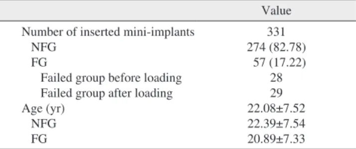
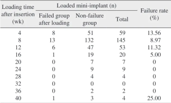
after 4, 8, 12, and 16 weeks, respectively.(Table 2, Fig. 2) In the failed mini-implant group, 75.44% of failures occurred within 16 weeks after insertion. The failure of mini-implants had a peak from 4 to 5 weeks.(Fig. 2) The mini-implants loaded during the first 4 weeks after insertion showed the highest failure rate.(Table 3, Fig. 3) The failure rates in re- lation to loading time after insertion were 13.56%, 8.97%, 11.32%, and 5.00% at 4, 8, 12, and 16 weeks, respectively.
Mini-implants loaded during the first week after insertion showed a 42.50% failure rate.(Fig. 3) Percentages of failed mini-implants after loading in this group were 13.79%, 24.14%, 20.69%, and 6.90% at 4, 8, 12, and 16 weeks, re- spectively.(Table 4, Fig. 4) Mini-implant failures after load- ing in this group occurred mostly during the first 12 weeks after loading. From 4 to 12 weeks after loading, the failure rate of mini-implants in this group was 58.62%. Failures were There was no significant difference in marginal bone loss
between immediate loading and delayed loading in such im- plants17.
However, the bone remodeling process involving bone resorption and apposition after insertion of a mini-implant needs time to heal in order to provide stable support of the mini-implant18.
Because of the high failure rates of orthodontic mini- implants, loading time is believed to be important. Analysis regarding the failure time of orthodontic mini-implant after insertion and loading could help guide clinical practice when using orthodontic mini-implants. This study was conducted to evaluate the pattern of failure time after insertion, failure rate according to loading time after insertion, and the pattern of failure after loading.
II. Materials and Methods
Subjects included 134 patients (mean age, 20.08±7.52 years) with 331 inserted mini-implants (Miangan; Bioma- terials Korea, Seoul, Korea) with a diameter of 1.2 mm and length of 7.0 mm. This study was done from July 2006 to June 2010 in dental clinic of Seoul National University Bun- dang Hospital (Seongnam, Korea). The surfaces of the mini- implants were machined but not treated. All mini-implants were installed on the buccal alveolar bone between the first premolar and the second molar of the maxilla and mandible.
Mini-implants were inserted by 1 operator using a self- drilling method after orthodontic leveling and alignment.
The mini-implants were classified into the failure group (FG) and non-failure group (NFG). The FG was divided into the failed group before loading (FGB) and failed group after loading (FGA). Orthodontic force was applied to both the NFG and FGA.
The failure rate was calculated for FG and NFG. To evaluate the pattern of failure time after insertion of mini- implant, the failed mini-implants were analyzed according to the failure time (weeks) after insertion in the FG.(Fig. 1)
Insertion Loading start Failure
Loading time after insertion Failed time after loading Failed time after insertion
Fig. 1. The diagram to explain ‘failed time after insertion’, ‘loading time after insertion’, and ‘failed time after loading’.
Jong-Wha Jeong et al: Analysis of time to failure of orthodontic mini-implants after inser- tion or loading. J Korean Assoc Oral Maxillofac Surg 2015
Table 1. Number of mini-implants and age in the non-failed group (NFG) and failure group (FG)
Value Number of inserted mini-implants
NFG FG
Failed group before loading Failed group after loading Age (yr)
NFG FG
331 274 (82.78) 57 (17.22)
28 29 22.08±7.52 22.39±7.54 20.89±7.33
Values are presented as number only, number (%), or mean±standard deviation.
Jong-Wha Jeong et al: Analysis of time to failure of orthodontic mini-implants after inser- tion or loading. J Korean Assoc Oral Maxillofac Surg 2015
IV. Discussion
The results of this study showed some initial failures after insertion. About 75% of failed mini-implants occurred during the 16 weeks after insertion.(Table 2) Specifically, when the observed intermittently until 44 weeks after loading.(Fig. 4)
Tables 2-4 illustrate the data in 4-week intervals. Fig. 2-4 present the data in 1-week intervals to provide greater detail.
Table 2. Failed mini-implants according to time after insertion Time after
insertion (wk)
Failed group
before loading Failed group
after loading Total Accumulated total 4
8 12 16 20 24 28 32 36 40 44 48 52
9 (32.14) 17 (60.71) 2 (7.14)
0 0 0 0 0 0 0 0 0 0
0 4 (13.79) 5 (17.24) 6 (20.69) 1 (3.45) 2 (6.90) 2 (6.90) 1 (3.45) 2 (6.90) 4 (13.79) 1 (3.45)
0 1 (3.45)
9 (15.79) 21 (36.84) 7 (12.28) 6 (10.53) 1 (1.75) 2 (3.51) 2 (3.51) 1 (1.75) 2 (3.51) 4 (7.02) 1 (1.75)
0 1 (1.75)
9 (15.79) 30 (52.63) 37 (64.91) 43 (75.44) 44 (77.19) 46 (80.70) 48 (84.21) 49 (85.96) 51 (89.47) 55 (96.49) 56 (98.25) 56 (98.25) 57 (100.00) Values are presented as number (%).
Jong-Wha Jeong et al: Analysis of time to failure of orthodontic mini-implants after inser- tion or loading. J Korean Assoc Oral Maxillofac Surg 2015
No.offailedmini-implants
4 10
9 8 7 6 5 4 3 2 1 0
Time (wk)
0 8 12 16 20 24 28 32 36 40 44 48
Failed group after loading Failed group before loading
Fig. 2. The number of failed mini- implants as to the time after insertion.
Jong-Wha Jeong et al: Analysis of time to failure of orthodontic mini-implants after insertion or loading.
J Korean Assoc Oral Maxillofac Surg 2015 Table 3. Failed mini-implants according to loading time after in- sertion
Loading time after insertion
(wk)
Loaded mini-implant (n)
Failure rate Failed group (%)
after loading Non-failure
group Total
4 8 12 16 20 24 28 32 36 40
8 13 6 1 0 0 0 0 0 1
51 132 47 19 7 9 4 0 2 3
59 145 53 20 7 9 4 0 2 4
13.56 8.97 11.32 5.00 0 0 0 0 0 25.00 Jong-Wha Jeong et al: Analysis of time to failure of orthodontic mini-implants after inser- tion or loading. J Korean Assoc Oral Maxillofac Surg 2015
Failurerate(%)
4 45.00
40.00 35.00 30.00 25.00 20.00 15.00 10.00 5.00 0
Time (wk)
0 2 6 8 10 12 14 16 18 20 22 24 26 28 30 32 34 36 38 40
Fig. 3. Failure rate as to the loading time after insertion.
Jong-Wha Jeong et al: Analysis of time to failure of orthodontic mini-implants after insertion or loading.
J Korean Assoc Oral Maxillofac Surg 2015
layed loading24. However, the failure rate in relation to load- ing time after insertion in the present study was highest dur- ing the first week.(Fig. 3) This might indicate that immediate loading after insertion activates bone resorption and results in mini-implant failure25.
Because mini-implants have a smaller diameter, shorter length, and less surface area contacting the bone compared to conventional implants, mechanical stability in the early stages could be affected and reduced by limited bone resorp- tion around the mini-implant26.
In this study, the failure of loaded mini-implants happened in case of the load was applied during the first 12 weeks after insertion except 37 weeks. This suggests that the load until 12 weeks after insertion affects the stability of a mini-implant, and loading is thus recommended at approximately 12 weeks after insertion.
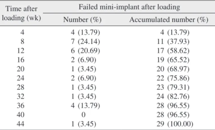
Failure after loading occurred frequently until 13 weeks (Fig. 4), with a high failure rate from 6 to 13 weeks. It is rec- ommended that the clinician should be careful until 3 months after loading to sustain the mini-implant because of failure.
Additionally, failure after loading was observed intermittent- ly until 35 weeks. This suggests that the stability of the mini- implant is affected by bone remodeling after tooth movement through the end of treatment. This might be the result of insufficient surface area of the mini-implant in contact with bone in order to obtain sufficient surrounding support27.
Failure of the mini-implant results in difficulties and affects the treatment plan. Although there are various causes of mini- implant failure, selection of loading time after insertion is an important factor for avoiding failure. Immediate loading is not recommended, and delayed loading of the mini-implant is suggested until 12 to 16 weeks after insertion.
For more stable use of mini-implants in clinics, further studies regarding surface treatment of mini-implants are loading time after insertion was less than 12 weeks, the fail-
ure rates of mini-implant were high. These results signify that the stability of the mini-implant is acquired 12 to 16 weeks after insertion. They indicate that the bone remodeling pro- cess after insertion occurs during the first 3 to 4 months after insertion. For implant success, loading time is important.
Time for bone healing of tissue damaged through the process of implantation is needed. Therefore, delayed loading has been suggested by other studies focusing on implant stabil- ity19,20.
Before loading, the failure rate during the first 4 to 5 weeks was the highest.(Fig. 2) It is possible that bone resorption in the bone remodeling process is most active around 4 weeks after insertion in humans, and that bone apposition is achieved 3 to 4 months after insertion.
Some studies have reported that immediate or early loading of mini-implants shows good stability21-23. A previous animal study has suggested no significant differences in success rate or histological findings between immediate loading and de-
Table 4. Failed mini-implants according to failed time after loading Time after
loading (wk)
Failed mini-implant after loading Number (%) Accumulated number (%) 4
8 12 16 20 24 28 32 36 40 44
4 7 6 2 1 2 1 1 4 1
(13.79) (24.14) (20.69) (6.90) (3.45) (6.90) (3.45) (3.45) (13.79) 0 (3.45)
4 11 17 19 20 22 23 24 28 28 29
(13.79) (37.93) (58.62) (65.52) (68.97) (75.86) (79.31) (82.76) (96.55) (96.55) (100.00) Jong-Wha Jeong et al: Analysis of time to failure of orthodontic mini-implants after inser- tion or loading. J Korean Assoc Oral Maxillofac Surg 2015
Fig. 4. The number of failed mini-im- plants as to the failed time after load- ing.
Jong-Wha Jeong et al: Analysis of time to failure of orthodontic mini-implants after insertion or loading.
J Korean Assoc Oral Maxillofac Surg 2015
No.offailedmini-implants
4 5
4
3
2
1
0
Time (wk)
0 8 12 16 20 24 28 32 36 40 44
6. Wiechmann D, Meyer U, Büchter A. Success rate of mini- and micro-implants used for orthodontic anchorage: a prospective clini- cal study. Clin Oral Implants Res 2007;18:263-7.
7. Kim JS, Choi SH, Cha SK, Kim JH, Lee HJ, Yeom SS, et al. Com- parison of success rates of orthodontic mini-screws by the insertion method. Korean J Orthod 2012;42:242-8.
8. Kim SH, Lee SJ, Cho IS, Kim SK, Kim TW. Rotational resistance of surface-treated mini-implants. Angle Orthod 2009;79:899-907.
9. Grunder U. Immediate functional loading of immediate implants in edentulous arches: two-year results. Int J Periodontics Restorative Dent 2001;21:545-51.
10. Kim YK, Kim BS, Yun PY, Mun SU, Yi YJ, Kim SG, et al. The seven-year cumulative survival rate of Osstem implants. J Korean Assoc Oral Maxillofac Surg 2014;40:68-75.
11. Ioannidou E, Doufexi A. Does loading time affect implant surviv- al? A meta-analysis of 1,266 implants. J Periodontol 2005;76:1252- 12. Catharino PC, Dominguez GC, Pinto Ddos S Jr, Morea C. Histo-8.
logic, histomorphometric, and radiographic monitoring of bone healing around in-office-sterilized orthodontic mini-implants with or without immediate load: study in rabbit tibiae. Int J Oral Maxil- lofac Implants 2014;29:321-30.
13. Robling AG, Niziolek PJ, Baldridge LA, Condon KW, Allen MR, Alam I, et al. Mechanical stimulation of bone in vivo reduces os- teocyte expression of Sost/sclerostin. J Biol Chem 2008;283:5866- 14. Klein-Nulend J, van der Plas A, Semeins CM, Ajubi NE, Frangos 75.
JA, Nijweide PJ, et al. Sensitivity of osteocytes to biomechanical stress in vitro. FASEB J 1995;9:441-5.
15. Forwood MR. Inducible cyclo-oxygenase (COX-2) mediates the induction of bone formation by mechanical loading in vivo. J Bone Miner Res 1996;11:1688-93.
16. Tatsumi S, Ishii K, Amizuka N, Li M, Kobayashi T, Kohno K, et al.
Targeted ablation of osteocytes induces osteoporosis with defective mechanotransduction. Cell Metab 2007;5:464-75.
17. Kim YK, Ahn KJ, Yun PY, Kim M, Yang HS, Yi YJ, et al. Effect of loading time on marginal bone loss around hydroxyapatite-coated implants. J Korean Assoc Oral Maxillofac Surg 2013;39:161-7.
18. Rebaudi A, Laffi N, Benedicenti S, Angiero F, Romanos GE. Mi- crocomputed tomographic analysis of bone reaction at insertion of orthodontic mini-implants in sheep. Int J Oral Maxillofac Implants 2011;26:1233-40.
19. Raghavendra S, Wood MC, Taylor TD. Early wound healing around endosseous implants: a review of the literature. Int J Oral Maxillofac Implants 2005;20:425-31.
20. Atsumi M, Park SH, Wang HL. Methods used to assess implant sta- bility: current status. Int J Oral Maxillofac Implants 2007;22:743- 21. Al-Sawai AA, Labib H. Success of immediate loading implants 54.
compared to conventionally-loaded implants: a literature review. J Investig Clin Dent 2015. doi: 10.1111/jicd.12152. [Epub ahead of print]
22. Romanos G, Grizas E, Laukart E, Nentwig GH. Effects of early moderate loading on implant stability: a retrospective investiga- tion of 634 implants with platform switching and morse-tapered connections. Clin Implant Dent Relat Res 2015. doi: 10.1111/
cid.12314. [Epub ahead of print]
23. Kim YK, Kim JH, Yi YJ, Kwon MJ, Yun PY. Prospective compara- tive study of tapered implants with SLA surfaces in the maxillary posterior area according to 3- and 6-month loading time. Int J Peri- odontics Restorative Dent 2015;35:271-6.
24. Ramazanzadeh BA, Fatemi K, Dehghani M, Mohtasham N, Jahan- bin A, Sadeghian H. Effect of healing time on bone-implant contact of orthodontic micro-implants: a histologic study. ISRN Dent 2014.
doi: 10.1155/2014/179037.
25. Lee SJ, Ahn SJ, Lee JW, Kim SH, Kim TW. Survival analysis of orthodontic mini-implants. Am J Orthod Dentofacial Orthop 2010;
needed because implant surface might influence osseointegra- tion time and implant failure. Additionally, if other factors, such as loading time, can be controlled in future studies, the more detailed results would be helpful to clinicians.
V. Conclusion
1. Approximately 75% of mini-implant failures occurred within 16 weeks of insertion. When the loading time after insertion was less than 12 weeks, the failure rate of the mini- implant was high. Proper stability of the mini-implant is ac- quired about 3 to 4 months after insertion.
2. The failure rate according to loading time after insertion was highest when the mini-implants were loaded during the first week after insertion. Immediate loading could cause fail- ure of a mini-implant.
3. Failure after loading was frequently observed in the mini- implant until 13 weeks. Therefore, attention to the stability of the mini-implant is necessary until 3 months after loading.
Conflict of Interest
No potential conflict of interest relevant to this article was reported.
ORCID
Jong-Wha Jeong, http://orcid.org/0000-0002-4080-2046 Jong-Wan Kim, http://orcid.org/0000-0002-2752-0116 Nam-Ki Lee, http://orcid.org/0000-0003-1505-2551 Young-Kyun Kim, http://orcid.org/0000-0002-7268-3870 Jong-Ho Lee, http://orcid.org/0000-0002-8843-545X Tae-Woo Kim, http://orcid.org/0000-0003-2824-8270
References
1. Suh HY, Lee SJ, Park HS. Use of mini-implants to avoid maxillary surgery for Class III mandibular prognathic patient: a long-term post-retention case. Korean J Orthod 2014;44:342-9.
2. Kanomi R. Mini-implant for orthodontic anchorage. J Clin Orthod 1997;31:763-7.
3. Lee HJ, Lee KS, Kim MJ, Chun YS. Effect of bite force on orth- odontic mini-implants in the molar region: finite element analysis.
Korean J Orthod 2013;43:218-24.
4. Herrmann I, Lekholm U, Holm S, Kultje C. Evaluation of patient and implant characteristics as potential prognostic factors for oral implant failures. Int J Oral Maxillofac Implants 2005;20:220-30.
5. Lee JH, Choo H, Kim SH, Chung KR, Giannuzzi LA, Ngan P. Re- placing a failed mini-implant with a miniplate to prevent interrup- tion during orthodontic treatment. Am J Orthod Dentofacial Orthop 2011;139:849-57.
27. Kim JW, Ahn SJ, Chang YI. Histomorphometric and mechanical analyses of the drill-free screw as orthodontic anchorage. Am J Or- thod Dentofacial Orthop 2005;128:190-4.
137:194-9.
26. Kim SH, Cho JH, Chung KR, Kook YA, Nelson G. Removal torque values of surface-treated mini-implants after loading. Am J Orthod Dentofacial Orthop 2008;134:36-43.