J. Exp. Biomed. Sci. 2013, 19(2): 118~123 pISSN : 1738-3226
Differential Effects of TNF-α on the Survival and Apoptosis of Human Granulocytes and the Human Myeloid Leukemia Cell Line
Eun Ju Yang and Jeong Hyun Chang
†Department of Clinical Laboratory Science, Daegu Haany University, Gyeongsan-si 712-715, Korea
Tumor necrosis factor-alpha (TNF-α) is a proinflammatory cytokine that mediates the inflammatory response and immune functions, and modulates the proliferation, differentiation and cell death of cancer cells. The differential functions of TNF-α in various human cells due to the formation of different stimulating pathway upon the binding of TNF-α to its receptors. In the present study, we examined the different effects of TNF-α on the survival and apoptosis between normal granulocytes and human myeloid leukemia HL-60 cells. Although TNF-α did not affect on the constitutive apoptosis of granulocytes, TNF-α strongly induced the apoptosis of HL-60 cells in a dose- and a time-dependent manner. TNF-α- induced apoptosis was occurred via the activation of caspase 8, caspase 9 and caspase 3/7 and the induction of ROS production in HL-60 cells. Also, BAY-11-7085, a NF-κB inhibitor, blocked the TNF-α-induced apoptosis in HL-60 cells. NF-κB may be involved in TNF-α-induced apoptotic signaling pathway in HL-60 cells. These results suggest that TNF-α activates apoptotic pathways and its process depends on cell type and many cellular factors. A better understanding of the differential effect of TNF-α on cell apoptosis and survival may provide important information that can be used to elucidate the specific inhibitory effect of TNF-α on the cancer dis.
Key Words: Apoptosis, Granulocyte, HL-60 cells, Survival, TNF-α
서 론
세포의 면역반응은 감염 등 다양한 세포외 자극에 의 해 세포 내 신호전달과정을 조절하는데 그 중 TNF-α에 의해 세포의 활성이 조절되는 기전도 있다 (Callard and Yates, 2005). TNF-α는 면역과 염증반응에 관여하는 대표 적인 염증 사이토카인 중 하나로 수용체에 결합하여 세 포 내로 자극을 전달한다 (Baud and Karin, 2001). 특히, TNF-α와 그 수용체의 결합은 주요 전사인자인 nuclear factor-kappa B (NF-κB)의 활성을 유도하여 caspase의 연 쇄반응을 조절함으로써 세포자멸사를 억제한다. 또한 TNF-α의 농도가 낮으면 세포자멸사 유도에는 관여하지
못하지만, 고농도의 TNF-α는 인체 내에서 급성 쇼크 등 의 증상을 일으킬 수 있다 (Wajant et al., 2003). 이전 연 구에서도 TNF-α의 양, 처리시간, 결합하는 수용체의 종 류 등 다양한 요소들에 의해 서로 다른 반응들을 유도한 다고 보고되고 있다 (Wallach, 1997; Wajant et al., 2003). 그 러므로 TNF-α는 활성화되는 면역반응에 조절에 의해 세 포의 죽음을 유도할 수도, 세포의 생존을 유도할 수도 있 는 양면적인 기능이 있는 물질로 여겨지고 있다. 본 연구 에서는 이를 토대로 세포의 생사에 있어서 TNF-α의 양면 적인 작용기전을 이용하여 정상 세포와 사람 골수구성 백혈병 세포주인 HL-60 세포의 생존에 TNF-α의 작용을 확인하고자 하였다.
HL-60 세포는 혈액과 골수에서 발생되는 암인 골수구 성 백혈병 연구에 주로 사용되는 세포로 특히 급성 골수 구성 백혈병 (acute myelogenous leukemia, AML)의 발병기 전 및 치료법을 위한 연구에 이용된다. 특히 AML은 외 과적 수술로 치료하기 어려운 암이기 때문에 주로 화학 요법을 통해 치료한다. 화학요법은 암세포에 특이적인 세 포독성을 가지고 있는 물질을 처리한 다음 암세포의 죽
*Received: June 27, 2013 / Revised: June 28, 2013 Accepted: June 28, 2013
†Corresponding author: Jeong Hyun Chang. Department of Clinical Laboratory Science, Daegu Haany University, Gyeongsan-si 712-715, Korea.
Tel: +82-53-819-1352, Fax: +82-53-819-1269 e-mail: [email protected]
○CThe Korean Society for Biomedical Laboratory Sciences. All rights reserved.
Original Article
음이 유발되는지 확인하여 항암효과를 확인하는 것으로 이때 암세포의 세포자멸사와 그와 관련된 작용기전을 연구하고 규명하는 것이 중요하다 (Johnstone et al., 2002).
AML의 치료제로는 주로 비타민 A의 파생물인 all-trans retionic acid (ATRA)를 이용해 백혈병 세포의 분화 및 세 포자멸사를 유도하여 치료에 사용해 왔지만, ATRA는 일부 호흡곤란, 발열, 저혈압 등을 동반하는 retionic acid syndrome (RAS)를 유발한다 (Tallman, 2002). 여전히 부작 용이 없으면서 효과적인 항암 치료제가 부족한 필요한 실정이고 더욱 새로운 치료물질을 발굴 및 개발하는 것 이 필요하다.
이에 본 연구에서 염증성 물질로 알려져 있지만 세포 의 종류, 처리용량 및 시간 등에 의해 세포의 활성을 유 도할 수도, 반대로 세포의 활성을 억제할 수도 있는 TNF-α를 이용하여 암세포주인 HL-60 세포에 특이적으 로 작용하여 제거할 수 있는지에 대해 확인해 보고자 하 였다 .
재료 및 방법
세포 배양
사람 골수구성 백혈병 세포주인 HL-60 세포 (American Type Culture Collection, Rockville, USA)는 10% heat- inactivated fetal bovine serum (FBS), 100 U/ml의 penicillin, 100 μg/ml의 streptomycin (Life technologies, Inc., USA)이 포 함된 RPMI 1640배지 (Life technologies, Inc.)에서 배양하 였다 . 세포는 37℃, 5% CO
2조건으로 배양하였다.
정상과립구는 먼저 정상인의 혈액을 채취한 다음 Ficoll-Hypaque (Amersham phamacia biotechnology, UK)를 이용하여 과립구층을 분리하였다. 분리된 과립구층에서 적혈구를 저장성 용액으로 용해시키고 얻은 과립구는 각 각 10% FBS, penicillin (100 U/ml), streptomycin (100 g/ml)을 함유한 RPMI 1640 배지에 3 × 10
6cells/ml의 농도로 부 유하였다 . 세포는 trypan blue 염색을 통해 생존율이 90%
이상인 것을 확인하였다. 본 실험 전 정상인에게서 동의 를 구한 다음 승인을 거쳐 시행하였다.
세포자멸사 측정
과립구 및 HL-60 세포를 모아 생리식염수 (Phosphate beffered saline, PBS)에 세포를 부유시킨 다음, 세포부유액 에 annexin V-FITC와 propidium iodide (PI) (BD bioscience, USA)를 각각 첨가하여 실온에서 15분간 방치한 후 염
색된 세포는 CellQuest software를 이용한 유세포 분석기 (BD bioscience)를 통해 측정하였다. Annexin V-FITC가 염 색된 모든 세포를 세포자멸사가 일어난 세포로 정의하고 각 샘플 당 총 10,000개의 세포를 분석하였다.
활성산소종 발현 측정
TNF-α 처리에 의한 활성산소종 (reactive oxygen species, ROS)의 발현 여부를 확인하기 위해 우선 물질 처리가 끝난 HL-60 세포를 모아 최종 5 × 10
5cells/100 μl의 농 도가 되게 37℃의 PBS에 부유시켰다. 그 다음 5 μM의 2',7'-dichlorodihydrofluorescein diacetate (2',7'-DCFDA)를 처 리하여 10분간 실온에서 반응시켰다. 이후 2',7'-DCFDA 가 염색된 세포는 즉시 유세포 분석기를 이용하여 측정 하였다 . 2',7'-DCFDA의 형광의 세기를 ROS가 생성된 양 에 비례한다고 판단하였다.
Caspase-3/7, 8, 9 활성 측정
세포자멸사에 작용하는 caspase를 확인하기 위해 caspase-Glo
®3/7 assay kit, caspase-Glo
®8 assay kit, caspas- Glo
®9 assay kit (Promega, USA)를 이용하여 caspase의 연 쇄반응을 측정하고자 하였다. 먼저 96-well plate에서 세 포를 분주하고 물질을 처리하여 배양하였다. 배양이 끝 나면 키트의 방법에 따라 반응시약과 기질을 넣어 반 응시킨 다음 luminometer TECAN GENios를 이용하여 chemiluminescence를 측정하였다.
통계처리
모든 실험 결과를 mean ± S.D로 표현하였다. 각 대조 군과 실험군 사이의 통계학적의 유의성은 SPSS statistical software package (Version 10.0, Chicago, IL)을 이용하여 student's t-test로 분석하였고 P value가 0.05 이하면 통계 학적인 유의성이 있다고 판단하였다.
결 과
TNF-α에 의한 골수구성 백혈병 세포의 세포자멸사 유도
효과
암세포인 골수구성 백혈병 세포에서 TNF-α의 효과를
확인하고 또한 정상 세포에서의 효과를 비교하기 위해
먼저 정상인의 말초혈액을 채취한 다음 과립구를 분리
하고 암세포주인 HL-60 세포를 배양하였다. 각 세포의
세포자멸사에 TNF-α의 효과를 농도별로 확인하기 위해
TNF-α 1, 10, 20 ng/ml을 각각 18시간 동안 처리하여 annexin V-FITC & PI 염색을 통해 유세포 분석기로 측정 하여 세포자멸사를 확인하였다. 과립구에 TNF-α를 농도 별로 처리한 결과 거의 변화를 보이지 않았다 (Fig. 1A).
반면 HL-60 세포에서는 TNF-α를 처리한 결과 1 ng/ml부 터 유의하게 세포자멸사를 유도하였고 20 ng/ml의 농도 에서 가장 많이 세포자멸사를 증가시켰다 (Fig. 1B). 이 후 모든 실험은 HL-60 세포에서 유의한 효과를 보인 20 ng/ml의 TNF-α를 처리하여 진행하였다.
또한 TNF-α 처리 후 각 시간별로 나타나는 효과를 확 인하고자 하였다. 과립구에서는 TNF-α 처리에 따라 시간 별로 세포자멸사에 큰 차이를 보이지 않았으나 (Fig. 2A), HL-60 세포에서는 TNF-α 처리 12시간부터 급격히 세포 자멸사가 증가함을 관찰하였다 (Fig. 2B).
이를 통해 TNF-α가 정상 세포와 암세포주에서 각각 다른 작용을 하는 것을 알 수 있었다. 더불어 과립구에 비해 HL-60 세포에서 TNF-α가 짧은 처리시간과 낮은 농 도에서 일찍 반응하는 것을 보고 HL-60 세포에 TNF-α가 더 크게 영향을 미치는 것을 알 수 있었다.
TNF-α에 의한 HL-60 세포 내의 caspase cascade의 활 성 유도 효과
세포의 죽음에는 다양한 caspase가 작용하게 되고 활 성화된 caspase 3는 최종적으로 작용하여 세포자멸사를 일으키게 된다. 본 연구에서도 HL-60 세포에서 TNF-α가 유도하는 세포자멸사에 다양한 caspase 활성의 변화가 있 는지 확인하고자 하였다 (Porter, 2006). 그 중 세포자멸사 초기에 작용하는 caspase 8, 미토콘드리아를 거쳐 발생하 는 세포자멸사에 작용하는 caspase 9, 최종 세포자멸사의 실행자로 알려진 caspase 3/7의 활성 변화를 확인하였다.
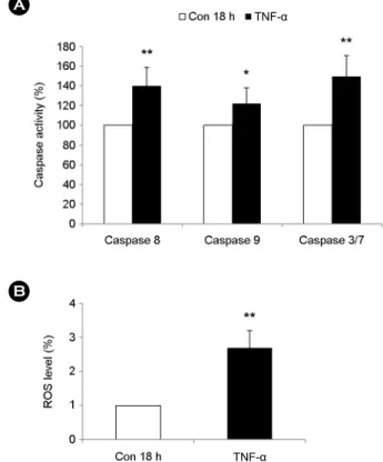
HL-60 세포에 TNF-α 20 ng/ml을 18시간 처리한 결과 caspase 8, 9, 3/7의 활성도가 처리하기 전에 비해 유의하 게 증가한 것을 확인하였다 (Fig. 3A). 이는 TNF-α에 의 한 HL-60 세포의 세포자멸사가 caspase 8, 9, 3/7을 통해 작용하는 것을 나타낸다.
TNF-α에 의한 HL-60 세포의 활성산소종 생산 유도
TNF-α에 의한 HL-60 세포의 세포자멸사가 세포독성이 있다고 알려진 활성산소종 (ROS)과 관련 있는지 파악하 고자 세포에 TNF-α 20 ng/ml을 처리한 다음 2'7'-DCFDA 염색을 실시하여 유세포 분석기로 측정하였다. 그 결과,
Fig. 1. The differential effect of TNF-α on the apoptosis of human granulocytes and human myeloid leukemia cell line in a dose-dependent manner. (A) Human granulocytes and (B) HL-60 cells were incubated for 18 h in the presence and absence of TNF-α in the indicated concentration (n=3). Apoptosis of these cells was analyzed by measuring the binding of annexin V-FITC and PI.Data are expressed as the means ± SD. *P < 0.05 and **P < 0.01 indicate a significant difference between the control (con 18 h) and TNF-α-treated groups.
A
B
TNF-α 1 ng/ml
TNF-α 10 ng/ml
TNF-α 20 ng/ml
TNF-α 20 ng/ml TNF-α
10 ng/ml TNF-α
1 ng/ml
TNF-α 처리 18시간 후 HL-60 세포에서 활성산소종의 생 산이 급격히 증가함을 관찰하였고 (Fig. 3B), TNF-α는 HL-60 세포 내에서 활성산소종의 생산을 유도하는 것을 확인하였다.
TNF-α에 의한 NF-κB의 활성이 HL-60 세포의 세포자멸 사에 미치는 영향
TNF-α에 의해 세포 내부로 자극이 전달되면 주로 NF- κB의 활성을 통해 세포의 생사를 조절한다 (Rangamani and Sirovich, 2006). TNF-α에 의한 HL-60 세포의 세포자멸 사가 NF-κB의 활성에 관계가 있는지 파악하기 위해 NF- κB의 신호전달 단백 특이 억제제인 BAY-11-7085 (BAY) 를 전처리하는 것에 따라 TNF-α의 세포자멸사의 변화를
관찰하였다 . 그 결과, BAY를 처리한 세포는 TNF-α에 의 해 증가한 세포자멸사가 유의하게 감소함을 나타내었다 (Fig. 4A). 또한 HL-60 세포에 TNF-α 처리 후 급격히 증 가한 caspase 중 caspase 8, 3/7이 BAY의 전처리 이후 감 소하였다. Caspase 9은 BAY 처리 후에 큰 영향을 나타나 지 않았다 (Fig. 4B). 이는 TNF-α에 의해 HL-60 세포 내 에서 활성화된 NF-κB는 caspase cascade 일부를 활성화시 켜 세포자멸사를 유도한다는 것을 보여준다.
고 찰
정상세포인 말초혈액으로부터 분리한 과립구와 암세 포주인 골수구성 백혈병 세포 HL-60 세포에서 차별화된
AB
Con TNF-α
Con TNF-α
A
Fig. 3. Caspase activities and ROS production are mediated by TNF-α in HL-60 cells. HL-60 cells were incubated for 18 h in the absence or presence of TNF-α (20 ng/ml). (A) Activities of caspase 8, 9 and 3/7 were determined by monitoring cleavage of each luminescence-labeled substrate peptide in HL-60 cells (n=3). Data are presented in relation to the control (Con 18 h), which are set at 100%. (B) ROS production was detected by 2',7'- DCFCA staining. Relative increase of ROS was calculated by comparing the mean fluorescence intensity (MFI) of 2',7'-DCFCA stained-treatment group to that of the control group (n=3). Data are expressed as the means ± SD. *P < 0.05 and **P < 0.01 indicate a significant difference between the control (con 18 h) and TNF-α-treated group.
A
B
TNF-α
TNF-α
Fig. 2. The differential effect of TNF-α on the apoptosis of human granulocytes and human myeloid leukemia cell line in a time-dependent manner. (A) Human granulocytes and (B) HL-60 cells were incubated for the indicated time with or without TNF-α (20 ng/ml) (n=3). Apoptosis of these cells was analyzed by measuring the binding of annexin V-FITC and PI. Data are expressed as the means ± SD. *P < 0.05 and **P < 0.01 indicate a significant difference between the control and TNF-α-treated groups at the same time.
TNF-α의 작용기전을 보았다. TNF-α는 20 ng/ml로 과립구 에 처리하였을 때와는 다르게 HL-60 세포에서 1 ng/ml의 낮은 농도인 TNF-α에 의해서도 세포자멸사를 처리시간 에 따라 유의하게 증가시켰다 (Fig. 1). TNF-α는 면역반응 에서 전형적으로 세포의 수용체와 결합하여 세포 내부의 다양한 신호전달과정을 활성 또는 억제하는 대표적인 염 증 사이토카인이기 때문에 세포 외적으로는 자극 수용체 에 따라, 내적으로는 세포 내 신호전달과정의 활성화에 따라 적용기전이 달라진다.
TNF-α는 주로 TNF receptor 1 (TNFR1)과 TNF receptor 2 (TNFR2)로 알려진 두 종류의 transmembrane receptor 와 결합하고 그 반응기전은 세포마다 다양하게 작용한다
(Parameswaran and Patial, 2010). TNFR1은 대부분의 세포 및 조직에 발현되어 있고, TNFR2는 면역반응 및 세포의 종류에 따라 제한적으로 발현되어 있으면서 두 receptor 모두 세포 증식, 생존, 분화, 세포자멸사 등에 관여한다.
그 중 TNFR1은 세포 밖에서 TNF-α와 결합하면 세포 내 부의 TNFR1과 결합되어 있는 TNF receptor-associated death domain (TRADD), receptor interacting protein-1 (RIP-1), TNFR-associated factor 2 (TRAF2)의 복합체가 떨어져 나 온다 (Hsu et al., 1995; Hsu et al., 1996; Takeuchi et al., 1996).
떨어져 나온 RIP-1은 mitogen-activated protein kinase kinase kinase (MEKK) 3에 작용하고 자극 받은 MEKK 3는 IKK, IκBα 순으로 활성화시키고 최종적으로 NF-κB의 활성을 유도한다 (Hayden and Ghosh, 2004; Vallabhapurapu and Karin, 2009). TRAF2 역시 NF-κB의 활성을 유도한다 (Devin et al., 2001). 이러한 TNF-α-TNFR1 pathway는 NF- κB의 활성 유도를 통해 다양한 세포 자극 및 억제에 작 용하게 되고 TNFR2 역시 NF-κB의 활성을 유도하여 세 포독성 뿐만 아니라 세포자멸사 조절에 관여하게 된다 (Tartaglia et al., 1991). 본 연구에서도 TNF-α 처리 후 과 립구와는 다르게 HL-60 세포에서 세포자멸사를 유도하 였고 이 작용은 NF-κB의 활성을 통해 이루어졌다 (Fig.
4). 그리고 이러한 NF-κB는 다양한 다른 신호전달과정을 통해 c-Jun N-terminal kinase나 활성산소 매개체의 증가에 관여한다 (Baker and Reddy, 1998; Schulze-Osthoff et al., 1998) (Fig. 3B). TNFR의 adaptor protein인 FADD와 IKK의 발현 차이에 의해서도 NF-κB의 활성을 조절하는 경우도 있다 (Rangamani and Sirovich, 2006).
TNF-α는 TNFR와 결합 후 분리되어 나간 FADD가 death-inducing signaling complex (DISC) 활성을 통해 procaspase 8을 자극하여 세포자멸사를 유도하기도 한다 (Micheau and Tschopp, 2003). 이렇게 활성화된 caspase 8은 caspase 9을 거치거나 또는 caspase 3/7을 연속적으로 자 극하여 세포자멸사를 실행한다 (Wallach et al., 1998; Yang and Chang, 2011). 본 연구 결과에서도 Fig. 3에서 나타난 바와 같이 TNF-α에 의한 HL-60 세포의 세포자멸사가 caspase 8, caspase 9, caspase 3/7의 활성 증가를 통해 유도 됨을 확인하였다. 또한 ROS 생산이 증가되었는데 (Fig.
3), 세포 내에서의 ROS 증가는 NF-κB의 활성화를 유도 하게 되고 활성화된 NF-κB는 세포자멸사 유도인자로 작용하여 caspase가 안정상태인 전구체에서 활성화 형태 로 분리되고 결국 세포자멸사를 실행시킨다 (Wang et al., 2002; Ha et al., 2007).
Fig. 4. Induction of apoptosis in response to TNF-α occurs through the activation of NF-κB in HL-60 cells. HL-60 cells were pretreated for 1 h with and without 1 μM of BAY-11-7085 (BAY) and the cells were incubated for 18 h in the presence and absence of TNF-α (20 ng/ml). (A) Apoptosis of the cell was analyzed by measuring the binding of annexin V-FITC and PI (n=3). (B) Activities of caspase 8, 9 and 3/7 were determined by monitoring cleavage of each luminescence-labeled substrate peptide in HL-60 cells (n=3). Caspase activity was presented in relation to the control (Con 18 h), which was set at 100%. Data are expressed as the means ± SD. *P < 0.05 and **P < 0.01 indicate a significant difference between the TNF-α-treated and the BAY-treated group.
TNF-α
TNF-α
A
B
TNF-α+BAY
TNF-α+BAY TNF-α
TNF-α
결론적으로, TNF-α가 세포의 종류와 세포 내 요인들에 따라 세포 생존과 죽음에 다르게 작용하는 것을 알 수 있었다 . 특히, 암세포에 특이적으로 작용하여 정상 세포 에는 영향을 주지 않은 채 죽음을 유도하는 작용효과와 직접적인 기전을 더욱 세밀하게 파악한다면 부작용을 최 소화면서 특이성이 높은 치료 물질의 개발에 기여할 것 으로 사료된다.
Acknowledgement
This research was supported by a grant from Daegu Haany University.
REFERENCES
Baker SJ, Reddy EP. Modulation of life and death by the TNF receptor superfamily. Oncogene. 1998. 17: 3261-3270.
Baud V, Karin M. Signal transduction by tumor necrosis factor and its relatives. Trends Cell Biol. 2001. 11: 372-377.
Callard RE, Yates AJ. Immunology and mathematics: Crossing the divide. Immunology. 2005. 115: 21-33.
Ha D-H, Choi Y-J, Yoo S-M. Effects of Vanillic Acid on the cell viability and melanogenesis in cultured human skin melanoma cells damaged by ROS-induced cytotoxicity. J Exp Biomed Sci. 2007. 13: 349-354.
Hayden MS, Ghosh S. Signaling to NF-kappaB pathway. Genes Dev. 2004. 18: 2195-2224.
Hsu H, Huang J, Shu HB, Baichwal V, Goeddel DV. TNF- dependent recruitment of the protein kinase RIP to the TNF receptor-1 signaling complex. Immunity. 1996. 4: 387-396.
Hsu H. Xiong J, Goeddel DV. The TNF receptor 1-associated protein TRADD signals cell death and NF-kappa B activation.
Cell. 1995. 81: 495-504.
Johnstone RW, Ruefli AA, Lowe SW. Apoptosis: a link between cancer genetics and chemotherapy. Cell. 2002. 108: 153-164.
Micheau O, Tschopp J. Induction of TNF receptor I-mediated apoptosis via two sequential signaling complexes. Cell. 2003.
114: 181-190.
Parameswaran N, Patial S. Tumor necrosis factor-α signaling in
Macrophages. Crit Rev Eukaryot Gene Expr. 2010. 20: 87 -103.
Porter AG. Flipping the safely catch of procaspase-3. Nature Chemical biology. 2006. 2: 509-510.
Rangamani P, Sirovich L. Survival and apoptotic pathways initiated by TNFα: Modeling and predictions. Biotechnology and Bioengineering. 2007. 97: 1216-1229.
Schulze-Osthoff K, Ferrari D, Los M, Wesselborg S, Peter ME.
Apoptosis signaling by death receptors. Eur J Biochem. 1998.
254: 439-459.
Takeuchi M, Rothe M, Goeddel DV. Anatomy of TRAF2. Distinct domains for nuclear factor-kappaB activation and association with tumor necrosis factor signaling proteins. J Biol Chem.
1996. 16: 19935-19942.
Tallman MS. Retinoic acid syndrome: a problem of the past?.
Leukemia. 202. 16: 160-161.
Tartaglia LA, Weber RF, Figari IS, Reynolds C, Palladino MA Jr, Goeddel DV. The two different receptors for tumor necrosis factor mediate distinct cellular responses. Proc Natl Acad Sci USA. 1991. 88: 9292-9296.
Vallabhapurapu S, Karin M. Regulation and function of NF- kappaB transcription factors in the immune system. Annu Rev Immunol. 2009. 27: 693-733.
Wajant H, Pfizenmaier K, Scheurich P. Tumor necrosis factor signaling. Cell Death Diff. 2003. 10: 45-65.
Wallach D. Cell death induction by TNF: A matter of self control.
Trends Biochem Sci. 1997. 22: 107-109.
Wallach D, Kovalenko AV, Varfolomeev EE, Boldin MP. Death- inducing functions of ligands of the tumor necrosis factor family: A Sanhedrin verdict. Curr Opin Immunol. 1998. 10:
279-288.
Wang S, Kotamaraju S, Konorev E, Kalivendi S, Joseph J, Kalyanaraman B. Activation of nuclear factor-κB during doxorubicin-induced apoptosis in endothelial cells and myocytes is pro-apoptotic: the role of hydrogen peroxide.
Biochem. J. 2002. 367: 729-740.
Yang EJ, Chang JH. Potassium cyanate induces apoptosis of human colorectal cancer cell via mitochondrial pathway. J Exp Biomed Sci. 2011. 17: 177-184.