골수성 백혈병 세포주에서의 Curcumin의 세포 사멸 효과
건국대학교 의과대학 내과학교실1, 서울대학교병원 암연구소2, 임상의학연구소3, 서울대학교 의과대학 내과학교실4, 삼성생명과학연구소 암연구센터5
조요한1․김영주2․박주원2․안광성2․김인호2,3,4
배은경5․박선양2,3,4․김병국2,3,4․윤성수2,3,4
Apoptosis Induction by Curcumin in Human Myelogenous Leukemia Cell Lines
Yo-Han Cho1, Young-Ju Kim2, Ju Won Park2, Kwang-Sung Ahn2, In ho Kim2,3,4, Eun Kyung Bae5, Seon yang Park2,3,4, Byoung Kook Kim2,3,4 and Sung-Soo Yoon2,3,4
1Department of Internal Medicine, Konkuk University College of Medicine, 2Cancer Research Institute, 3Clinical Research Institute, Seoul National University Hospital, 4Department of Internal Medicine, College of Medicine, Seoul National University,
5Cancer Research Center, Samsung Biomedical Research Institute, Seoul, Korea
Background: Curcumin, a naturally occurring biologically active compound extracted from rhizomes of
Curcuma species, has been shown to possess potent anti-inflammatory, anti-tumor and anti-oxidative properties. The effects and possible mechanism of this agent were investigated on 2 human myelogenous leukemic cell lines.Methods: K562 and KG-1 cells were the two cell lines selected. The MTT assay and flow cytometry
were used to assess the cytotoxicity and for cell cycle analysis, respectively. The protein expressions were analyzed by Western blotting; the caspase activity was also checked.Results: Both cell lines showed dose-dependent susceptibility to curcumin, and the cell cycle analysis
showed an increased sub-G1 phase in the KG-1 cells. In the K562 cell, curcumin down regulated the expressions of PCNA (proliferating cell nuclear antigen) and cyclins D1 and B1. The expression of Akt was also down-regulated, but caspase-3 was activated to induce cleaved PARP (polyadenosine ribose polymerase) and apoptosis. However, the expression of phospho-Erk was unaffected. Co-treatment of cyclosporin A (CsA) with curcumin resulted in an attenuation of apoptosis in the K562 cells, implying curcumin-induced apoptosis is dependent on the release of cytochrome c from the mitochondria.Conclusion: Curcumin induced cell cycle arrest and apoptosis in both human myelogenous leukemic cell
lines, with the apoptosis appearing to be dependent on the release of cytochrome c from the mitochondria.(Korean J Hematol 2005;40:75-81.)
Key Words: Curcumin, Myelogenous leukemia, Apoptosis
75
접수:2005년 4월 7일, 수정:2005년 5월 23일 승인:2005년 6월 23일
교신저자:윤성수, 서울시 종로구 연건동 28
}110-744, 서울대학교 의과대학 내과학교실 Tel: 02-2072-3079, Fax: 02-762-9662
E-mail: [email protected]
본 연구는 서울대학교병원 임상지정진료 신진연구비(05- 2001-002-0)의 지원을 받았음.
Correspondence to:Sung-Soo Yoon, M.D.
Department of Internal Medicine, Seoul National University College of Medicine
28 Yeongeon-dong, Jongno-gu, Seoul 110-744, Korea Tel: +82-2-2072-3079, Fax: +82-2-762-9662 E-mail: [email protected]
들이 보고되고 있다. 이러한 작용의 기전들로 curcu- min이 protein kinase C9)나 IκB kinase,4) EGF receptor tyrosine kinase10)를 억제하고 c-jun, c-fos, c-myc 등 암 유전자를 억제한다고 제시되었으나,11) 자세한 기전은 아직 완전히 밝혀지지 않았다.
이에 저자들은 골수구성 백혈병 세포주에서 curcu- min의 증식 억제 효과를 확인하고 그 특징과 기전을 알아보고자 본 연구를 시행하였다.
재료 및 방법 1. 세포주 및 세포 배양
만성골수성백혈병 세포주 K-562와 급성골수성백혈 병 세포주 KG-1을 10% 우태아 혈청과 항생제가 첨가 된 RPMI-1640 (Biowhittacker, USA) 배양액을 사용하 여, 5% CO2, 37oC의 항온 배양기에서 배양하였고, 배양 액은 3 내지 4일 간격으로 교체하였으며 4~5일 간격 으로 분주하여 사용하였다.
2. Cytotoxicity 분석
K-562와 KG-1 세포주를 96 well corning plates에 각 각 5×10³/well씩 분주하고 200ul의 배양액에 curcu- min을 각기 다른 농도와 시간으로 배양하였다. 배양액 제거 후 Tetrazolium Salt MTT 용액을 각 well마다 10ul 첨가하고 37℃에서 4시간 배양시킨 후 540nm에서 측 정하였다.
3. 유세포 측정
K562 세포주와 KG-1 세포주를 1×Phosphatebuf- fered saline (PBS, PH7.4)로 수세하고, 차가운 70% 에 탄올로 고정하고, 다시 1×PBS로 수차례 수세 후 Propidium iodide 10μg/mL (PI; 0.5%, Tween-20; 0.1%
4. Western blotting
세포들을 SDS sample buffer를 이용하여 용해시킨 뒤 10~15% gel로 SDS-PAGE를 실시하였다. 분리된 단백 을 1시간 동안 전기적 방법으로 nitrocellulose mem brane으로 옮긴 후, membrane을 blocking buffer (1×
TBS, 0.1% Tween-20, 5% skin milk)에 넣고 상온에서 2시간 반응시킨 후 각각의 일차 항체를 처리하여 4oC 에서 밤새도록 반응시키고 HRP 결합을 가진 anti- rabbit과 anti-mouse 2차 항체를 사용해 다시 1시간 동 안 반응시킨 뒤 ECL (Amersham, USA)시스템을 이용 하여 발색시키고 필름에 감광시켰다.
5. Caspase activity 측정
K562 세포주를 96 well microplate에서 24시간 동안 배양한 후 세포를 용해시켜 얻은 cell lysate에 cleavage aspartate residues의 특이 peptide sequence에 반응하는 DEVD를 기질로 혼합하여 5% CO2 배양기에 4시간 동 안 둔 후 luminescence spectrophotometer (Perkin-Elmer LS-50B)를 사용하여 7-amino-4-trifluoromethyl couma- rin (AFC)의 생성을 측정하였다.
결 과 1. Curcumin의 세포 독성 효과
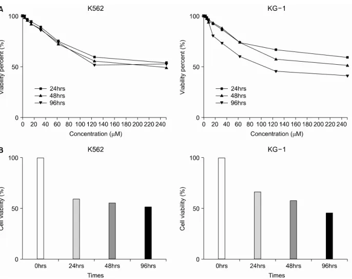
K562와 KG-1 세포주에 curcumin을 0~240μM까지 농도별로 처리했을 때 양 세포 모두 120μM까지는 curcumin 농도의 증가에 따라 세포 독성이 증가하였으 나 그 이상의 농도에서는 증가하는 정도가 둔화되었 다. Curcumin 노출 시간을 24, 48, 96시간으로 달리 하 였을 때 K562의 경우는 시간에 따른 차이가 없었으나 KG-1은 노출 시간이 길어짐에 따라 세포 독성이 증가 하는 소견이 관찰되었다(Fig. 2A). 이를 바탕으로 cur-
Fig. 2. Dose and tim e dependency of curcumin effect on the leukem ic cell line K562 and KG-1. Curcum in was treated at various concentrations and tim es (A) and then its concentration was fixed at 25μM (B). Curcum in at 110~130μM concentration reduced cell viability by 45~50% and KG-1 showed m ore tim e-dependent pattern of cytotoxicity to curcum in.
cumin 농도를 25μM로 고정하고 약물 처리 시간에 따 른 세포 독성 효과를 조사한 후 이를 막대그래프를 사 용하여 나타내었다(Fig. 2B). K562 세포주는 저농도에 서 시간에 따른 특별한 경향을 보이지 않았으나 KG-1 은 처리 시간이 길어질수록 세포 독성이 증가하였다.
즉, curcumin은 KG-1에서 시간 의존적인 세포 독성을 보였다.
2. Curcumin에 의한 세포 주기 변화 분석과 cyclin D, A, B의 변화
유세포 측정법을 이용하여 curcumin에 의한 세포 주 기 변화를 분석하였다. 대조군으로 curcumin을 처리하 지 않은 세포주를 사용했으며, curcumin 농도를 10μM 에 고정한 후 각각 24, 48, 72시간 후의 세포 주기를 관찰하였다. 이전 실험 결과를 바탕으로 예상한 것과
같이 KG-1이 curcumin 10μM의 낮은 농도에서 대조군 보다 G1기가 24시간 후 3배 이상 증가했으며 48시간 후와 72시간 후에서도 1.5배 이상 증가했다(Fig. 3). 그 러나 K562 세포주는 10μM의 curcumin 농도에서는 큰 변화가 없었다.
이어서 curcumin을 10μM로 고정시킨 후 cyclin D의 12, 24, 48시간의 발현 양상을 살핀 결과 KG-1의 경우 는 12시간과 24시간에는 감소하다 48시간 후에는 약간 증가하였고 K562에서는 대조군에 비해 시간이 지남에 따라 증가하는 양상을 보였다. cdk4 (cyclin-dependent kinase4)는 curcumin에 의해서 아무런 영향을 받지 않 는 것으로 나타났다(Fig. 4). 이로 보아 KG-1의 경우, curcumin이 12시간 이내에 G1/S기의 세포주기를 늘릴 때 cyclin D/cdk4의 접합체 중 cdk4보다는 cyclin D를 통해서 세포 주기를 정지시킨다고 생각했다.
Fig. 4. W estern blot results of cyclin D1, cdk4 at curcum in 10μM . The expressions of cdk4 and cyclin D1 were not decreased in K562 cells at this concentration.
Fig. 3. The effect of curcumin on the cell cycle progression. K562 and KG-1 were PI stained at 24hrs, 48hrs, 72hrs after treatm ent with curcum in 10μM . KG -1 cells in sub-G 1 phase were increased at 24hrs, but K562 cells appeared not affected.
Fig. 5. Curcum in effect on cell cycle and apoptosis. curcu- m in caused decreased expression of cyclin D1, B1, and PCNA but not cyclin A at after treatm ent of curcum in at 100 μM for 6hours in K562 cells. Apoptosis was induced by curcum in exposure of either 100μM for 6hours or 25μM for 24hrs.
K562의 경우 curcumin 농도를 높인 후 농도와 시간 을 달리하여 다시 cyclin A, B, D 및 PCNA (prolifer- ating cell nuclear antigen)를 Western blot으로 검사해 본 결과 cyclin D1, B와 PCNA가 주로 농도에 의존하여 감소하는 소견을 볼 수 있었다. 그러나, cyclin A는 변 화가 없었다(Fig. 5).
3. Erk와 Akt, Bcl-2의 발현
K562와 KG-1 세포주에서 curcumin의 또 다른 가능 한 성장 억제 작용 기전을 알아보기 위해 Erk (extra- cellular signal-regulated kinase)와 Akt 그리고 세포사 멸의 억제인자인 Bcl-2의 발현을 Western blot 방법을 통해 알아보았다. Curcumin 25μM을 두 세포주에 15 분, 30분, 1시간 처리 후의 발현 또는 활성화 양상을 살핀 결과 curcumin을 처리하기 전부터 이미 Erk가 활 성화되어 있었고(phospho-Erk) 1시간이 지난 후에도 Erk의 인산화는 감소하지 않고 오히려 약간 증가하는 형태를 보였다. 즉, 두 세포주는 이미 활성화된 Erk 경 로를 보이고 있었고 curcumin은 여기에 영향을 주지
않는 것으로 보였다.
Akt의 경우는 curcumin 25μM의 처리 30분 후부터 감소하는 것이 관찰되었다. 아울러 Akt와 관련되어 있 는 항 세포사멸 인자인 Bcl-2는 KG-1에서 빠른 발현과 동시에 30분 후 사라짐이 관찰되었다(Fig. 6).
4. Curcumin의 세포사멸 유도
세포사멸을 알아보기 위해 K562에서 procaspase-3와
Fig. 6. The expression of phospho-Erk Akt, bcl-2 after treatm ent with curcum in at 25μM Akt was decreased within an hour and bcl-2 was also reduced rapidly in KG -1 cell.
However, phospho-Erk was not affected by curcum in.
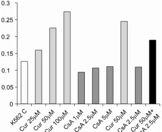
Fig. 7. Activity of caspase 3 treated with various con- centrations of curcum in, cyclosporin A (CsA), and cyclos- porin A+ curcum in in K562 cell.
cleaved-PARP (polyadenosine ribose polymerase)를 측 정하였다.12) Caspase-3가 최대로 활성화 되는 순간 (100 μM-6시간, 25μM-24시간)에서 cleaved-PARP의 85kDa 의 분리가 관찰되었다(Fig. 5). K562에서 세포사멸은 curcumin의 시간-농도에 비례하여 증가하는 것으로 보 였다.
이러한 세포사멸의 기전을 좀더 명확히 알고자 cy- closporin A (CsA)를 사용하였다. CsA는 calcineurin을 억제시키는 기전으로 mPTP (mitochondrial membrane permeability transition pore)을 비활성화시켜 cytoch- rome c의 세포질 내 방출을 막는다.13,14) K562 세포주 에 curcumin 50μM과 CsA 2.5μM을 동시에 24시간 동 안 처리했을 때 처리하지 않은 대조군에 비해 caspase 3의 활성도가 둔화되는 것을 볼 수 있었고(Fig. 7) 이는 curcumin이 K562 세포주에서 미토콘드리아 의존적인 경로를 통해 세포사멸을 일으키는 것을 시사한다.
고 찰
Curcumin은 만성백혈병 세포주와 급성백혈병 세포 주 모두에서 세포의 성장을 억제하였으며 본 실험에서 살펴본 결과 세포 주기의 조절과 세포사멸의 유도가 주요한 기전으로 생각한다. 암세포에서 과발현되는 PCNA를 curcumin이 감소시킨 것은 PCNA가 초기 G1/
S기에 관여하는 세포주기 관련 요소이므로 curcumin 이 세포 주기에 영향을 준다는 것을 시사한다.15) Cy- clin D는 cdk4와 복합체를 형성해 활성을 갖는데 bcr- abl 단백에 의해 과발현되어 세포의 분열을 촉진한 다.16) 본 실험에서 curcumin은 cdk4에는 아무런 영향 을 주지 않았고 cyclin D의 발현을 억제함으로써 G1/S 기의 arrest를 일으키는 것으로 보인다. Cyclin D의 발
현억제는 상위 조절 요소 NF-κB나 STAT3에 연차적 으로 작용하는 것도 생각할 수 있고,17) 최근에는 cur- cumin이 cyclin D의 유전자 발현을 직접적으로 억제한 다는 보고도 있다.18)
Akt는 세포의 분열, 증식과 생존에 필수적인 역할을 하는 중요 단백으로 많은 암에서 활성화되어 있다.
PI3K/Akt 경로는 cyclin D를 안정화시키며 p21Cip1 단 백을 조절하여 세포주기를 활성화시킨다. 또한 Akt는 Bad를 인산화시켜 bcl-2를 유리시킴으로써 세포의 생 존에 기여한다. 이 외에도 Akt에 의한 IκB kinase 활성 화, procaspase-9의 억제 등이 세포사멸을 억제하는 기 전으로 알려져 있다.19)
K562와 KG-1에서 curcumin에 의한 cyclin D의 감소 나 caspase의 활성화에 의한 세포사멸의 진행 등이 Akt 의 감소와 연관되어 보인다. Akt의 빠른 감소와 KG-1 에서 bcl-2가 30분 내로 사라짐은 이를 뒷받침하고 있 다. Bad, Bax가 증가할 것이라는 예상을 갖고 동일한 조건 하에 Bad의 발현 유무를 관찰한 결과 curcumin에 의해 Bad가 증가하는 양상을 나타내었다(결과는 제시 하지 않음). Caspase 3와 cleaved-PARP의 발현으로부 터 curcumin에 의한 세포사멸을 확인할 수 있었는데 일 반적으로 세포사멸의 유도에는 미토콘드리아로부터 cytochrome c의 유리가 일어나는 내재적 경로와 FADD (Fas-associated death domain)과 연관되는 외재적 경로 가 알려져 있다. CsA 병용처리 결과로부터 curcumin에 의한 세포사멸의 주요 기전이 미토콘드리아-관련 내재 경로임을 추측할 수 있고 이는 이전에 보인 Akt의 감
배경: 자연계에 존재하는 페놀계 화합물인 curcumin 은 심황의 뿌리에서 분리된 화합물로서 항염증 효과와 항암, 항산화효과가 있음이 알려지고 있다. 저자들은 골수성 백혈병 세포주인 K562와 KG-1에서 curcumin 의 효과와 그 기전을 알고자 하였다.
방법: 세포 독성 평가를 위해 MTT 방법을 사용하였
고, 세포 주기 평가는 유세포 측정법을 사용하였다. 그 외 각종 단백 발현 분석을 위해 Western blotting을 사 용하였고 caspase 활성을 측정하였다.
결과: 두 세포주는 curcumin에 의해 농도-의존적인 세포독성을 보였다. KG-1 세포주는 sub G1세포주기의 증가를 보였고 K562 세포주에서는 curcumin에 의해 cyclin D1, B1, PCNA (proliferating cell nuclear anti- gen)의 발현이 감소하였다. 또한 Akt의 감소와 함께 caspase-3의 활성화, PARP (polyadenosine ribose poly- merase)의 분리가 관찰되었다. 그러나 Erk 경로는 관 련이 없어 보였다. Cyclosporin A 병용 처리는 cur- cumin의 세포사멸 유도 효과를 약화시켰고 이는 cur- cumin에 의한 세포사멸에 있어서 미토콘드리아로부터 cytochrome c의 유리가 일어나는 내재적 경로가 중요 함을 시사하였다.
결론: Curcumin은 골수성 백혈병 세포주에서 세포분
열 정지와 세포사멸을 유도하였다. 그리고 이러한 세 포 사멸은 미토콘드리아에서의 cytochrome c의 유리에 의존하는 것으로 보였다.
참 고 문 헌
1) Liao S, Lin J, Dang MT, et al. Growth suppression of hamster flank organs by topical application of catechins, ali zarin, curcumin, and myristoleic acid.
Arch Dermatol Res 2001;293:200-5.
on bladder cancer cell line EJ in vitro. Zhong Yao Cai 2004;27:848-50.
6) Zhang J, Qi H, Wu C. Research of anti-proliferation of curcumin on A549 human lung cancer cells and its mechanism. Zhong Yao Cai 2004;27:923-7.
7) Duvoix A, Morceau F, Schnekenburger M, et al. Cur- eumin-induced cell death in two leukemia cell lines:
K562 and Jurkat. Ann N Y Acad Sci 2003; 1010:389- 92.
8) Han SS, Keum YS, Seo HJ, Surh YJ. Curcumin suppresses activation of NF-kappaB and AP-1 indu- ced by phorbol ester in cultured human promye- locytic leukemia cells. J Biochem Mol Biol 2002;
35:337-42.
9) Lin JK. Suppression of protein kinase C and nuclear oncogene expression as possible action mechanisms of cancer chemoprevention by curcumin. Arch Pha- rm Res 2004;27:683-92.
10) Korutla L, Kumar R. Inhibitory effect of curcumin on epidermal growth factor receptor kinase activity in A431 cells. Biochim Biophys Acta 1994;1224:
597-600.
11) Lin JK, Pan MH, Lin-Shiau SY. Recent studies on the biofunctions and biotransformations of curcu- min. Biofactors 2000;13:153-8.
12) Lazebnik YA, Kaufmann SH, Desnoyers S, Poirier GG, Earnshaw WC. Cleavage of poly (ADP-ribose) polymerase by a proteinase with properties like ICE.
Nature 1994;371:346-7.
13) Hansson MJ, Persson T, Friberg H, et al. Powerful cyclosporin inhibition of calcium-induced perme- ability transition in brain mitochondria. Brain Res 2003;960:99-111.
14) Ghribi O, DeWitt DA, Forbes MS, Arad A, Herman MM, Savory J. Cyclosporin A inhibits Al-induced cytochrome c release from mitochondria in aged rabbits. J Alzheimers Dis 2001;3:387-91.
PCNA acts as a stationary loading platform for tran- siently interacting Okazaki fragment maturation proteins. Nucleic Acids Res 2005;33:3521-8.
16) Andreu EJ, Lledo E, Poch E, et al. BCR-ABL induces the expression of Skp2 through the PI3K pathway to promote p27Kip1 degradation and proliferation of chronic myelogenous leukemia cells. Cancer Res 2005;65:3264-72.
17) Kwon YK, Jun JM, Shin SW, Cho JW, Suh SI.
Curcumin decreases cell proliferation rates through
cells. Int J Oncol 2005;26:1597-603.
18) Chen A, Xu J. Activation of PPAR{gamma} by cur- cumin inhibits Moser cell growth and mediates suppression of gene expression of cyclin D1 and EGFR. Am J Physiol Gastrointest Liver Physiol 2005;
288:G447-56.
19) Chang F, Lee JT, Navolanic PM, et al. Involvement of PI3K/Akt pathway in cell cycle progression, apo- ptosis, and neoplastic transformation: a target for cancer chemotherapy. Leukemia 2003;17:590-603.