40(2) : 118 122 (2009)
118
와송의 HL60백혈병세포의 Apoptosis유도 효과
오찬호1
*
·배진범1·김남석1·전권훈2·한광수3·이문준4·권권진51우석대학교식품생명공학과
,
2우석대학교약학대학,
3우석대학교대체의학과,
4전북대학교환경생명자원대학
,
5한국재활복지대학의료보장구과Effect of Orostachys japonicus A. Berger on Apoptosis Induction of Human Leukemia HL60 Cells
ChanHo Oh
1* , JinBeom Bae1, NamSeok Kim
1, Hoon Jeon
2, KwangSoo Han
3, MoonJun Lee
4 and Jin Kwon
5
1
Department of Food & Biotechnology, Woosuk University, Wanju 565-701
2
College of Pharmacology, Woosuk University, Wanju 565-701
3
Department of Alternative Medicine, Woosuk University, Wanju 565-701
4
College of Environmental & Bioresource Sciences, Chonbuk National University, Jeonju 561-756
5
Department of Prosthetics and Orthotics, Korea National College of Rehabilitation and Welfare, Pyongtaek 459-070
Abstract −
Methanol extracts of Orostachys japonicus A. Berger (OAB) were found to exhibit apoptosis induction of HL60 human acute promyelocytic leukemia cells. Treatment of OAB exerted strong cytotoxicity against HL60 cells. OAB induced DNA fragmentation of HL60 cells in a dose dependent manner. Nitric oxide production were also increased in OAB-treated RAW264.7 macrophage cell lines. Treatment of OAB increased the expression of p53 and iNOS gene and the expression of p53, NF-
κB and iNOS protein in cultured HL60 and RAW264.7 cells. These results suggest that OAB are effective on strong anti-cancer properties and can be useful as a chemo-preventive agents.
Key words −
Orostachys japonicus A. Berger, HL60 leukemia cell, apoptosis, p53, NF-
κB
와송
(
Orostachys japonicusA. Berger)
은岩松또는屋松으로불리우며
,
돌나물과에속한다년생초본으로서오래된기와지붕또는깊은산중의바위 중에서생육하는식물이 다
.
예로부터알려진주된약리효능으로는혈관수축,
호흡흥분및장관의긴장도증강작용1)등이보고되어있으며한 방에서는해열
,
지혈,
소종,
이뇨등에응용되고있고민간요법으로항암요법제로많이사용되고있지만자세한작 용기전에대한연구보고는거의없는실정이다
.
2,3)본연구자등은이미와송이면역체계에미치는영향등을관찰하 여발표4)를한바가있으며와송추출물의각종암세포에미 치는항암효능에관한연구보고가아직미흡한실정이어서 여기에서는와송의주로인간백혈병세포에미치는아폽토 시스유도효과를검토하고약간의지견을얻었기에보고하 고자한다
.
재료 및 방법
실험재료및추출 − 본실험에사용한와송은울산광명 당제약에서구입하여
100 g
을95%
의메탄올로추출한후,
여과한여액을
rotary evaporator
로농축한다음, freeze dryer
로동결건조하여
(
이하OAB
라함)
멸균PBS
에용해시켜사용하였다
.
계대배양세포주 − 본실험에사용한세포주로서는한국 세포주은행
(KCLB)
에서 분양받은HL60 (human acute promyelocytic leukemia cell)
인간급성전골수성백혈병세포주
, 293T (human embryonic kidney epithelial cell)
인간신장상피세포주및
RAW264.7 (mouse monocyte/macrophage cell)
대식세포주를사용하였다.
시약 및 기구 − 실험에사용한시약은
RPMI1640
배지, fetal bovine serum (FBS), phosphate buffered saline (PBS), propidium iodide (PI)
등은Sigma, Nonidet NP-40
*교신저자(E-mail):[email protected] (Tel):063-290-143
1
(Amresco), Taq DNA polymerase, M-MLV reverse transcriptase, Oligo (dT), PCR marker (Promega), dNTP set (Amersham), p53/iNOS/NF-
κB rabbit polyclonal IgG antibody, goat anti-rabbit IgG-HRP antibody, Coomassie Blue R-250(Santa Cruz)
등이며사용기기로는ELISA reader (Molecular Device, VERSAmax), Flow cytometer (Coulter, EPICS-XL), PCR system (Takara, PCR Thermal cycler Dice),
전기영동장치(Mupid-21)
등을사용하였다.
세포생존율측정 −
MTT
법5)에의해HL60
세포및293T
세포배양계에각농도의
OAB
를첨가하여48
시간동안37
oC
의
CO
2배양기(5%-CO
2, 95%-air)
내에서배양하였다.
배양종료
4
시간전에5 mg/ml
농도로DPBS-A(pH 7.4)
에희석한MTT
용액20
µl
를각well
에첨가하고, 0.1 N HCl
에녹인10% SDS 100
µl
로용해시켜18
시간동안은박지로빛을차단하였다
.
발색된각well
의흡광도를ELISA reader
를이용해서
570 nm
에서측정하고대조군의흡광도와비교하여세포생존율을백분율로환산하였다
.
Flow Cytometer
에의한Apoptosis
측정 −HL60
세포에각농도의
OAB
를첨가한후, 24
시간동안배양한다음,
세포를수집
,
세정(×3
회, 1,500 rpm, 5
분)
한후,
침전시킨세포분획에
PI(10
µg/ml)
를20
µl/1×10
6세포의농도로염색(4
oC, 30
분간반응)
한다음flow cytometer (Coulter, EPICS XL; excitation: 488 nm, emission: 620 nm)
를 이용해서Sub-G1 peak
를정량하였다.
6)Nitric oxide (NO)
측정 −Griess
방법7)에 준하여RAW 264.7
세포를5×10
5세포/ml
로조정하여12 well plate
의 각well
에부착시킨후,
검체를농도별로처리하였다. 37
oC
의CO
2 배양기내에서24
시간동안배양한후NO
를측정하였다
. NO
측정은Griess
시약(1% sulfanilamide, 0.1% N- naphtyl-ethylenediamine
·2HCl/2.5% phosphoric acid)
을사용하여
570 nm
에서microplate reader
로흡광도를측정하고미리작성한
NaNO
2의검량선에의해NO
2-의농도를환산 하였다.
RT-PCR
−HL60
세포및RAW264.7
세포에OAB (1~100
µ
g/ml)
를첨가한후, 24
시간동안배양한후,
세포로부터RNA
를분리하여p53
및iNOS
유전자를확인하였다. Total RNA
는Trizol reagent
를이용하며제조회사의방법에준하였다
. cDNA
는M-MLV reverse transcriptase
와oligo dT
를primer
로이용하여합성하고, Taq DNA polymerase
를이용하여
Takara PCR thermocycler (DICE)
에서30 cycle
동안증폭하였다
.
각각의cycle
은95
oC
에서30
초간denaturation
시킨후
, 55
oC
에서30
초간annealing
시키고, 72
oC
에서30
초간
extension
시켰다. PCR
산물은1% agarose gel
상에서전기영동하고
, ethidium bromide
로염색하여확인하였다.
8)Western Blotting
−HL60
세포 및RAW264.7
세포에OAB (1~100
µg/ml)
를첨가한후, 24
시간동안배양하고1%
NP-40 lysis buffer (250 mM NaCl, 25 mM Tris-HCl pH 7.5, 5 mM EDTA pH 8.0, 1% NP-40, 0.6 mM PMSF, 10
µg/ml aprotinin, 1
µg/ml pepstatin A)
로단백질을분리하였으며
, Coomassie blue R-250
을이용한Bradford
법을사용해정량하였고
, Western blot analysis
를하기위해단백질20
µg
을12.5% SDS-PAGE
를 실행한 다음, nitrocellulose membrane
으로transfer
한후, 5% Non-Fat Dry Milk
로18
시간 동안
blocking
하고, p53/NF-
κB/iNOS rabbit polyclonal IgG antibody (1:1000 dilution)
로1
차염색하여4
oC
에서1
시간동안반응시키고
, goat anti-rabbit IgG-HRP conjugated antibody
로2
차염색하여동일한방법으로반응시켜X-ray
필름을
enhanced chemiluminescence (ECL)
법으로감광하였다.
9)통계학적처리 − 모든
data
는mean±S.D.
로나타내었고통계처리는
Student's t-test
를실시하여, p<0.05
이하를유의성이있는것으로판정하였다
.
10)결과 및 고찰
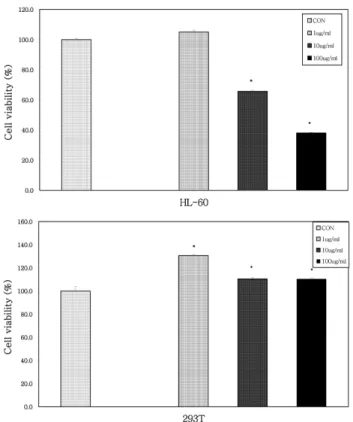
와송
(OAB)
의항암효과를주로아폽토시스와관련하여검토하고자계대배양한인간급성전골수성백혈병세포주인
HL60
세포 배양계에메탄올로추출한OAB
를 농도별(1-
100
µg/ml)
로첨가해서백혈병세포의생존율에미치는효과를살펴보았다
. MTT
법으로측정한결과에서OAB
는HL60
백혈병세포의생존율을농도의존적으로감소시키고있음을 확인하였다
(Fig. 1).
또한 대조군으로사용한정상293T (human kidney epithelial cell)
세포에서는 세포생존율에별다른영향이없었다
.
이러한결과는OAB
가백혈병세포의증식을억제시켜항암활성을나타내는것으로추정되며동 일한농도에서정상의신장상피세포에서는세포독성이없 음을확인하였다
. (Fig. 1)
다음에는
HL60
세포의아폽토시스유도에미치는효과를검토하기위하여
HL60
세포배양계에OAB(1~100
µg/ml)
를첨가해서
Flow cytometer
를이용해서Sub G1 peak
를측정한결과
, HL60
세포의apoptosis
를농도의존적으로유의성있게촉진시키고있음을확인하였는데
(Fig. 2),
이결과는상기의세포생존율을억제시키는결과와일맥상통하는결과 이고
, OAB
가L1210
및U937
백혈병세포의아폽토시스를증가시켜서항암작용을가진다는이전의연구보고4)와도일 치하는결과라할수있으며
,
또한과산화수소로유도한신경세포의아폽토시스를
OAB
에의해보호한다는연구결과11)에비추어보면
OAB
는세포에따라서선택적으로작용하고있는것으로사료되어매우의미있는결과라추정된다
. (Fig. 2)
또한상기의결과들을뒷받침하는작용기전을확인하기 위하여이번에는
NO(Nitric oxide)
생성에미치는와송의효과를살펴보고자
,
대식세포주인RAW264.7
세포배양계에OAB
를농도별로처리하고NO
생성에미치는효과를살펴본결과
, OAB 10
및100
µg/ml
의농도에서대조군에비하여유의성있는
NO
생성의증가가관찰되었다(Fig. 3).
이결과는
OAB
가대식세포에서생성되는NO
를증가시킴으로써면역계의활성을조절하여결과적으로암세포의아폽토 시스를 유도하는 것이라 추정된다
. NO
는 생체내에서vasodilating agent, neurotransmitter
로서의작용이외에세균이나암세포에대한비특이적효능물질로서잘알려져있 고특히생쥐에서는대식세포등의탐식작용을지니는세
포에서분비되는물질이다
.
12,13)상기의결과는항암성분인flavone-8-acetic acid
와xantherone-4-acetic acid
가대식세포를활성화시키며
NO
생성이촉진되어항암작용을한다는이전의연구보고14)와유사한결과라할수있다
. (Fig. 3)
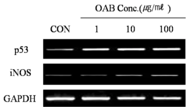
상기의결과들을재확인하기위하여항암유전자인
p53
및NO
생합성효소(iNOS)
유전자발현에미치는효과를검토해보았다
.
결과는HL60
세포및RAW264.7
세포 배양계에OAB(1~100
µg/ml)
를첨가하여배양한결과,
대조군에비하여현저한
p53
유전자및iNOS
유전자의발현이촉진되었음이관찰되었다
(Fig. 4).
이러한결과는OAB
가NO
생합성효소인
iNOS
유전자의활성을증강시키고결과적으로는종양억제유전자인
p53
유전자를활성화시킨결과,
백혈병세포에대한항암활성을나타내고있음을뒷받침하는중요한결 과라시사된다
. (Fig. 4)
최종으로상기의백혈병세포의아폽토시스를유도하는활 성을단백질발현패턴으로확인하고자
HL60
세포에OAB (1-100
µg/ml)
를첨가해서24
시간동안배양한 다음p53, NF-
κB
단백질발현과RAW264.7
세포에OAB
를첨가해서iNOS
단백질발현을Western blot
으로확인해본결과,
대조군에비하여
OAB
를첨가한군에서상기단백질들의발현이농도의존적으로증가되었다
(Fig. 5). p53
단백질은암세포Fig. 1.
Effect of the OAB on the proliferation of HL60 (human acute promyelocytic leukemia), and normal 293T (human embryonic kidney epithelial) cell lines in vitro . Sample was added into the cultured different cell lines at indicated concentration and cultured for 48 hours at 37
oC. The proliferation of the cells was assayed by MTT method. Each bar represents the mean±S.D. of three determinations.
*: Significantly different from control group(*p<0.05)
Fig. 2.
Effect of the OAB on the DNA fragmentation of HL60 leukemia cell line. Sample were treated to cultured HL60 leukemia cell lines, incubated for 24 hours, and then cells were collected and the DNA fragmentation (Sub G
1peak) was measured by a flow cytometer staining with propidium iodide.
The data represents the mean±S.D. of 3 experiments.
*: Significantly different from control group(*p<0.05)
Fig. 3.
Effect of the OAB on the nitric oxide production from RAW264.7 macrophage cell line. Sample was added into the cultured RAW264.7 macrophage cell line and incubated in a 5% CO
2incubator at 37
oC for 24 hours. The OD of each well was measured at 570 nm with a ELISA reader. Nitric oxide standard curve were measured with NaNO
2. Each bar represents the mean±S.D. of three determinations.
CON(-): Non-treatment, CON(+): LPS-treatment
*: Significantly different from control group(*p<0.05)
의아폽토시스를비롯하여광범위하게종양억제에관여하 는단백질로알려져있으며
, NF-
κB
는면역,
염증스트레스,
세포증식및아폽토시스를조절하는데관여하는전사인자
로알려져있다
.
15-19)본실험결과에서OAB
는HL60
세포에서의
p53
등항암관련단백질과NF-
κB(p50)
의induction
을농도의존적으로증가시키며또한
NO
생합성효소인iNOS
단백질발현을농도의존적으로증가시켜암세포의아폽토 시스를유도하는항암활성을보유하고있는것으로추정된 다
. (Fig. 5)
결 론
와송
(
Orostachys japonicusA. Berger; OAB)
메탄올추출물의
HL60
백혈병세포에대한항암작용을연구하기위하여
HL60(
인간급성전골수성백혈병세포주)
세포의아폽토시스유도에미치는효과를관찰한결과다음과같은결론 을얻었다
.
와송메탄올추출물
(OAB)
의세포독성을확인하기위하여MTT assay
를수행한결과HL60
백혈병세포의증식을농도의존적으로억제시켰으며대조군으로사용한정상
293T(
신장상피세포주
)
에서는고농도에서도세포독성이없음을확인하였다
. OAB
는HL60
세포의apoptosis
를유의성있게증가시켰고
,
대식세포주인RAW264.7
세포에서의NO
생성을농도의존적으로촉진시켰다
.
또한RT-PCR
에의한 아폽토시스관련유전자의
mRNA
발현을확인한결과, HL60
세포에서종양억제인자인
p53
유전자및RAW264.7
세포에서NO
생합성효소인
iNOS
유전자의발현을농도의존적으로촉진시켰다
.
상기의결과를최종확인하고자HL60
세포에서p53, NF-
κB
단백질발현과RAW264.7
세포에서iNOS
단백질발현을관찰하기위하여
Western blot analysis
를수행한결과, p53, NF-
κB
및iNOS
단백질의발현이농도의존적으로증가되었다
.
이상의실험결과에서와송은대식세포에서생성되는
NO
를경유해서급성전골수성백혈병세포의apoptosis
를유도하는항암활성을보유하는것으로추정되며
,
장래와송의항암병용요법제로서의개발가능성을시사하는결과 이다
.
사 사
“
이논문은2009
년우석대학교교내연구비지원및교육과학기술부
(
지역거점연구단육성사업/
헬스케어기술개발사업단
)
로부터지원받아수행된연구임” 인용문헌
1.
鄭普燮(1990)
鄕藥大事典,
永林社, p. 600.
2.
박희준(1992)
와송의화학성분및항돌연변이활성에관 한연구,
한국생명과학회지,
2(1): 69-71.
3.
양서현(1989)
靈芝.
山慈姑,
仙鶴草,
瓦松이흰쥐의 자연 살해세포에 미치는 영향,
대한 한의학회지,
10(2): 103- 114.
4.
권진,
한광수(2004)
瓦松추출물이면역체계에 미치는영 향,
한국약용작물학회지,
12(4): 315-320.
5. Mosmann, T. (1983) Rapid colorimetric assay for cellular growth and survival application to proliferation and cytotoxic assays. J. Immunol. Methods,
65: 55-63.
6. Oh, S. H. and Oh, C. H. (2003) Brown rice extracts with enhanced levels of GABA stimulate immune cells. Food Sci.
Biotechnol .
12: 248-252.
7. Albina, J. E., Cui, S., Mateo, R. B. and Reichner, J. S. (1993) Nitric oxide-mediated apoptosis in murine peritoneal mac-
Fig. 4.
Effect of OAB on the p53 and iNOS gene expression in HL60 cell and RAW264.7 cell. Treatment of OAB for 24 hours leads to increase the p53 gene expression in HL60 leukemia cell and iNOS gene expression in RAW264.7 cell.
Poly(A
+)RNA isolated from untreated cells(CON) and cells treated with OAB for 24 hours.
Fig. 5.
Western blot analysis of OAB on the expression of p53, NF-
κB in HL60 leukemia cell and iNOS in RAW264.7 cell. Treatment of OAB for 24 hours leads to increase the expression of p53 and NF-
κB protein in HL60 cell and iNOS expression in RAW264.7 cell.
Lane 1; CON(negative control), Lane 2; OAB 1
µg/ml, Lane
3; OAB 10
µg/ml, Lane 4; OAB 100
µg/ml
rophages. J. Immunol.
150: 5080-5085.
8. Miyashita, T. and Reed, J. C. (1995) Tumor suppressor p53 is a direct transcriptional activator of the human bax gene.
Cell,
80: 293-299.
9. Towbin, H., Staehelin, T. and Gordon, J. (1992) Electro- phoretic transfer of proteins from polyacrylamide gels to nitrocellulose sheets: procedure and some applications. Bio- technology ,
24: 145-149.
10. Dowdy, S. and Wearden, S. (1983) Statistics for research, Wiley, New York, p. 262.
11. Yoon, Y. S., Kim, K. S., Hong, S. G., Kang, B. J., Lee, M. Y.
and Cho, D. W. (2000) Protective effects of Orostachys japonicus A. Berger(Crassulaceae) on H
2O
2-induced apop- tosis in GT1-1 mouse hypothalamic neuronal cell line. J. Eth- nopharmcol.
69(1) : 73-78.
12. Bredt, D. S. and Snyder, S. H. (1994) Nitric oxide: a phys- iologic messenger molecule. Annu. Rev. Biochem .,
63: 175- 13. Panossian, A. G., Oganessian, A. S., Ambartsumian, M., Gab- 195.
rielian, E. S., Wagner, H. and Wikman, G. (1999) Effects of heavy physical exercise and adaptogens on nitric oxide con- tent in human saliva. Phytomedicine ,
6(1): 17-26.
14. Thomsen, L. L., Ching, L. M. and Baguley, B. C. (1991) Evi-
dence for the production of nitric oxide by activated mac- rophages treated with the antitumor agents flavone-8-acetic acid and xantherone-4-acetic acid. Cancer Res .,
51: 6073- 6078.
15.
전인성,
허민석,
최항문,
이삼선,
최순철(2000) DMBA
유 도햄스터협낭암발생과정에서방사선조사가증식세포 핵항원(PCNA)
의 발현과apoptosis
유발에 미치는 영향,
대한구강악안면방사선학회지
,
30(3): 207-216.
16. Floryk, D and Huberman, E. (2005) Differentiation of andro- gen-independent prostate cancer PC-3 cells is associated with increased NF-
κB activity, Cancer Res .,
65(24): 11588-11596.
17. Dai Y, Rahmani M. and Grant S. (2003) An intact NF-
κB pathway is required for histone deacetylase inhibitor-induced G1 arrest and maturation in U937 human myeloid leukemia cells, Cell Cycle ,
2: 467-472.
18. Kim J. Y., Ahn M. H., Jun H. S., Jung J. W., Ryu J. S., Min D. Y. (2006) Toxoplasma gondii inhibits apoptosis in infected cells by caspase inactivation and NF-
κB activation. Yonsei med. Journal,
47(6): 862-869.
19. Kastan, M. B., Canman, C. E., and Leonard, C. J. (1995) p53, cell cycle control and apoptosis: implications for cancer. Can- cer Metas. Rev,
14(1): 3-15.
(2009년 5월 9일 접수)