저작자표시-비영리-변경금지 2.0 대한민국 이용자는 아래의 조건을 따르는 경우에 한하여 자유롭게 l 이 저작물을 복제, 배포, 전송, 전시, 공연 및 방송할 수 있습니다. 다음과 같은 조건을 따라야 합니다: l 귀하는, 이 저작물의 재이용이나 배포의 경우, 이 저작물에 적용된 이용허락조건 을 명확하게 나타내어야 합니다. l 저작권자로부터 별도의 허가를 받으면 이러한 조건들은 적용되지 않습니다. 저작권법에 따른 이용자의 권리는 위의 내용에 의하여 영향을 받지 않습니다. 이것은 이용허락규약(Legal Code)을 이해하기 쉽게 요약한 것입니다. Disclaimer 저작자표시. 귀하는 원저작자를 표시하여야 합니다. 비영리. 귀하는 이 저작물을 영리 목적으로 이용할 수 없습니다. 변경금지. 귀하는 이 저작물을 개작, 변형 또는 가공할 수 없습니다.
1
A THESIS
FOR THE DEGREE OF MASTER OF SCIENCE
Piceatannol enhances TRAIL-induced
apoptosis in human leukemia THP-1 cells
through Sp1- and ERK-dependent DR5
up-regulates
Kang Chang-Hee
Department of Marine Life Science
GRADUATE SCHOOL
JEJU NATIONAL UNIVERSITY
2
Piceatannol enhances TRAIL-induced apoptosis
in human leukemia THP-1 cells through Sp1- and
ERK-dependent DR5 up-regulation
Chang-Hee Kang
(Supervised by Professor Gi-Young Kim)
A thesis submitted in partial fulfillment of the requirement
For the degree of Master of Science
2012. 02.
This thesis has been examined and approved.
Thesis director, Seungheon Lee, Prof. of Marine Life science
Young Hyun Choi, Prof. of Dongeui University College of Oriental Medicine
Gi-Young Kim, Prof. of Marine Life science
.
Department of Marine Life Science
GRADUATE SCHOOL
3
CONTENTS
ABSTRACT... 5. 1. INTRODUCTION... 7. 2. MATERIALS AND METHODS... 9.
2.1. Reagents and antibodies
2.2. Cell culture and treatment with drugs 2.3. Flow cytometric analysis
2.4. In vitro caspase-3 activity assay 2.5. DNA fragmentation assay 2.6. Western blot analysis 2.7. RT-PCR analysis 2.8. Luciferase assays
2.9. Chromatin immunoprecipitation (ChIP) assay 2.10. Statistical analysis
3. RESULTS... 14. 3.1. PIC induces apoptosis in THP-1 cells via mitochondrial dysfunction
3.2. PIC triggers TRAIL-induced apoptosis in THP-1 cells
3.3. PIC enhances PIC/TRAIL induced-apoposis via up-regulation of DR5 expression 3.4. PIC up-regulates DR5 expression via Sp1 binding on DR5 promoter region 3.5. Activation of ERK is required for the enhanced DR5 expression
4. DISCUSSION... 35. 5. REFERENCES... 39.
4
LIST OF FIGURES
Fig. 1. PIC reduces a dose-dependent viability in THP-1 cells.
………. 17. Fig. 2. PIC sensitizes TRAIL-induced apoptosis in human leukemia cell.
………. 22. Fig. 3. PIC sensitizes TRAIL-induced-apoptosis via up-regulation of DR5.
………. 26. Fig. 4.PIC enhances DR5 expression via activation of Sp1.
………. 30. Fig. 5. PIC-induced pDR5/-605 promoter activation is mediated by the ERK signal pathway.
5 ABSTRACT
Although piceatannol (PIC) is known to mediate anti-cancer, anti-inflammatory, and
anti-oxidant activities, little is known about the mechanism of PIC in terms of tumor
necrosis factor-related apoptosis-inducing ligand (TRAIL)-induced apoptosis. In this
study, we examined whether combined treatment with PIC and TRAIL
synergistically induces apoptosis in THP-1 leukemia cells. Results indicate that PIC
substantially enhances TRAIL-induced cell death including DNA fragmentation and
poly(ADP-ribose) polymerase cleavage. Consistent with TRAIL-induced apoptosis,
PIC significantly increased the mRNA and protein expression levels of DR5, a death
receptor of TRAIL. Further, PIC enhanced DR5 promoter activity via Sp1 activation.
Interestingly, the DR5 chimera antibodies significantly suppressed PIC and
TRAIL-mediated apoptosis. The inhibitor of ERK also decreased PIC and TRAIL-induced
apoptosis by blocking DR5 expression. In conclusion, our results suggest that PIC
sensitizes TRAIL-induced apoptosis via Sp1- and ERK-dependent DR5
up-regulation.
6
ABSTRACT IN KOREAN
Piceatannol (PIC)의 항 암, 항 염증, 항 산화 효과에 대해서는 알려져 있으나 PIC와 TRAIL의 병행 처리에 대한 mechanism은 거의 밝혀져 있지 않기 때문에 이번 연구를 진행 하였다. 이 연구에서 우리는 PIC와 TRAIL의 병행 처리가 사람의 백혈병 세포주인 THP-1 세포에서 synergistically 하게 세포사멸 유도 효과를 갖고 있음을 확인 하였다. 결과들을 보면, PIC는 TRAIL이 유도 하는 DNA fragmentation과 PARP의 cleavage의 형성을 아주 강하게 증가시켰음을 알 수 있다. 이러한 효과는 PIC가 TRAIL 수용체인 death receptor 5 (DR5)의 발현을 mRNA에서부터 발현을 증가 시켰기 때문이다. 이것은 PIC에 의해서 DR5의 주요 전사인자인 Sp1이 핵 안으로 translocation 활성이 증가하여 DNA 결합 활성이 증가 함에 따라 DR5의 promoter활성이 증가에 의한 것이었다. 흥미로운 것은, DR5와 TRAIL의 결합을 방해 하기 위하여 DR5 결합 항체를 제작 하여 처리 하게 되면 PIC와 TRAIL이 유도 하던 세포 사멸 효과가 강하게 억제가 되었다. 이에 따라 DR5의 증가가 세포사멸에 강하게 영향을 미치고 있음을 증명하였다. 뿐만 아니라, MAPK 중 하나인 ERK의 활성 억제제를 처리 하였을 때 역시 세포사멸 유도 효과가 억제 됨을 확인 하였고 이는 ERK 억제제가 DR5의 발현을 억제 하였기 때문이었다. 결론적으로 우리의 결과는 PIC가 Sp1과 ERK 의존적으로 DR5의 발현을 증가시키기 때문에 TRAIL의 세포 사멸 유도 효과를 강하게 한다는 것을 증명하였다.
7
1. INTRODUCTION
Tumor necrosis factor-related apoptosis-inducing ligand (TRAIL), a member of
the TNF superfamily members, has been established to induce apoptosis in various
human cancer cells in vivo and in vitro, without inducing any side-effects on normal
cells (Wiley et al., 1995; Kischkel et al., 2000; Van Geelen et al., 2004). Previous
studies have demonstrated that TRAIL induced-apoptosis is mediated by two cellular
membrane proteins, TRAIL receptor, DR4 or DR5 (Sheridan et al., 1997). Binding of
TRAIL to DR4 or DR5 leads to the activation of Fas-associated death domain and
procaspase-8 (Chaudhary et al., 1997; Pan et al., 1997a; Pan et al., 1997b), which
then forms death-inducing signaling complex, thereby inducing death signaling and
the apoptosis pathway (Muzio et al., 1996; Sprick et al., 2000). Nevertheless, a recent
study has demonstrated that many tumor cells acquire resistance to TRAIL-induced
apoptosis through down-regulation of their death receptors (Jin et al., 2006). Thus, it
is important to develop an agent that could regulate the DR4 and/or DR5 expression
and overcome TRAIL resistance. Recent evidence suggests that treating leukemia
cells with a combination of TRAIL and dihydroflavonol BB-1 or honokiol enhances
TRAIL-induced apoptosis (Hasegawa et al., 2006; Raja et al., 2008). Consequently, a
8
a novel tool in developing anti-cancer therapy.
Piceatannol (PIC; 3,5,3’,4’-tetrahydroxytrans-stilbene), a phenolic compound and
an analogue of resveratrol naturally occurring in grapes and red wine (Roupe et al.,
2006), has been shown to possess anti-cancer properties in leukemia, melanoma, and
other cancer cell lines. The anti-cancer activity of PIC appears to occur primarily
through reduction of anti-apoptotic protein Bcl-2 and inhibitors of apoptosis (IAP)
family expression, and through caspase activation (Kuo and Hsu 2002; Larrosa et al.,
2004; Chowdhury et al., 2005; Kim et al., 2008b). In addition, PIC has also been
shown to possess anti-inflammatory activity via down-regulation of nuclear
transcription factor (NF-κB) (Islam et al., 2004; Jin et al., 2006). However, the
precise mechanism of PIC on TRAIL-mediated cancer cell death has not been fully
elucidated thus far.
In this study, we examined whether the combined treatment of PIC and TRAIL
(PIC/TRAIL) induces apoptosis in human leukemia THP-1 cells. We found that the
combined treatment with PIC/TRAIL increases cell death via up-regulated DR5
expression through activation of extracellular signal-regulated protein kinase (ERK)
and Sp1. Together, findings suggest that PIC is an ideal candidate for
9
2. MATERIALS AND METHODS
2.1. Reagents and antibodies
PIC, 3-(4,5-dimethylthiazol-2-yl)-2,5-diphenyltetrazolium bromide (MTT), and
propidium iodide (PI) were purchased from Sigma (St. Louis, MO). PIC was
dissolved in DMSO (vehicle control). PD98059, SB203580, and SP600125 were
obtained from Calbiochem (San Diego, CA). RPMI 1640 medium and fetal bovine
serum (FBS) was purchased from Invitrogen Corp. (Carlsbad, CA) and GIBCO-BRL
(Gaithersburg, MD), respectively. Antibodies against caspase-3, caspase-8, caspase-9,
cytochrome-c, DR4, DR5, and PARP were purchased from Santa Cruz
Biotechnology (Santa Cruz, CA). Antibodies against Sp1, nucleoline, p-JNK, JNK,
p-ERK, ERK, p-p38 and p38 were purchased from Cell Signaling (Beverly, MA).
Antibody against β-actin was obtained from Sigma. Peroxidase-labeled rabbit and
mouse polyclonal immunoglobulins were purchased from KOMA Biotechnology
(Seoul, Republic of Korea).
2.2. Cell culture and treatment with drugs
Human monocytic leukemia cells (THP-1 and U937) and human acute
10
Collection (Manassas, VA). Cells were cultured in RPMI 1640 supplemented with
10% FBS and antibiotics (Sigma). Cells were seeded at 1 × 10⁵ cells/ml and treated
with the indicated concentrations of PIC, TRAIL or PIC/TRAIL. After treatment for
24 h, cell viability was determined by MTT assays and by under microscope,
respectively.
2.3. Flow cytometric analysis
Cells were fixed with 1 U/ml RNase A (DNase free) and 10 µg/ml propidium
iodide (Sigma) overnight at room temperature in the dark. A FACSCalibur flow
cytometer (Becton Dickenson; San Jose, CA) was used to analyze the level of
apoptotic cells containing sub-G1 DNA content. For annexin-V staining, live cells
were incubated with annexin-V (R&D Systems, Minneapolis, MN) according
manufacturer’s instructions.
2.4. In vitro caspase-3 activity assay
A caspase-3 activation kit was used according to the manufacturer’s protocol (R&D systems, Minneapolis, MN) to measure the activity of caspase-like protease.
11
Cells were lysed in a buffer containing 10 mM Tris (pH 7.4), 150 mM NaCl, 5
mM EDTA, and 0.5% Triton X-100 for 30 min on ice. Lysates were vortexed and
cleared by centrifugation at 10,000 g for 20 min. Fragmented DNA in the supernatant
was extracted with an equal volume of phenol/chloroform/isoamyl alcohol mixture
(25:24:1) and analyzed.
2.6. Western blot analysis
Total cell extracts were prepared using the PRO-PREP protein extraction solution
(iNtRON Biotechnology; Sungnam, Republic of Korea). Total cell extracts were
separated on polyacrylamide gels and standard procedures were used to transfer them
to the nitrocellulose membranes. The membranes were developed using an ECL
reagent (Amersham; Arlington Heights, IL).
2.7. RT-PCR analysis
Total RNA was extracted from THP-1 cells using the TRIzol reagent (Invitrogen;
Carlsbad, CA). RT-PCR was performed following the manufacturer’s protocol
(One-Step RT-PCR Premix; Daejeon, Republic of Korea). The sense primer 5’-GTC TGC
12
CTA TG-3’ were used to amplify human DR5 mRNA. The sense primer 5’- CTG
AGC AAC GCA GAC TCG CTG TCC AC-3’ and the anti-sense primer 5’- TCA
AAG GAC ACG GCA GAG CCT GTG CCA T-3’ were used to amplify human
DR4 mRNA. For glyceraldehyde-3-phosphate dehydrogenase (GAPDH), the sense
primer 5’-CGT CTT CAC CAT GGA GA-3’ and the anti-sense primer 5’-CGG CCA
TCA CGC CCA CAG TTT-3’ were used.
2.8. Luciferase assays
The pDR5/-605 plasmid [containing DR5 promoter sequence (-605/+3)] was
kindly provided by Dr. T. Sakai (Kyoto Prefectural University of Medicine, Kyoto,
Japan) (Yoshida et al., 2001). Briefly, THP-1 cells were seeded onto 6 well plates at
a density of 5 ´ 104 cells/ml and grown overnight. The cells were co-transfected with
1 µg of pDR5/-605 plasmid construct and 0.2 µg of the pCMV-β-galactosidase
plasmid by the LipofectAMINE Plus reagent (Life Technologies), in accordance with
the manufacturer’s instructions. Luciferase activity was normalized by
β-galactosidase activity in cell lysates and expressed as an average of three
13
2.9. Chromatin immunoprecipitation (ChIP) assay
The ChIP assay was performed using the EZ-ChipTM assay kit according to the
manufacturer’s protocol (Upstate Biotechnology; Lake Placid, NY). The primers
used for the amplification of the Sp1 binding site of DR5 promoter region were as
follows: 5’-GCC AGG GCG AAG GTT A-3’ (sense) and 5’-GGG CAT CGT CGG
TGT AT-3’ (antisense) (276-bp DNA product).
2.10. Statistical analysis
All data were derived from at least three independent experiments. The images
were visualized with Chemi-Smart 2000 (Vilber Lourmat, Marine; Cedex, France).
Images were captured using Chemi-Capt (Vilber Lourmat) and transported into
Photoshop. All bands were quantified by Scion Imaging software
(http://www.scioncorp.com). Statistical analyses were conducted using SigmaPlot
software (version 6.0). Values were presented as mean ± SD. Significant differences
between the groups were determined using the unpaired Student’s t-test. Statistical
14 3. RESULTS
3.1. PIC induces apoptosis in THP-1 cells via mitochondrial dysfunction
In order to investigate whether PIC induces anti-proliferative activity, we treated
the indicated concentrations of PIC in THP-1 cells for 24 h. Treatment with PIC
resulted in dose-dependent inhibition of cell viability (Fig. 1A). PIC also inhibited
cell growth in HL60 and U937 leukemia cells (data not known). These results were
further confirmed by light microscopic and FACS analysis. We found that cell
shrinkage (upper panel), annexin-V+ populations (middle panel), and sub-G1 phases
(lower panel) were significantly increased at more over 30 μM PIC (Fig. 1B).
Furthermore, we attempted elucidate the putative apoptotic effect of PIC by
mitochondrial dysfunctions using Western blot analysis. Treatment with PIC resulted
in a significant increase in the cleavage of capase-3, -8, -9, and PARP (Fig. 1C). In
consistent with the cleavage of effector molecules, PIC highly increased caspase-3
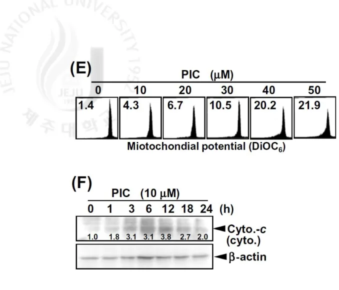
activity (Fig. 1D). PIC treatment also decreased mitochondrial potential in a
dose-dependent manner (Fig. 1E). Thus, we also found that cytochrome-c is released into
cytosol by treatment with PIC (Fig. 1F). These results indicate that PIC triggers
17
Fig. 1. PIC reduces a dose-dependent viability in THP-1 cells. Cells were seeded at 1
´ 105 cells/ml and treated with the indicated concentrations of PIC for 24 h. (A) Cell
viability was determined by MTT assay. (B) The cellular morphology of cells with or
without PIC for 24 h was examined under light microscopy (´ 400) (upper panel).
Annexin-V+ population (middle panel) and sub-G
1 cell distribution (lower panel) of
THP-1 cells were analyzed using flow cytometer. (C) For analyzing expression of
apoptotic proteins, cells were treated with PIC for 24 h and lysed. Samples (30 µg
protein/lane) were subjected to 10% SDS-PAGE, transferred to nitrocellulose
18
determined following the manufacturer’s protocol. (E) Mitochondrial membrane
potential was measured by DiOC6 dye using flow cytometry. (F) The translocation of
cytochrome-c into cytosol was analyzed by Western blot analysis. β-Actin was used
as a loading control. Data are representative of three independent experiments. Each
point represents the mean ± SD of three independent experiments. The significance
was determined by Student’s t-test (*, p < 0.05 vs. vehicle control). Cas.-8,
19
3.2. PIC triggers TRAIL-induced apoptosis in THP-1 cells
To investigate the effects of PIC on TRAIL-mediated apoptosis, THP-1 cells were
treated with sub-cytotoxic 10 μM PIC or 20 ng/ml TRAIL alone, and PIC/TRAIL.
We did not find any cytotoxicity in treatment with PIC or TRAIL alone (Fig. 2A).
However, treatment with both PIC and TRAIL remarkably triggered THP-1 cell
death to approximate 50%. During this process, the combined treatment significantly
increased caspase-3 activity (Fig. 2B). Next, we examined the cytotoxic effects in
combination with PIC and TRAIL in other human leukemia cells U937 and HL60. In
consistent with the date in THP-1 cells, PIC or TRAIL alone did not significantly
induce cytotoxicity, however the cytotoxicity was remarkably increased by treatment
with a combination of PIC and TRAIL (Fig. 2C). To assess the effects more detail,
we analyzed the cell populations of annexin-V+ and sub-G
1 phase using flow
cytometer. Treatment with PIC or TRAIL alone induced limited cell death (< 10%) at
24 h (Fig. 2D). However, PIC/TRAIL significantly increased the accumulation of
annexin-V staining (upper panel) and sub-G1 phase (lower panel) in THP-1 cells.
Western blot analysis also revealed that PIC/TRAIL increases PARP cleavage to a
greater extent than do PIC or TRAIL alone (Fig. 2E). Furthermore, DNA
20
fragmentation treated with PIC/TRAIL, but not in cells treated with PIC or TRAIL
alone (Fig. 2F). These findings suggest that PIC treatment enhances apoptosis
22
Fig. 2. PIC sensitizes TRAIL-induced apoptosis in human leukemia cell. THP-1 cells
were treated with 10 µM PIC alone, 20 ng/ml TRAIL alone, or PIC/TRAIL for 24 h.
(A and C) Cell viability was determined by MTT assay [(A); THP-1 and (C); U937
and HL60]. (B) Caspase-3 activity was determined following the manufacturer’s
protocol. (D) Annexin-V+ and sub-G
1 populations were analyzed using flow
23
lysates were resolved by SDS-PAGE, transferred to nitrocellulose, and probed with
specific antibodies to PARP. β-Actin was used as an internal control. (F) After the
indicated treatment for 24 h, fragmented DNAs were extracted from the treated cells
and analyzed on 1.5% agarose gel. Data are representative of three independent
experiments. Each point represents the mean ± SD of three independent experiments.
24
3.3. PIC enhances PIC/TRAIL induced-apoptosis via up-regulation of DR5 expression
Given that TRAIL is known to induce apoptotic signals through two types of
death receptors, DR4 and DR5, we examined whether PIC treatment regulates the
expression levels of DR4 and/or DR5. Treatment with PIC increased expression
levels of DR5 in a time-dependent manner, but does not affect the levels of DR4 in
THP-1 cells (Fig. 3A). FACS analysis also showed that the PIC-induced surface
expression of DR5 was significantly increased in THP-1 cells (Fig. 3B).Consistent
with these results,PIC treatment also increased DR5 protein levels in other human
cancer cell lines, U937 and HL60 (Fig. 3C). Next, we assessed whether DR5
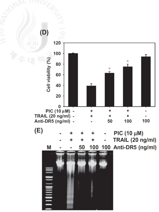
expression is associated with PIC/TRAIL-induced apoptosis. Treatment with DR5
chimera antibodies restored PIC/TRAIL-induced apoptosis in THP-1 cells (Fig. 3D).
Similarly, addition of DR5 chimera antibodies decreased the DNA fragmentation
induced by PIC/TRAIL treatment (Fig. 3E). These results indicate that PIC-induced
26
Fig. 3. PIC sensitizes TRAIL-induced apoptosis via up-regulation of DR5. (A)
27
isolated and Western blot analysis for DR4 and DR5 was performed. β-Actin was
used as an internal control. (B) Flow cytometry was used to analyze the surface
expression of DR5. Black shade histograms, untreated control; black line, treated
cells with PIC. (C) U937 and HL60 cells were treated with 10 µM PIC for the
indicated times and cell extracts were prepared for Western blot analysis for DR5.
β-Actin was used as an internal control. (D and E) THP-1 cells were pre-treated with
10 µM PIC and 20 ng/ml TRAIL in the presence of the indicated concentrations of
DR5 chimera antibodies. After treatment for 24 h, cell viabilities were measured by
MTT assay (D) and fragmented DNA was extracted and analyzed on 1.5% agarose
gel containing EtBr (E). Data are representative of three independent experiments.
Each point represents the mean ± SD of three independent experiments. The
28
3.4. PIC up-regulates DR5 expression via Sp1 binding on DR5 promoter region
To access whether PIC-induced DR5 up-regulation is tightly controlled at
transcriptional level, we analyzed expression of DR5 mRNA by RT-PCR. We found
that PIC treatment increases DR5 mRNA expression in a time-dependent manner, but
not DR4 mRNA (Fig. 4A). Next, the effects of PIC on the promoter activities of
reporter constructs containing 605-bp fragments of the DR5 gene promoter region
(pDR5/-605) in THP-1 cells were examined by luciferase assay. Results indicated
that PIC significantly increases the promoter activities of pDR5/-605 in a
time-dependent manner (Fig. 4B). In particular, 10 µM PIC enhanced the DR5 promoter
activity approximately three-fold greater than control values at 24 h, supporting the
idea that PIC-induced DR5 up-regulation is controlled at the transcriptional level. To
further confirm the precise mechanism by which PIC regulates DR5 expression,
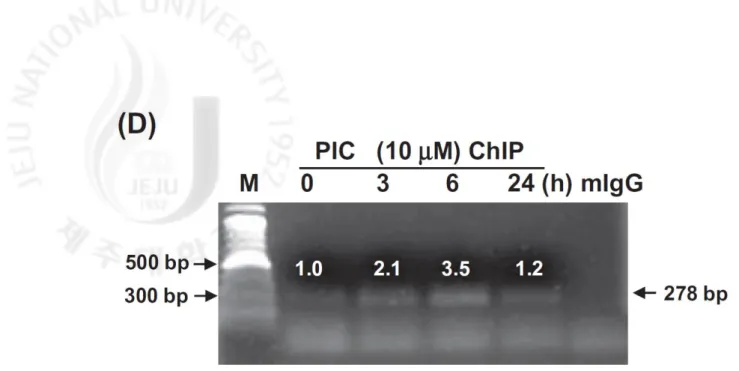
Western blot and ChIP analysis was performed to quantify Sp1 activity on the DR5
promoter regions. Nuclear translocation of Sp1 protein increased from at 3 h and
reclined at 12 h in the presence of PIC (Fig. 4C). PIC also increased Sp1 binding to
the promoter regions of DR5 and significant Sp1 binding levels were observed at 6 h
after PIC treatment (Fig. 4D). These results show that PIC at least up-regulates DR5
30
Fig. 4. PIC enhances DR5 expression via activation of Sp1. (A) THP-1 cells were
treated with 10 μM PIC for the indicated time points. (A) Total RNA was isolated
and RT-PCR analysis for DR4 and DR5 was performed. GAPDH was used as an
internal control. (B) pDR5/–605 promoter was transfected into THP-1 cells, which
were than treated with 10 μM PIC for the indicated time points, lysed and assayed
luciferase activity. (C) Nucleus protein was prepared and Western blotting analysis of
Sp1 was conducted. Nucleolin was used as an internal control. (D) ChIP assay was
performed using antibodies against Sp1. Negative controls were performed using
antibody against rabbit IgG. Data are expressed as overall mean ± SD from three
independent experiments. Statistical significance was determined by Student’s t-test
31
3.5. Activation of ERK is required for the enhanced DR5 expression
Recent studies have revealed that MAPKs are considered important regulators in
DR5 expression as the TRAIL sensitizing signal pathway. To investigate the role of
MAPKs in PIC-induced DR5 expression, Western blot analysis was used to assess
the change in phosphorylations of JNK, ERK, and p38. PIC time-dependently
increased the phosphorylations of JNK, ERK, and p38 (Fig. 5A). Thus, THP-1 cells
were incubated with PIC in the presence of PD98059, SB239063 and SP600125 to
investigate the functional roles of these phosphorylations. Only inhibition of ERK by
PD98059 significantly restored induction of sub-G1 population induced by treatment
with PIC and TRAIL (Fig. 5B). Pre-treatment with SB203580 or SP600125 slightly
increased sub-G1 population under the same experimental conditions. We also
investigated whether ERK phosphorylation directly contributes to the increase in
DR5 expression. Consistent with the decrease in sub-G1 populations, pre-treatment
with PD98059 significantly decreased PIC-induced DR5 up-regulation (Fig. 5C).
Therefore, we assessed the effect of PD98059 on DR5 promoter activity.
Pre-treatment with PD98059 significantly decreased PIC-induced DR5 promoter activity
suggesting that PIC up-regulates DR5 expression via ERK-dependent activation (Fig.
32
33
Fig. 5. PIC-induced pDR5/–605 promoter activation is mediated by the ERK signal
pathway. (A) THP-1 cells were treated with 10 µM PIC for the indicated times.
34
to nitrocellulose membranes, and probed with antibodies against JNK, JNK,
p-ERK, p-ERK, p-p38 and p38. (B) Flow cytometric analysis of sub-G1 population by
PIC (10 μM)/TRAIL (20 ng/ml) after pre-treatment with 20 µM PD98059 (PD), 10
µM SB203580 (SB) or 20 µM SP600125 (SP) for 1 h was performed using flow
cytometry. (C) PD98059 (20 µM) was treated 1 h before treatment with PIC. Western
blot analysis was performed using specific antibody against DR5 and ERK. β-Actin
was used as an internal control. (D) pDR5/–605 was transfected into THP-1 cells,
which were then pretreated with 20 µM PD98059 for 1 h and further treated with 10
35 4. Discussion
PIC has been previously known as an active component of Melaleuca
leucadendrom (white tea tree), Cassia garretiana (Asian legume), and Rheum undulatum (Korean rhubarb), which are considered to be useful in traditional herbal medicine (Roupe et al., 2006). Recent studies have demonstrated that PIC induces
anti-cancer activity in several cancer cell lines (Kuo and Hsu 2002; Larrosa et al.,
2004; Chowdhury et al., 2005; Kim et al., 2008b). Ashikawa et al. (2002) also
investigated whether PIC sensitizes TNF-induced apoptosis through inhibition of
IKK activity, thereby blocking NF-κB activation. However, the information on the
effect of PIC in TRAIL-induced apoptosis is limited. Here, we demonstrated that PIC
may be an ideal candidate for novel TRAIL enhancer in some human leukemia cells.
Further, on the basis of these findings, we propose that various other cancer cells will
be tested for elucidating the molecular mechanisms as a TRAIL sensitizer.
Several studies have suggested that TRAIL may be a potential candidate for
treating different types of cancer cells, because it lacks the ability to induce side
effects in normal cells (Wiley et al., 1995; Kischkel et al., 2000; Van Geelen et al.,
2004). Although the mechanism by which cancer cells successfully develop
36
acquire resistance against TRAIL-induced apoptosis (Dida et al., 2008). Therefore, it
is essential to discover TRAIL sensitizers to overcome this resistance. Many
flavonoids have been proposed as candidate novel strategies to induce synergistic
activity with TRAIL but have minimal toxicity. A number of studies have reported
that flavonoids, especially curcumin and resveratol, significantly sensitize
TRAIL-induced apoptosis in its resistant cancer cells through DR5 up-regulation
(Andrezejewski et al., 2008; Ivanov et al., 2008). Regulation of DR5 expression by
flavonoids may be an important target for sensitizing TRAIL-induced apoptosis. As
has been reported previously, PIC enhanced TRAIL-induced apoptosis via DR5
up-regulation expression in this study. These effects with PIC were significantly blocked
by DR5 chimera antibodies. Further studies will be needed to precisely elucidate the
molecular mechanisms of DR5 expression by PIC.
Many reports have investigated that overexpression of p53 transactivates DR4 and
DR5 expression, confirming that DR5 is regulated by p53 (Wu et al., 1997; Takimoto
and El-Deiry 2008). However, our results rule out this possibility because all types of
cells used in this study are p53-deficeint. Recent studies have suggested that binding
of Sp1 transcription factor in the promoter regions tightly regulates DR5
37
demonstrated that Sp1 activation by TRAIL sensitizers increases sensitivity in its
resistant cancer cells through enhancement of DR5 expression (Sun et al., 2008).
Thus, Sp1 up-regulation is a useful target in TRAIL-resistant cancer cells to augment
its sensitivity through DR5 up-regulation. In this study, we also found that PIC
sensitizes up-regulation of DR5 via activation of Sp1. Additionally, Sp1 regulators
have been also investigated because of their importance in Sp1 up-regulation in DR5
expression. Previous studies suggest that a JNK inhibitor completely abrogates bile
acid-induced DR5 expression via involvement of Sp1, whereas ERK and p38 MAPK
inhibitors failed to block DR5 induction (Higuchi et al., 2004). In contrast, recent
studies demonstrated that ERK or p38 MAPK are also associated with
TRIAL-induced apoptosis via up-regulation of DR5 (Kim et al., 2008a). Our data showed
that PIC induces DR5 up-regulation for TRAIL sensitivity via Sp1 activation which
can be regulated by ERK. Nevertheless, further studies are needed to confirm how
Sp1 activity is regulated by the ERK signal pathway. In addition, the role of other
transcriptional factors in the regulation of DR5 expression remains to be determined,
because NF-κB (Ammann et al., 2009) and CHOP (Yamaguchi and Wang 2004) have
been implicated in regulating DR5 expression. Additionally, it was recently reported
38
2006; Chen et al., 2010), though many antioxidants trigger TRAIL-induced apoptosis
through inhibition of NF-κB or activation of CHOP to up-regulate DR5 expression
(Lirdprapamongkol et al., 2010; Kim et al., 2010). Nevertheless, we also found that
ROS is not important to regulate DR5 expression (Moon et al., 2010). To settle this
discrepancy, further studies are needed more details how antioxidant PIC enhances
TRAIL-induced apoptosis.
Taken together, this study demonstrates that PIC/TRAIL potentiates the triggering
of an apoptotic cascade via up-regulation of DR5 expression in THP-1 cells. We also
showed that DR5 expression is tightly regulated by Sp1 activity in the promoter
39 References
Ammann, J.U., Haag, C., Kasperczyk, H., Debatin, K.M., Fulda, S., 2009.
Sensitization of neuroblastoma cells for TRAIL-induced apoptosis by NF-κB
inhibition. International Journal of Cancer 124, 1301–1311.
Andrezejewski, T., Deeb, D., Gao, X., Danyluk, A., Arbab, A.S., Dulchavsky, S.A.,
Gautam, S.C., 2008. Therapeutic efficacy of curcumin/TRAIL combination
regimen for hormone-refractory prostate cancer. Oncology Reports 17, 257–267.
Ashikawa, K., Majumdar, S., Banerjee, S., Bharti, A.C., Shishoda, S., Aggarwal,
B.B., 2002. Piceatannol inhibits TNF-induced NF-κB activation and
NF-κB-mediated gene expression through suppression of IκBα kinase and p65
phosphorylation. Journal of Immunology 169, 6490–6497.
Chaudhary, P.M., Eby, M., Jasmin, A., Bookwalter, A., Murray, J., HOOD, L., 1997.
Death receptor 5, a new member of the TNFR family, and DR4 induce
FADD-dependent apoptosis and activate the NF-κB pathway. Immunity 7, 821–830.
Chen, C.Y., Tai, C.J., Zheng, J.J., Chen, Y.Z., Liu, T.Z., Yiin, S.J., Chern, C.L., 2010.
6-dehydrogingerdione sensitizes human hepatoblastoma Hep G2 cells to
TRAIL-induced apoptosis via reactive oxygen species-mediated increase of DR5.
40
Chowdhury, S.A., Kishino, K., Satoh, R., Hashimoto, K., Kikuchi, H., Nishikawa, H.,
Shirataki, Y., Sakagami, H., 2005. Tumor-specificity and apoptosis-inducing
activity of stilbenes and flavonoids. Anticancer Research 25, 2055–2063.
Dida, F., Li Y., Iwao, A., Deguchi, T., Azuma, E., Komada, Y., 2008. Resistance to
TRAIL-induced apoptosis caused by constitutional phosphorylation of Akt and
PTEN in acute lymphoblastic leukemia cells. Experimental Hematology 36,
1343–1353.
Hasegawa, H., Yamada, Y., Komiyama, K., Hayashi, M., Ishibashi, M., Yoshida, T.,
Sakai, T., Koyano, T., Kam, T.S., Murata, K., Sugahara, K., Tsuruda, K.,
Akamatsu, N., Tsukasaki, K., Masuda, M., Takasu, N., Kamihira, S., 2006.
Dihydroflavonol BB-1, an extract of natural plant Blumea balsamifera,
abrogates TRAIL resistance in leukemia cells. Blood 107, 679–688.
Higushi, H., Grambihler, A., Canbay, A., Bronk, S.F., Gores, G.J., 2004. Bile acids
up-regulate death receptor 5/TRAIL-receptor 2 expression via a c-Jun
N-terminal kinase-dependent pathway involving Sp1. Journal of Biological
Chemistry 279, 51–60.
Islam, S., Hassan, F., Mu, M.M., Ito, H., Koide, N., Mori, I., Yoshida, T., Yokochi,
41
(NO) production and nuclear factor (NF)-κB activation by inhibiting IκB kinase
(IKK). Microbiology and Immunology 48, 729–736.
Ivanov, V.N., Partridge, M.A., Johnson, G.E., Huang, S.X., Zhou, H., Hei, T.K.,
2008. Resveratrol sensitizes melanomas to TRAIL through modulation of
antiapoptotic gene expression. Experimental Cell Research 314, 1163–1176.
Jin, C.Y., Moon, D.O., Lee, K.J., Kim, M.O., Lee, J.D., Choi, Y.H., Park, Y.M., Kim,
G.Y., 2006. Piceatannol attenuates lipopolysaccharide-induced NF-κB activation
and NF-κB-related proinflammatory mediators in BV2 microglia.
Pharmacological Research 54, 461–467.
Jin, C.Y., Park, C., Kim, G.Y., Lee, S.J., Kim, W.J., Choi, Y.H., 2009. Genistein
enhances TRAIL-induced apoptosis through inhibition of p38 MAPK signaling
in human hepatocellular carcinoma Hep3B cells. Chemico-Biological
Interactions 180, 143–150.
Kim, H., Kim, E.H., Eom, Y.W., Kim, W.H., Kwon, T.K., Lee, S.J., Choi, K.S.,
2006. Sulforaphane sensitizes tumor necrosis factor-related apoptosis-inducing
ligand (TRAIL)-resistant hepatoma cells to TRAIL-induced apoptosis through
reactive oxygen species-mediated up-regulation of DR5. Cancer Research 66,
42
Kim, M.O., Moon, D.O., Kang, C.H., Kwon, T.K., Choi, Y.H., Kim, G.Y., 2010.
Β-Ionone enhances TRAIL-induced apoptosis in hepatoma cellular carcinoma cells
through Sp1-dependent upregulation of DR5 and downregulation of NF-κB
activity. Molecular Cancer Therapeutics, 9, 833–843.
Kim, Y.H., Lee, D.H., Jeong, J.H., Guo, Z.S.. Lee, Y.J., 2008a. Quercetin augments
TRAIL-induced apoptotic death: involvement of the ERK signal transduction
pathway. Biochemical Pharmacology 75, 1946–1958.
Kim, Y.H., Park, C., Lee, J.O., Kim, G.Y., Lee, W.H., Choi, Y.H., Ryu, C.H., 2008b.
Induction of apoptosis by piceatannol in human leukemic U937 cells through
down-regulation of Bcl-2 and activation of caspases. Oncology Reports 19, 961–
967.
Kischkel, F.C., Lawrence, D.A., Chuntrarapai, A., Schow, P., Kim, K.J., Ashkenazi,
A. 2000. Apo2L/TRAIL-dependent recruitment of endogenous FADD and
caspase-8 to death receptors 4 and 5. Immunity 12, 611–620.
Kuo, P.L., Hsu, Y.L., 2008. The grape and wine constituent piceatannol inhibits
proliferation of human bladder cancer cells via blocking cell cycle progression
and inducing Fas/membrane bound Fas ligand-mediated apoptosis pathway.
43
Larrosa, M., Tomás-Barberán, F.A., Espín, J.C., 2004. The grape and wine
polyphenol piceatannol is a potent inducer of apoptosis in human SK-Mel-28
melanoma cells. European Journal of Nutrition 43, 275–284.
Lirdprapamongkol, K., Sakurai, H., Suzuki, S., Koizumi, K., Prangsaengtong, O.,
Viriyaroj, A., Ruchirawat, S., Svasti, J., Saiki, I., 2010. Vanillin enhances
TRAIL-induced apoptosis in cancer cells through inhibition of NF-κB activation.
In Vivo 24, 501–506.
Moon, D.O., Kim, M.O., Choi, Y.H., Kim, G.Y., 2010. Butein sensitizes human
hepatoma cells to TRAIL-induced apoptosis via extracellular signal-regulated
kinases/Sp1-dependent DR5 upregulation and NF-κB inactivation. Molecular
Cancer Therapeutics, 9, 1583–1595.
Muzio, M., Chinnaiyan, A.M., Kischkel, F.C., O’rourke, K., Shevchenko, A., Ni, J.,
Scaffidi, C., Bretz, J.D., Zhang, M., Gentz, R., Mann, M., Krammer, P.H., Peter,
M.E., Dixit, V.M., 1996. FLICE, a novel FADD homologous ICE/CED-3-like
protease, is recruited to the CD95 (Fas/APO-1) death-inducing signaling
complex. Cell 85, 817–827.
Pan, G., Ni J., Wei, Y.F., Yu, G., Gentz, R., Dixit, V.M., 1997a. An antagonist
44
815–818.
Pan, G., O’rourke, K., Chinnaiyan, A.M., Gentzy, R., Ebner, R., Ni, J., Dixit, V.M.,
1997b. The receptor for the cytotoxic ligand TRAIL. Science 276, 111 –113.
Raja, S.M., Chen, S., Yue, P., Acker, T.M., Lefkove, B., Arbiser, J.L., Khuri, F.R.,
Sun, S.Y., 2008. The natural product honokiol preferentially inhibits cellular
FLICE-inhibitory protein and augments death receptor-induced apoptosis.
Molecular Cancer Therapeutics 7, 2212–2223.
Roupe, K.A., Remsberg, C.M., Yanez, J.A., Davies, N.M., 2006. Pharmacometrics of
stilbenes: segueing towards the clinic. Current Clinical Pharmacology 1, 81–101.
Ssheridan, J.P., Marsters, S.A., Pitti, R.M., Gurney, A., Skubatch, M., Baldwin, D.,
Ramakrishnan, L., Gray, C.L., Baker, K., Wood, W.I., Gooddard, A.D.,
Godowski, P., Ashkenazi, A. 1997. Control of TRAIL-induced apoptosis by a
family of signaling and decoy receptors. Science 277, 818–821.
Sprick, M.R., Wwigand, M.A., Riester, E., Rauch, C.T., Juo, P., Blenis, J.,
Krammer, P.H., Walczak, H. 2000. FADD/MORT1 and caspase-8 are recruited
to TRAIL receptors 1 and 2 and are essential for apoptosis mediated by TRAIL
receptor 2. Immunity 12, 599–609.
8-chloro-45
adenosine-upregulated death receptor 5 expression in human hepatoma cells.
Oncology Reports 19, 177–185.
Takimoto, R., EL-DEIRY, W.S., 2008. Wild-type p53 transactivates the
KILLER/DR5 gene through an intronic sequence-specific DNA-binding site.
Oncogene 19, 1735–1743.
Van Geelen, C.M., De Vries, E.G., De Jong, S., 2004. Lessons from
TRAIL-resistance mechanisms in colorectal cancer cells: paving the road to
patient-tailored therapy. Drug Resistance Updates 7, 345–358.
Wiley, S.R., Schooley, K., Smolak, P.J., Din, W.S., Huang, C.P., Nicholl, J.K.,
Sutherland, G.R., Smith, T.D., Rauch, C., Smith, C.A., 1995. Identification and
characterization of a new member of the TNF family that induces apoptosis.
Immunity 3, 673–682.
Wu, G.S., Burns, T.F., McDonald 3rd E.R., Jiang, W., Meng, R., Krantz, I.D., Kao, G.,
Gan, D.D., Zhou, J.Y., Muschel, R., Hamilton, S.R., Spinner, N.B., Markowitz,
S., Wu, G., El-Deiry, W.S., 1997. KILLER/DR5 is a DNA damage-inducible
p53-regulated death receptor gene. Nature Genetics 17, 141–143.
Xu, J., Zhou, J.Y., Wei, W.Z., Philipsen, S., Wu, G.S., 2008. Sp1-mediated TRAIL
46
Yamaguchi, H., Wang, H.G., 2004. CHOP is involved in endoplasmic reticulum
stress-induced apoptosis by enhancing DR5 expression in human carcinoma
cells. Journal of Biological Chemistry 279, 45495–45502.
Yoshida, T., Maeda, A., Tani, N., Sakai, T., 2001. Promoter structure and
transcription initiation sites of the human death receptor 5/TRAIL-R2 gene.
47
감사의
글
사실 공부와는 거리가 멀었던 제가 대학원에 진학을 하고 이렇게 석사과정을 마친다는 것이 아직 실감이 나지 않습니다. 아직 부족하고 배워야 할 것이 많지만 그런 것들은 박사 과정에 들어가서 더욱 열심히 하여 채우도록 하겠습니다. 이토록 제가 석사 과정을 마치고 더 나아가 박사 과정을 밟을 수 있도록 많은 격려와 도움을 주신 모든 분들에게 지면으로 나마 감사의 마음을 전하고 싶습니다. 부족한 저를 지금까지 믿고 이끌어 주시며, 항상 좋은 말들로 격려와 자극을 통해 저를 지도해주신 김기영 교수님, 그리고 처음 실험실에 들어와 교수님이 미국에 가 계셔 자리를 비우셨을 때 그 자리를 채우며 많은 가르침을 주신 문동오 교수님, 문옥이 누나 에게 진심으로 감사 드립니다. 또한 철없던 학부생활부터 저에게 많은 가르침을 주신 이기완 교수님, 송춘복 교수님, 허문수 교수님, 이제희 교수님, 여인규 교수님, 최광식 교수님, 이영돈 교수님, 이경준 교수님, 정준범 교수님, 그리고 대학원 생활 이 후부터 가르침을 주신 이승헌 교수님, 정석근 교수님께 진심으로 감사 드립니다. 그리고 많은 실험을 가르쳐 준 상혁이형, 내가 힘들 때 마다 격려의 말을 아끼지 않았던 경용이 형과 진이 누나에게 감사하다는 말을 하고 싶습니다. 또한 얘기는 잘 통하지 않지만 그래도 나를 믿고 따라 주는 프라사드, 딜루에게 고맙다는 말을 전합니다. 이번 제 석사 논문 심사를 위해 부산에서 내려와 제 발표와 연구내용을 보시고 많은 조언을 해주신 최용현 교수님과 세포들뿐만 아니라 실험 기기들을 아낌없이 지원해주신 동의대의 박철 선생님, 성윤 선생님, 민호형, 부산대에 진우형에게도 감사의 말을 전합니다.48 그리고 이름을 모두 열거 할 수는 없지만 항상 격려를 해주며 많은 도움을 주신 각 실험실 선배님들께도 진심으로 감사 드립니다. 그리고 내가 힘들거나, 외롭거나, 아프거나, 기분이 좋을 때 항상 옆에서 많은 얘기를 나누고 나를 다독이고 힘이 되어 주던 영민이, 병훈이, 민철이, 일태, 용덕이 에게 고맙다는 말을 전합니다. 그리고 언제나 내 옆에 있어주며 힘든 얘기를 들어 준 우종이, 자주 보지 못하고 연락 할 수 없지만 항상 내 편이 되어주고 내 힘이 되어주는 준보, 정욱이, 주범이, 세호 등등 고등학교 친구들에게도 고맙다는 말을 전합니다. 그리고 바쁘다는 핑계로 일을 도와 드리지 못해 항상 죄송하고, 이렇게 제가 공부를 할 수 있게 많은 도움을 주는 우리 부모님과 수희 누나, 정화 누나에게 진심으로 고맙다는 말을 하고 싶습니다. 모든 분들에게 항상 건강하시고 웃는 일들만 가득하길 바라며 언제나 행복 했으면 좋겠습니다. 그리고….. 사랑합니다.