* Corresponding author
Phone: +82-51-850-7413, Fax: +82-51-853-4036 E-mail: [email protected]
인체백혈병세포의 증식에 미치는 불등가사리 메탄올 추출물의 영향
최우영1,2․김기영3․이원호2․배송자4․최영현1*
1
동의대학교 한의과대학 생화학교실, 대학원 바이오물질제어학과 및 한의학연구소
2
부산대학교 자연과학대학 생물학과
3
제주대학교 해양과학대학 해양과학부
4
신라대학교 의생명과학대학 식품영양학과
Induction of Apoptosis by Methanol Extract of
Gloiopeltis furcata
in Human Leukemia Cell Line U937. Woo Young Choi1,2, Cheol Park1,2, Gi Young Kim3, Won Ho Lee2, Song-Ja Bae4 and Yung Hyun Choi1,*. 1Department of Biochemistry, College of Oriental Medicine and Department of Biomaterial Control, Dongeui University Graduate School and Institute of Oriental Medicine, Dongeui University, Busan 614-052,
2Department of Biology, Pusan National University, Busan 609-735,
3Faculty of Applied Marine Science, Cheju National University, Jeju 690-756,
4Department of Food and Nutrition, and Marine Bio- technology Center for Bio-Functional Material Industries, Silla University, Busan 617-736, Korea Abstract
Epidemiological studies have indicated that the ubiquitous consumption of seaweeds is a protective factor against some types of cancer. Previous results showed that the admin- istration of seaweed powder or extract reduced the incidence rate of chemically induced tumori- genesis using in vivo animal model. Recently, we reported that the extracts of Gloiopeltisfurcata, a kind of Korean edible seaweed, caused he cell growth inhibition of various human
cancer cell lines, among them methanol extract exhibited a relatively strong antiproliferative activity. However, the molecular mechanisms of this seaweed in malignant cells have been poorly studied until now. To elucidate this problem, we investigated the effects of methanol extract of G. furcata (MEGF) on the growth inhibition in several human cancer cell lines, and further we analyzed the effects of this extract were tested on the activity of apoptosis induction in human leukemic cells. The results demonstrated that MEGF treatment resulted in the morphological changes and the growth inhibition in a dose-dependent manner.Furthermore, MEGF potently suppresses the growth of human leukemic U937 cells by induction of apoptosis, which was associated with induction of cyclin-dependent kinase inhibitor p21(WAF1/CIP1) in a tumor suppressor p53-independent fashion and up-regulation of Fas/FasL system. Further studies will be needed to identify the active compounds that confer the anti- cancer activity of MEGF. Once such compounds are identified, the mechanisms by which they exert their effects can begin to be characterized.
Key words : Gloiopeltis furcata, human cancer cells, apoptosis, p21, Fas/FasL
서 론
암은 치료상의 급진적이고 기술적인 진보에도 불 구하고 여전히 정복되지 못하고 있는 질병 중의 하 나이며, 한국에서도 암은 전체 사망률의 가장 많은 원인 중의 하나로 지적되고 있다. 장기별에 따른 암
의 발생빈도는 인종 및 지역적 차이에 따라 발생되
는 부위가 다르며, 이는 생활습관 및 주변 환경과도
관련되어 있다고 알려져 있다. 대개 서양에서는 폐
암·대장암·유방암 등이 잘 발생하며 한국을 비롯한
동남아시아 지역에서는 위암·간암·자궁경부암 등이
잘 발생한다. 하지만 생활습관 등이 변화함에 따라
서 발생 빈도가 높은 암의 종류도 점차 달라지고 있 는 추세이다[1,14,19]. 따라서 많은 화학적·생리학적 인 암치료/예방제의 개발과 발견이 이루어지고 있으 나, 그 부작용이나 제한적인 사용 등의 여러 이유에 의해 다른 대체 요법이나 정상세포에 유해한 선도물 질의 개발이 필요시 되고 있다. 이에 따라 우리 주위 에서 흔히 볼 수 있으며, 우리의 생활과 밀접하게 연관되어 있는 천연물질의 이용이 주목받고 있고 많 이 연구되고 있다[10,15,16].
우리의 생활과 밀접한 연관이 있는 많은 천연물들 이 항암작용이 있는 것으로 알려져 있지만 최근 육 상자원의 한계를 극복하기 위하여 해양생물 유래 천 연 물질에 대한 관심이 매우 높아지고 있다[2]. 그 중에서도 식용이 가능한 해조류는 강력한 항산화 효 과가 있다고 잘 알려져 있으며, 혈중 콜레스테롤을 내려주며, 고혈압과 혈액의 점도를 낮추고 궤양을 예방하고, 항균작용을 가지는 등의 다양한 효과가 있는 것으로 보고되어지고 있다[3,8,22].
한편 암의 효과적인 치료와 예방에 있어서 비정상 적인 세포나 암세포의 apoptosis 유발은 많은 치료제 의 표적이 되고 있으며, apoptosis는 개체보존의 수준 에서 손상된 세포들의 제거를 위한 세포의 사멸 현 상으로서 중요한 수단이기도 하다[4]. Apoptosis의 유발에 종양억제유전자 p53 등과 같은 다양한 유전 자가 관여한다는 사실이 알려지면서 apoptosis와 연 관된 분자적 기전이 최근 많이 밝혀지고 있으며 아 직도 많은 연구가 진행되고 있으며, apoptosis는 특히 암세포의 생장, 증식의 억제와 암세포 파괴의 한 방 법으로써 널리 연구되고 있다[11,18].
본 연구에서는 우리나라를 포함한 세계적으로 널 리 분포하고 있으며, 항돌연변이 효능이 우수하여 항암활성효능이 높을 것으로 예상되는 해조류의 한 종류인 홍조식물 풀가사리과의 불등가사리(G.loiopeltis
furcata)[7,8,13,20]에 함유된 항암/암예방 후보물질의 도출을 위하여 불등가사리의 메탄올 추출물이 인 체 암세포의 증식에 미치는 영향을 조사하였으며, 특히 불등가사리 메탄올 추출물에 높은 감수성을 보 이는 인체백혈병세포를 대상으로 암세포 증식 억제 효과가 apoptosis 유발과 연관성이 있는지의 여부를 조사하였다.
재료 및 방법
실험 재료 및 세포배양
본 실험에 사용한 인체 전립선암세포인 PC-3, 대 장암세포인 HT-29, 폐암세포인 A549, 위암세포인 AGS 그리고 백혈병세포인 U937은 생명공학연구소 (KRIBB, Taejeon, Korea)에서 분양 받았으며, 세포의 배양을 위해 10%의 우태아혈청(fetal bovine serum, FBS, Gibco BRL, Grand Island, NY, USA)과 1%의 penicillin-streptomycin 등이 포함된 RPMI-1640 및 DMEM 배지(Gibco BRL)를 사용하여 37 ℃, 5% CO
2조건하의 CO
2incubator에서 배양하였다. 불등가사 리는 전라남도 무안군에 위치한 (주)삼일물산에서 제공받았으며, 메탄올 추출물(methanol extracts of G.
furcata, MEGF)을 얻기 위하여 건조 후 메탄올을 첨
가하고 37 ℃에서 진탕한 다음 4시간 동안 3회 반복 추출하고 회전식 진공 농축기로 감압 농축시킨 후 동결 건조하였다[13].
암세포의 형태 변화 관찰
세포 배양용 6 well plate에 각 암세포의 성장 속도 에 따라 적정량 분주하여 24시간 동안 안정화시킨 다음 MEGF를 적정 농도로 배지에 희석 처리하여 다시 48시간 동안 배양한 후, 위상차 현미경(inverted microscope, Carl Zeiss, Germany)을 이용하여 200배 의 배율로 각 암세포주의 농도에 따른 형태의 변화 를 관찰하였다.
MTT assay에 의한 세포 성장억제 조사
세포 배양용 6 well plate에 각 암세포주를 적정량 분주하여 24시간 동안 안정화시킨 다음 MEGF를 적 정량 처리하여 48시간 동안 재배한 후 각 well의 배 지를 깨끗하게 제거한 후 tetrazolium bromide salt (MTT, Amresco, Solon, OH, USA)를 0.5 mg/ml 농도 가 되게 희석하여 2 ml씩 분주하고 3시간 동안 CO
2incubator에서 빛을 차단하여 반응시킨 다음 MTT 시
약을 제거하고 DMSO로 각 well에 생성된 formazin
을 각각 동량으로 녹인 후 ELISA reader (Molecular
Devices, Sunnyvale, CA, USA)를 이용하여 540 nm에
서의 흡광도를 측정하였다. 측정은 모두 세 번을 하
였으며, 그에 대한 평균값과 표준 오차를 Sigma Plot
4.0 프로그램(SPSS Ins.)으로 구하였다.
Table 1. Gene-specific primers for RT-PCR
Gene name Sequence
p53 sense
antisense
5'-GCT CTG ACT GTA CCA CCA TCC-3' 5'-CTC TCG GAA CAT CTC GAA GCG-3'
p21 sense
antisense
5'-CTC AGA GGA GGC GCC ATG-3' 5'-GGG CGG ATT AGG GCT TCC-3'
Fas sense
antisense
5'-TCT AAC TTG GGG TGG CTT TGT CTT C-3' 5'-GTG TCA TAC GCT TTC TTT CCA T-3'
FasL sense
antisense
5'-GGA TTG GGC CTG GGG ATG TTT CA-3' 5'-AGC CCA GTT TCA TTG ATC ACA AGG-3'
GAPDH sense
antisense
5'-CGG AGT CAA CGG ATT TGG TCG TAT-3' 5'-AGC CTT CTC CAT GGT GGT GAA GAC-3' DNA flow cytometry 분석
정상 및 MEGF을 처리한 배지에서 48시간 동안 배양시킨 각 암세포주를 2,000 rpm에 5분간 원심분 리하여 상층의 배지를 버리고 세포 pallet만 모아서 PBS 1 ml로 충분히 재부유 시킨 후 2,000 rpm으로 5분간 원심분리 한 후 상층의 PBS만 버리고 남은 세포 pallet에 CycleTEST PLUS DNA REAGENT KIT (Becton Dickinson, San Jose, CA, USA)를 사용하여 고정 및 propidium iodide (PI, concentration, 50ug/ml;
Sigma) 염색액을 처리하여 암실, 4 ℃에서 15분 동안 염색하였다. 염색 후 DNA flow cytometry (Becton Dickinson, San Jose, CA, USA)에 대조군 및 MEGF 처리군을 적용시켜 형광반응에 따른 histogram을 ModiFit LT (Becton Dickinson) program으로 분석하 였다.
DAPI staining에 의한 세포핵의 형태 관찰
MEGF 처리에 의한 암세포의 apoptosis 유발 여부 확인을 위한 핵의 형태적 변화를 관찰하기 위하여 정상 및 MEGF가 처리된 배지에서 48시간 동안 배양 된 세포를 모은 다음 37% formaldehyde 용액과 PBS 를 1 : 9의 비율로 섞은 fixing solution을 모아진 세포 에 500 ㎕ 첨가하여 잘 섞어준 후, 실온에서 10분 동안 고정하였다. 2,000 rpm으로 5분간 원심 분리한 후 상층액을 제거하고 PBS 200 ㎕ 를 넣어서 충분히 섞은 후, slide glass 위에 120 ㎕ 정도 떨어뜨려 1200 rpm에서 5분간 cytospin하였다. PBS로 2 ~ 3회 wash- ing하고 slide glass를 건조시킨 후 4’,6-diamidino-2-
phenylindole (DAPI, Sigma, St. Louis, MO, USA) 용액 을 세포가 고정된 slide glass 위에 적당량을 떨어뜨 린 후 빛을 차단하고 실온에서 염색시켰다. 약 10분 가량 염색시킨 후, 형광 현미경(Carl Zeiss)을 이용하 여 400배의 배율로 각 MEGF 처리 농도에 따른 인체 백혈병 세포의 핵의 형태 변화를 관찰하였다.
Reverse transcription-polymerase chain re- action 분석
상기와 동일한 조건에서 준비된 세포를 PBS로 세
척하고 TRIzol reagent (Invitrogen Co., Carlsbad, CA,
USA)를 4 ℃에서 1시간 동안 처리하여 total RNA를
각 처리 농도 별로 분리하였다. 분리된 RNA를 정량
한 후, 관련된 primer, DEPC water 그리고 ONE-STEP
RT-PCR PreMix Kit (Intron, Korea)를 넣고 Mastercycler
gradient (Eppendorf, Hamburg, Germany)를 이용하여
증폭하였다. 각 PCR 산물들을 양적 차이를 확인하
기 위하여 1x TAE buffer로 1% agarose gel을 만들고
well 당 각각의 primer에 해당하는 PCR 산물에 6x
agarose gel loading buffer (BIONEER, Korea)를 섞어
서 20 μl씩 well에 loading 한 후 100 V로 전기영동을
하였다. 전기영동으로 DNA 분리가 끝난 gel을 ethi-
dium bromide (EtBr, Sigma)을 이용하여 염색한 후
UV 하에서 확인하고 Picture works' photo enhancer를
이용하여 사진 촬영을 하였다. RT-PCR을 이용하여
분석을 시도한 유전자의 종류는 Table 1에 나타낸
바와 같으며, 이때 housekeeping 유전자인 glycer-
aldehyde-3-phosphate dehydrogenase (GAPDH) 유전
자를 internal control로 사용하였다.
Fig. 1. Morphological changes of several human cancer cell lines after treatment with methanol extracts of Gloiopeltis furcata
(MEGF). Cells were plated and incubated for 24 h. The cells were incubated with variable concentrations of MEGF. After 48 h incubation cells were sampled and examined under light microscopy. Magnification, X200.Western blotting 분석
정상 및 MEGF가 처리된 배지에서 자란 암세포들 을 적당량의 lysis buffer [25 mM Tris-Cl (pH 7.5), 250 mM NaCl, 5 mM EDTA, 1% NP-40, 1 mM phenyme- thylsulfonyl fluoride (PMSF), 5 mM dithiothreitol (DTT)]로 용해한 후, 고속원심분리기를 이용하여 14,000 rpm에 15 분간 세포내 잔사물을 분리시킨 다 음 동량의 단백질을 sodium dodecyl sulphate (SDS)- polyacrylamide gel을 이용하여 전기영동으로 분리하 였다. 분리된 단백질을 nitrocellulose membrane (Schleicher and Schuell, Keene, NH, USA)으로 elec- troblotting에 의해 전이시킨 후, 특정 단백질에 대한 항체와 그에 대한 이차 항체 반응을 실시한 후 암실 에서 Enhanced Chemiluminoesence (ECL) 용액 (Amersham Life Science Corp., Arlington Heights, IL, USA)을 처리시킨 다음 X-ray film에 감광시켜 특정 단백질의 양을 분석하였다. 본 실험에 사용된 항체 들은 Santa Cruz Biotechnology Inc. (Santa Cruz, CA, USA) 및 Calbiochem (Cambridge, MA, USA)에서 구 입하였으며, 2차 항체로 사용된 peroxidase-labeled donkey anti-rabbit 및 peroxidase-labeled sheep an-
ti-mouse immunoglobulin은 Amersham Life Science에 서 구입하였다.
결과 및 고찰
다양한 암세포에서 MEGF의 증식억제 효과
몇 가지 인체암세포의 증식에 미치는 불등가사리
메탄올 추출물(MEGF)의 영향을 비교하기 위하여
인체 전립선암세포(PC-3), 대장암세포(HT-29), 폐암
세포(A549), 위암세포(AGS) 및 인체 백혈병세포(U937)
에 MEGF를 다양한 농도(20~150 μg/ml)로 48시간동
안 처리한 후 각 암세포들의 형태를 정상배지에서
자란 암세포와 먼저 비교하였다(Fig. 1). 먼저 전립선
암세포 PC-3과 폐암세포 A549에서는 세포의 모양이
MEGF의 처리 농도가 증가할수록 수상돌기와 같은
형태가 점점 신장되기 시작하면서 마지막 처리 농도
에서는 세포의 부착력 상실하는 등의 심한 형태적인
변형을 보였으며 세포의 밀도 역시 처리 농도 의존
적으로 줄어들었다. 그리고 대장암세포인 HT-29 및
위암세포인 AGS에서는 MEGF 처리 농도가 증가할
수록 부착력의 상실이 증가함에 따라 세포가 성장 배
지에 부유하여 죽어가며 따라서 그 밀도가 점차 감소 하는 것을 확인할 수 있었다. 또한 백혈병세포인 U937에서는 세포밀도가 정상 배지에서 자란 세포와 비교하였을 때 MEGF의 처리 농도 의존적으로 상당 히 감소하는 것을 관찰하였으며, 세포의 사멸에 의한 잔존물들이 배양된 플라스크 상에서 처리 농도 의존 적으로 점차 늘어나는 것을 확인할 수 있었다(Fig. 1).
동일 조건에서 MTT assay를 이용하여 정상 배지 에서 자란 암세포와 MEGF를 희석 처리한 배지에서 자란 암세포의 상대적인 성장률을 비교하였을 때, Fig. 2의 결과에서와 같이 PC-3, HT-29 및 A549 암세 포에서보다 AGS와 U937 암세포에서 상대적인 성장 률이 매우 감소한 것을 확인할 수 있었으며, MEGF 를 150 μg/ml 농도로 처리한 실험군에서는 대조군과 비교하였을 때 상대적인 성장률이 실험된 모든 암세 포주에서 50% 미만으로 감소한 것을 관찰할 수 있었 다. 이와 같이 MEGF는 다양한 암세포에서 약간씩의 감수성에 따른 차이를 보이지만 처리 농도가 증가함 에 따라서 세포의 증식억제효과를 가지는 것을 실험 결과 알 수 있었으며, 본 연구에 사용한 암세포 중 백 혈병세포주인 U937 세포에서 가장 감수성이 크다는 것 역시 확인할 수 있었으나, 동일 조건에서 정상 간세 포의 경우 상대적인 성장률은 큰 변화가 없었다[13].
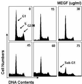
MEGF의 처리에 의한 apoptosis의 유발
MEGF의 처리농도에 따라 가장 증식억제 효과가
Fig. 2. Anti-proliferative effects of MEGF in several human
cancer cell lines. After cells were seeded as described in ma- terials and methods, and treated with MEGF. After 48 h in- cubation with MEGF, MTT assay was performed. Results are expressed as averages +/- SD form separate experiments.크게 나타났던 인체 백혈병 세포인 U937을 이용하 여서 그 증식 억제 효과가 apoptosis 유발과 상관성이 있는지를 조사하였다. Fig. 3 및 Fig. 4B의 flow cy- tometry 결과에서 나타난 것과 같이 정상 배지에서 자란 U937 세포의 주기와 비교하였을 경우 MEGF를 처리한 세포에서 처리 농도 의존적으로 세포의 사멸 이 일어난 집단으로 알려진 sub-G1기가 상당량 증가 하는 것을 확인하였으며, 각 세포 주기의 감소는 sub-G1기의 증가와 관련지어 보면 별다른 변화는 관 찰되지 않았다. 이와 같은 sub-G1기의 증가가 MEGF 처리에 의한 apoptosis에 의한 것인지 직접 조사하기 위하여 DAPI staining을 실시한 결과는 Fig. 4A에 나 타낸 바와 같다. 결과에서 알 수 있듯이 정상 배지에 서 배양된 U937 세포에서는 핵의 형태가 뚜렷하고 밝게 염색되었지만, MEGF를 처리한 배지에서 배양 된 세포는 핵 내의 chromatin이 응축에 의한 apoptotic body의 출현이 MEGF 처리 농도의존적으로 증가됨 을 확인할 수 있었다. 이와 같은 결과는 MEGF의 처 리에 의한 백혈병세포에서의 증식 억제 효과가 apop- tosis의 유발과 직접적인 연관성이 있음을 의미하여 주는 것이다.
Fig. 3. Effect of MEGF on the cell cycle distribution in U937
human leukemia cells. U937 cells were treated with MEGF for 48 h. Cells were fixed and then stained with PI. The per- centages of each fraction were determined by DNA flow cytometry. Data are means average of two separate ex- periments.Fig. 4. Induction of apoptosis by MEGF in U937 cells. (A)
After treated with MEGF for 48 h, cells were fixed and stained with DAPI. The nuclear morphology was photo- graphed with a fluorescence using blue filter. Magnification, X400. (B) Cells treated for 48 h with MEGF and then the cells were collected and stained with PI for flow cytometry analysis. The fraction of apoptotic sub-G1 cells is indicated.Data are presented as the mean values obtained from two independent experiments.
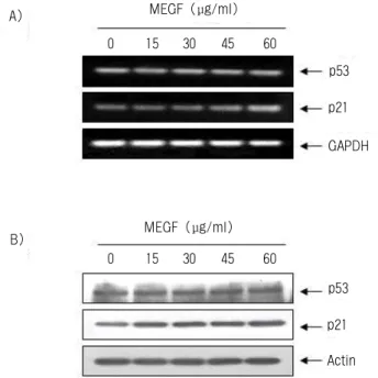
세포 성장 조절인자에 미치는 MEGF의 영향
MEGF의 처리에 의한 백혈병세포의 증식억제 현 상이 종양억제 유전자 또는 세포주기 조절 억제인자 들의 발현 변화와 상관성이 있는지의 여부를 조사하 기 위하여 현재까지 알려진 종양억제 유전자 중 가 장 중요한 p53 및 전체적인 세포주기 조절에 중요한 역할을 하는 cyclin-dependent kinase (Cdk) inhibitor p21(WAF1/CIP1)의 발현[5,12]에 미치는 MEGF의 영 향을 조사하였다. Fig. 5A 및 B의 결과는 RT-PCR 및 Western blotting에 의한 p53 및 p21의 발현에 미치 는 MEGF의 영향을 조사한 것으로서, p53의 상대적 인 발현량은 정상 배지에서 배양된 경우와 MEGF 처리군에서 전사 및 번역 수준에서 큰 변화가 관찰 되지 않았으나, p21의 경우는 전사 및 번역 수준에서
A)
B)
MEGF (μg/ml)
MEGF (μg/ml)
0 15 30 45 60
0 15 30 45 60
p53 p21
GAPDH
p53 p21 Actin
Fig. 5. Induction of Cdk inhibitor p21 by MEGF treatment
in U937 cells. Cell were incubated with or without MEGF for 48 h. (A) Total RNAs were isolated and reverse- transcribed. The resulting cDNAs were subjected to PCR with p53 and p21 primers, and the reaction products were sub- jected to electrophoresis in 1% agarose gel and visualized by EtBr staining. GAPDH was used as an internal control.(B) The cells were lysed and then cellular proteins were sepa- rated by SDS-polyacrylamide gels and transferred onto nitro- cellulose membranes. The membranes were probed with the anti-p53 and anti-p21 antibodies. Proteins were visualized us- ing an ECL detection system. Actin was used as an internal control.
MEGF 처리 농도 의존적으로 다소 증가되었다. 이는 MEGF 처리에 의한 백혈병세포의 증식억제가 p53 비의존적인 p21의 발현 증가와 연관성이 있으며, MEGF가 p53 유전자가 결손된 암세포에서도 항암효 능이 매우 높을 것이라는 것으로 의미하며 이에 관 한 보다 구체적인 연구가 요구된다.
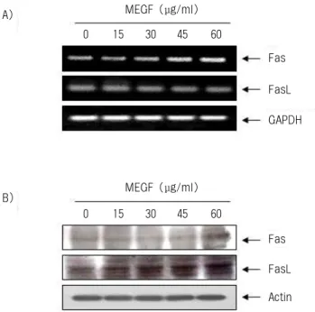
Fas/FasL system에 미치는 MEGF의 영향
현재까지 apoptosis의 유발 경로는 mitochondria를 경유하는 mitochondrial pathway와 death receptor에 의해 death signal을 전달하는 death receptor pathway 로 크게 나누어지며, 두 경로에서의 주요 인자들 사 이의 cross-talking 또한 apoptosis 유발에 매우 중요한 역할을 한다[6,9]. 그 중 death receptor pathway에서는 Fas (CD95/Apo1) 및 이와 결합하는 ligand인 FasL가 널리 알려져 있다[17,21]. 따라서 본 연구에서는
A)
B)
0 15 30
45 60 75
MEGF (μg/ml)
80 70 60 50 40 30 20 10 0
0 15 30 45 60 75
MEGF (μg/ml)
MEGF 처리에 의한 백혈병세포의 apoptosis 유발에 이러한 death receptor를 경유할 가능성의 여부를 조 사하였다. 이를 위하여 Fas 및 FasL 두 유전자의 mRAN 및 단백질 발현에 미치는 MEGF의 영향을 조사한 결과는 Fig. 6에 나타낸 바와 같다. 먼저 mRNA 의 발현에 미치는 MEGF의 결과는 Fig. 6A에 나타낸 바와 같이 Fas의 발현량은 MEGF의 처리 농도 의존 적으로 대조군에 비해 증가하였지만 FasL의 발현량 은 큰 변화가 없었다. 그러나 단백질 발현에서는 MEGF의 처리 농도 의존적으로 Fas 및 FasL 모두 매우 증가하는 것을 확인하였다. 즉 MEGF의 처리에 의한 인체 백혈병 세포 U937에서의 증식억제효과는 번역 수준에서 Fas/FasL death receptor를 경유하는 apoptosis의 유발과 상관관계가 있음을 의미한다.
A) MEGF (μg/ml)
0 15 30 45 60
Fas
GAPDH FasL
B) MEGF (μg/ml)
0 15 30 45 60
Fas
Actin FasL
Fig. 6. Effects of MEGF on Fas/FasL system expression in
U937 cells. (A) After 48 h incubation with MEGF, total RNAs were isolated and reverse-transcribed. The resulting cDNAs were subjected to PCR with Fas and FasL primers, and the reaction products were subjected to electrophoresis in 1% agarose gel and visualized by EtBr staining.GAPDH was used as an internal control. (B) The cells were lysed and then cellular proteins were separated by 10% SDS-polyacrylamide gels and transferred onto nitro- cellulose membranes. The membranes were probed with the anti-Fas and anti-FasL antibodies. Proteins were vi- sualized using an ECL detection system. Actin was used as an internal control.
요 약
본 연구에서는 다양한 인체암세포의 증식에 미치 는 등불가사리 메탄올 추출물(MEGF)의 영향을 조 사하였다. MEGF는 처리 농도의존적으로 암세포의 형태적변이 및 증식억제가 효과를 보여주었으며, 특 히 백혈병세포에서 가장 높은 감수성을 보여주었다.
따라서 백혈병세포의 증식억제 효과가 apoptosis 유 발과 연관성이 있는지를 flow cytometry 분석 및 DAPI staining 법으로 조사한 결과, MEGF 처리에 의 한 백혈병세포의 증식억제는 세포주기 교란과 무관 한 apoptosis 유발에 의한 것임을 sub-G1기 세포의 빈도 증가 및 apoptotic body 형성의 증가 등으로 확 인하였다. 또한 MEGF 처리에 의한 백혈병세포의 증 식억제 및 apoptosis 유발은 p53 비의존적인 p21의 발현 증가 및 Fas/FasL system의 발현 증가와 연관성 이 있음을 알 수 있었다. 이상의 결과들은 인체 암세 포, 특히 백혈병세포에서 MEGF의 항암작용을 이해 하는데 중요한 자료가 될 것이고 나아가 MEGF 내 함유된 생리활성 물질의 분리 및 항암적용 연구를 위한 중요한 기초 자료로 활용될 것이다.
감사의 글
본 연구는 2005년도 BTP 산학연구개발사업에 의 해 수행되었습니다.
참 고 문 헌
1. Ajani, J. A. and Takiuchi, H. 1999. Recent developments in oral chemotherapy options for gastric carcinoma.
Drugs 58, 85-90.
2. Aneiros, A. and Garateix, A. 2004. Bioactive peptides from marine sources: pharmacological properties and iso- lation procedures. J. Chromatogr B Analyt. Technol.
Biomed. Life Sci. 803, 41-53.
3. Ara, J., Sultana, V., Qasim, R. and Ahmad, V. U. 2002.
Hypolipidaemic activity of seaweed from Karachi coast.
Phytother. Res. 16, 479-483.
4. Barisic, K., Petrik, J. and Rumora, L. 2003. Biochemistry of apoptotic cell death. Acta Pharm. 53, 151-164.
5. Gartel, A. L. and Tyner, A. L. 2002. The role of the cyclin-dependent kinase inhibitor p21 in apoptosis. Mol.
Cancer Ther. 1, 639-649.
6. Ghobrial, I. M., Witzig, T. E. and Adjei, A. A. 2005.
Targeting apoptosis pathways in cancer therapy. CA
Cancer J. Clin. 55, 178-194.
7. Ham, S. S., Lee, S. Y., Choi, M. and HwangBo, H. J.
1998. Antimutagenicity and cytotoxicity effect of Woorimil wheat flour extracts added with wild herb and seaweed powder. J. Kor. Soc. Food Sci. Nutr. 27, 1177-1182.
8. Heo, S. J., Park, E. J., Lee, K. W. and Jeon, Y. J. 2005.
Antioxidant activities of enzymatic extracts from brown seaweeds. Bioresource Technol. 96, 1613-1623.
9. Jin, Z. and El-Deiry, W. S. 2005. Overview of cell death signaling pathways. Cancer Biol. Ther. 4, 139-163.
10. Khalil, M. L. 2006. Biological activity of bee propolis in health and disease. Asian Pac. J. Cancer Prev. 7, 22-31.
11. Liu, S. and Seidel-Dugan, C. 2006. In search of p53 tar- get genes for the therapeutic manipulation of cancer.
Curr. Opin. Drug Discov. Devel. 9, 176-183.
12. Morgunkova, A. A. 2005. The p53 gene family: control of cell proliferation and developmental programs.
Biochemistry 70, 955-971.
13. Park, S. Y., Jung, B. M., Choi, Y. H. and Bae, S. J.
2005. Growth inhibition effects of cancer cell lines by
Gloiopeltis furcata fractions in vitro, J. Kor. Soc. Food Sci. Nutr. 34, 771-775.
14. Randi, G., Franceschi, S. and La Vecchia, C. 2006.
Gallbladder cancer worldwide: geographical distribution
and risk factors. Int. J. Cancer 118, 1591-1602.
15. Reddy, L., Odhav, B. and Bhoola, K. D. 2003. Natural 16. Rogan, E. G. 2006. The natural chemopreventive com-
pound indole-3-carbinol: state of the science. In Vivo 20, 221-228.
17. Scholz, M. and Cinatl, J. 2005. Fas/FasL interaction: a novel immune therapy approach with immobilized biologicals. Med. Res. Rev. 25, 331-342.
18. Schultz, D. R. and Harrington, Jr. W. J. 2003. Apoptosis:
programmed cell death at a molecular level. Semin.
Arthritis Rheum. 32, 345-369.
19. Sim, H. G. and Cheng, C. W. 2005. Changing demog- raphy of prostate cancer in Asia. Eur. J. Cancer 41, 834-845.
20. Tokudome, S., Kuriki, K. and Moore, M. A. 2001.
Seaweed and cancer prevention. Jpn. J. Cancer Res. 92, 1008-1009.
21. Wajant, H. 2006. CD95L/FasL and TRAIL in tumour surveillance and cancer therapy. Cancer Treat Res. 130, 141-165.
22. Yuan, Y. V. and Walsh, N. A. 2006. Antioxidant and antiproliferative activities of extracts from a variety of edible seaweeds. Food. Chem. Toxicol. 44, 1144-1150.
products for cancer prevention: a global perspective.