만성 골수성 백혈병 세포주에서
As2O3가 세포주기 및 세포고사에 미치는 영향
안산1대학 임상병리과
심 문 정
The Effects of Arsenic Trioxide on Cell Cycle and Apoptosis in Chronic Myelogenous Leukemia Cell Line
Moon-Jung Shim
Department of Clinical Laboratory Science, Ansan College, Ansan 426-701, Korea
Leukemia arises in hematopoietic progenitor cells and is characterized by impaired or blocked differentiation, uncontrolled proliferation and resistance to apoptosis. Molecular mechanisms underlying cellular functions by As2O3, however, have been poorly investigated. The consensus of several reports is that As2O3 induces apoptosis in leukemia cells by activating genes for apoptosis. The present study aimed to investigate the effects of As2O3 on the cell cycle and its morphological change and a relationship between the caspase-3 and As2O3-induced apoptosis. Caspase-3 is involved in As2O3-induced apoptosis in K562 cells.
In this study, to address whether As2O3-induced apoptosis is mediated by caspase-3 activity, the same samples were probed with a specific antibody. The pretreatment of 25 μM Z-VAD-fmk, a specific inhibitor of caspase, decreased As2O3-induced cytotoxicity. And As2O3 significantly increased the percentages of the cells accumulated in the G2/M phase of the cell cycle in a time- and dose-dependent manner. Chromatin condensational changes were observed with Hoechst 33258 staining after treatment of As2O3. It was shown that As2O3-induced apoptosis is controlled through caspase-3 activation. These results may provide a useful rationale for CML treatment.
Key Words : CML, Apoptosis, Cell cycle, K562 cell, As2O3, Caspase 대한임상검사학회지 : 38권 제2호, 82-86, 2006
1)I. 서 론
만성골수성백혈병(CML)은 모든 성숙단계의 골수구계 세포의 증식과 세포유전학으로 필라델피아 염색체를 특 징적으로 나타내는 조혈모세포 질환으로 여러 자극에 대 해 세포고사(apoptosis)에 높은 내성을 가지고 있어 치료
교신저자 : 심문정, (우)426-701 경기도 안산시 상록구 일동 752 안산1대학 임상병리과
Tel : 031-400-6938, HP : 017-202-7020 E-mail : [email protected]
에 또한 어려움이 많은 질환 중의 하나이다. CML 세포주 인 K562 세포는 bcr-abl 융합유전자에 의한 tyrosine kinase의 높은 활성에 의해 발생되는 백혈병 세포이다.
세포고사는 세포증식과 세포사멸 사이의 균형을 유지 하여 조직 내 항상성을 유지하는 데 중요한 과정으로, 유 전적으로 손상받은 세포의 제거나, 개체방어 기작, 배발 생 과정과 종양이나 자가면역병, 바이러스 감염 등의 병 의 진전을 조절하는 데 나타나고 있다. 세포고사는 세포 사 중 괴사(necrosis) 과정과는 대별되는 과정으로, 세포
고사 개시동안은 여러 종류의 proapoptic, antiapoptic 단 백질의 발현이 조절되며, 이에 따라서 caspase가 활성화 되고, 세포 수축, 세포막 수축, 염색질 응축, DNA 분절 현상이 야기되며, 세포고사 후반기에 apoptic body를 형 성하여 세포는 파괴된다(Hale 등, 1996). 세포고사를 일으 키는 신호로는 여러 가지가 있으나, 최근 As2O3에 의하여 나타나는 세포고사 기작에 관하여 많이 연구되고 있다.
이에 관해서는 이미 Shim 등(2002)의 논문에서 arsenic trioxide에 의한 세포고사와 관련된 형태변화와 DNA 분 절현상, caspase-3 활성 등에 관하여 보고된 바 있다.
이에 본 연구에서는 arsenic trioxide가 만성 골수성 백 혈병 세포주인 K562 세포에서 As2O3에 의한 형광현미경 적 형태변화와 caspase-3 억제제가 세포독성에 미치는 영 향, 그리고 세포주기에 세포고사와 관련해서 어떠한 영향 을 미치는지를 알아보고자 하였다.
II. 재료 및 방법
1. 세포배양
만성 골수성 백혈병 환자의 골수세포에서 유래된 K562 세포를 10% FBS (Gibco BRL, USA)가 포함된 RPMI 1640(Gibco BRL, USA) 성장배지로 37℃, 5% CO2
항온 항습기(NAPCO 6001, USA)에 배양하였으며, 3~4 일에 한 번씩 계대유지 하였다.
2. Caspase 억제제 처리
K562 세포를 1~2일 배양하여 자란 세포에 caspase 억 제제인 Z-VAD-fmk(Enzyme System Products, USA) 200 μM을 포함하는 배양액 75 μL를 처리하여 1시간 배양한 후, As2O3를 각각 10, 20, 30 μM 처리하여 세포를 24시간 동안 배양하였고, 배양 후, 세포독성 측정은 MTT 법을 이용하였다.
3. 형광현미경을 이용한 세포의 형태변화 관찰
배양된 세포에 Hoechst 33258(Sigma Chemical Co., USA) 5 μg /mL을 넣고 37℃에서 30분간 배양한 후, 세포를 슬라이드에 옮겨 coverslip으로 고정하고, 형광현미경으로 DNA를 관찰하였다. Hoechst 33258로 염색하여 염색질이
응축되었거나, 핵이 분절된 세포를 고사세포로 간주하고, 각 그룹 당 200개 이상의 세포를 관찰하였다.
4. 세포분열 주기 측정
세포분열 주기는 세포의 DNA 양을 PI(propidium iodide) 염색법을 이용하여 유세포분석기로 측정하였다.
세포에 As2O3를 처리하고 일정기간 배양한 후, 배양액을 제거하고 세포를 분리하였고, 분리된 세포는 PBS 용액으 로 2회 세척 후, 75% ethanol을 첨가하여 -20℃에서 세 포를 고정시켰다. 고정시킨 세포는 원심분리하여 ethanol 을 제거하고 다시 PBS로 세척 후 1.12% sodium citrate buffer(pH 8.4) 250 μL와 RNase 12.5 μg을 첨가한 후 37
℃에서 30분간 방치하였다. PI(50 μg/mL) 250 μL로 30분 간 암소에서 세포내 DNA를 염색하였다. 이렇게 염색된 세포의 DNA량을 유세포분석기(FACScan flow cytometer (Becton Dickinson Co., USA))로 측정하고, 이를 G0/G1, S, G2/M의 세포주기로 구분하여 분석하였다.
III. 결 과
1. As2O3 처리 후 K562 세포에서의 형태변화
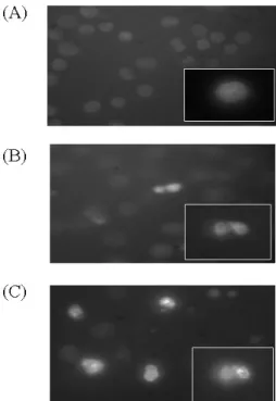
As2O3가 K562 세포주에 세포고사를 유도하는지 확인 하기 위해 형광물질인 Hoechst 33258을 이용해 관찰하였 다. 그 결과 Fig. 1과 같이 As2O3 처리 10 μM 이상에서부 터 핵 염색질의 응축과 핵의 절편이 관찰되었다. 이 결과 는 이미 Shim 등(2002)의 연구에서 보고된 세포고사에 의한 형태변화를 뒷받침해 주는 결과였다.
2. 세포독성에 미치는 Caspase 억제제의 영향
Caspase-3의 활성은 세포고사와 밀접한 관계가 있다고 이미 보고된 바 있으며, 이에 K562 세포주에서 As2O3가 나타내는 세포독성에 caspase-3 억제제가 미치는 영향에 대하여 조사하였다. As2O3에 의한 세포고사에 있어서 caspase-3의 역할을 규명하고자 caspase-3의 억제제인 Z-VAD-fmk 25 μM 을 미리 처리한 후 MTT 법을 실시한 결과 Fig. 2와 같이 As2O3만 처리한 군에 비해 caspase-3 억제제를 처리한 군에서 세포독성이 감소됨을 보였다. 이 결과를 통해 caspase-3가 As2O3에 의한 세포고사에 주된
Time % Cells
G0/G1 S G2/M
0 46.0 50.9 3.2
12 46.2 50.6 3.3
24 48.8 43.4 7.8
48 28.2 56.3 15.5
72 14.0 54.7 31.3
Table 1. Time-dependent alteration in cell cycle induced by As2O3
Fig. 1. Fluorescence microscopic appearance of Hoechst 33258- stained nuclei of As2O3-treated cells. The cells were treated with a control vehicle (A), 10 mM As2O3 for 24 h (B), 20 mM As2O3 for 24 h (C), stained with Hoechst 33258, and analyzed under a fluorescence microscope (x100).
역할을 한다고 알 수 있었다.
Fig. 2. Effect of caspase inhibitor, Z-VAD-fmk, on cytotoxicity in As2O3-treated K562 cells. The cells were treated with 25 mM Z-VAD-fmk for 1 h and then treated with the indicated concentrations of As2O3 for 24 h. Cytotoxicity was measured by MTT assay. *The datum in presence of Z-VAD-fmk is significantly different from that in the absence of Z-VAD-fmk (p<0.05).
3. 세포주기에 미치는 영향
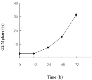
세포고사와 관련된 신호전달체계가 세포주기의 arrest- ed phase와 관계가 있다는 보고(Molnar 등, 1997)가 있어 K562 세포주의 세포주기에 As2O3가 영향을 미치는가 알 아보았다. As2O3를 처리한 후 Table 1에서 보는 바와 같 이 0시간에서 3.2%, 48시간 후엔 15.5%, 72시간 후엔 31.3%로 시간이 경과함에 따라 G2/M 세포의 비율이 높 게 나타났으며(Fig. 3, 4), 또한 농도별 처리 결과 역시 농 도가 증가함에 따라 G2/M phase 세포들이 증가함을 알 수 있었다(Fig. 5).
Fig. 3. FACScan analysis of K562 cells treated with As2O3. The cells were treated with 10 mM for indicated times, and then the cells were fixed 70% ethanol, stained with PI, and DNA content was measured by flow cytometry.
Fig. 4. Time-dependent alteration in G2/M phase induced by As2O3. Cells were incubated with 10 mM of As2O3 for indicated times, stained with propidium iodide, and assayed by flow cytometry.
Fig. 5. Dose-dependent alteration in cell cycle induced by As2O3. The cells were incubated with the indicated concentration of As2O3 for 48 h, stained with propidium iodide, and assayed by flow cytometry.
IV. 고 찰
세포고사는 세포내에서 단백질과 genomic DNA를 분 해하는 caspase와 nuclease의 활성화를 통해 일어나는 세 포사의 한 형태로, 대부분의 암치료제는 세포고사를 유도 한다고 보고되고 있다. 세포고사의 유발인자는 세포 증식 조절과 세포 생존조절의 균형에 의해 결정되며, 이 세포 생존조절은 계속적인 생존에 필수적인 신호의 철회와 세
포고사를 유발하는 신호의 균형에 의해 결정되어진다.
As2O3 역시 세포고사와 관계된 유전자들을 활성화시킴으 로써 백혈병 세포주에서 세포고사를 유도한다는 연구가 보고된 바 있다(Chen 등, 1996; Soignet 등, 1998).
Caspase family는 세포고사에 중요한 역할을 하고 있으 며, 특히 caspase-3는 세포고사 동안 여러 세포 종류에서 활성화된다고 하였다(Datta 등, 1999). 본 연구에서는 As2O3 처리에 의한 caspase-3의 활성화를 확인하기 위해 caspase-3 억제제를 함께 처리한 결과 As2O3만 처리한 군 에 비해 세포독성이 감소되었으며, 이를 통해 caspase-3가 세포고사의 중개자로서 작용한다는 것을 확인하였다. 그 리고 형광현미경을 통해 세포 형태변화를 살펴 본 결과 염색체 응축 등 세포고사로 진행되는 세포의 형태학적 변화를 관찰하였다.
세포주기는 세포의 진행을 촉진시키는 인자와 억제기 전에 관여하는 인자 사이의 균형에 의해 조절된다. 일반 적으로, 종양은 비정상 세포의 증식 뿐 아니라 세포고사 의 억제에 의한 세포의 감소된 죽음이 원인이 된다. K562 세포는 bcr-abl 융합유전자를 가지고 있으며, 종양 억제유 전자인 p53, p15, p16 등의 활성화를 억제시킨다고 알려 져 있다(Gale과 Canaani, 1984; Law 등, 1993; Otsuki 등, 1995). 또한 세포고사에 미치는 세포신호전달체계 중 MAPK(mitogen-activated protein kinase)의 영향에 대해 많이 연구되어지고 있다. MAPK에는 크게 3종류가 존재 하는데 ERK1/2와 JNK1/2, 그리고 p38 kinase이며 (Robinson과 Cobb, 1997), 3종류의 MAPK는 모두 serine/threonine kinase로 상위 kinase에 의해 tyrosin과 threonine residue가 인산화되어 활성화된다고 한다(Wu 등, 1993). 이에 As2O3와 관련된 연구에서는 p38 kinase가 관련된다는 보고가 있다. 세포고사는 적어도 일부분 세포 주기의 변화로부터 야기되며, 비정상적인 상태에서의 세 포성장의 자극은 여러 종류의 세포에서 세포고사를 유도 할 수 있다. Nishii 등(1996)의 연구에 의하면 bcr-abl을 transfection시킨 BaF3세포에 방사선을 쪼였더니 G2세포 가 증가하였으며, 이 세포분열의 증가는 세포고사와 관련 된 여러 특징을 보였다고 한다. 이에 대해 본 연구에서 As2O3가 세포주기에 영향을 미치는지 알아보고자 시간 별, 농도별로 처리하여 유세포 분석을 통해 세포주기 변 화를 측정한 결과 G2/M 세포의 비율이 높게 나타남을 확 인하였으며, 이는 세포고사와 관련되어 있다고 생각되었 다.
위 연구를 종합해 볼 때, K562 CML 세포주는 As2O3에
의해 염색질 응축 등의 세포고사로 진행되는 형태변화를 보였으며, 이 과정에서의 caspase-3의 활성증가를 caspase- 3 억제제를 이용한 실험에서 확인하였으며, G2/M phase arrest를 유도함으로써 세포독성을 가지고 있다는 것을 알 수 있었다. 따라서 As2O3에 의한 세포고사기전을 응용함 으로써 CML에 있어서 새로운 치료제의 가능성에 관한 정보의 제공과 함께 임상적 응용에도 도움을 줄 것으로 사료되며, 앞으로 이에 대해 지속적인 연구가 필요하리라 생각된다.
참 고 문 헌
1. Chen YR, Wang X, Templeton D, Davis RJ, Tan TH.
The role of c-Jun N-terminal kinase (JNK) in apoptosis induced by ultraviolet C and radiation. J Biol Chem 271:31929-31936, 1996.
2. Datta SR, Brunet A, Greenberg ME. Cellular survival:
a play in three Akts. Genes Dev 13:2905-2927, 1999.
3. Gale RP, Canaani E. An 8-kilobase abl RNA transcript in chronic myelogenous leukemia. Proc Natl Acad Sci USA 81:5648-5652, 1984.
4. Hale AJ, Smith CA, Sutherland LC, Stoneman VE, Longthorne VL, Culhane AC, Williams GT.
Apoptosis: molecular regulation of cell death. Eur J Biochem 236:1-26, 1996.
5. Law JC, Ritke MK, Yalowich JC, Leder GH, Ferrell RE. Mutational inactivation of p53 gene in the human erythroid leukemic K562 cell line. Leuk Res 17:1045-1050, 1993.
6. Molnar A, Threodoras AM, Zon LI, Kyriakis JM.
Cdc42Hs, but not Rac1, inhibits serum-stimulated cell cycle progression at G1/S through a mechanism requiring p38/RK. J Biol Chem 20:13229-13235, 1997.
7. Nishii K, Kabarowski JH, Gibbons DL, Griffiths SD, Titley I, Wiedemann LM, Greaves MF. BCR-ABL kinase activation confers increased resistance to genotoxic damage via cell cycle block. Oncogene 21:2225-2234, 1996.
8. Otsuki T, Clark HM, Wellmann A, Jaffe ES, Raffeld M. Involvement of CDKN2 (p16INK4A/MST1) and p15INK4B/MST2 in human leukemias and lymphomas.
Cancer Res 55:1436-1440, 1995.
9. Robinson MJ Cobb MH. Mitogen-activated protein kinase pathways. Curr Opin Cell Biol 9(2):180-186, 1997.
10. Shim MJ, Kim HJ, Yang SJ, Lee IS, Choi HI, Kim TU. Arsenic trioxide induces apoptosis in chronic myelogenous leukemia K562 cells: Possible involve- ment of p38 MAP kinase. J Biochem Mol Biol 35(4):
377-383, 2002.
11. Soignet SL, Maslak P, Wang ZG, Jhanwar S, Calleja E, Dardashti LJ, Corso D, DeBlasio A, Gabrilove JL, Scheinberg DA, Pandolfi PP, Warrell RP. Complete remission after treatment of acute promyelocytic leukemia with arsenic trioxide. N Engl J Med 339:1341-1348, 1998.
12. Wu J, Dent P, Jelinek T, Wolfman A, Weber MJ, Sturgill TW. Inhibition of the EGF-activated MAP kinase signaling pathway by adenosine 3',5'-mono- phosphate. Science 12:988-990, 1993.