The Cytotoxic and Apoptotic Effect of Pseudomonas aeruginosa Exotoxin A on Human Leukemia K-562 Cells
Jeong-Hyun Chang and Heun-Young Kwon
Department of Clinical Laboratory Science, College of Health Sciences, Catholic University of Pusan, Busan 609-757, Korea
After reports on regression of cancer in humans and animals infected with microbial pathogens date back more than 100 years, much effort has been spent over the years in developing a wild type or attenuated bacterial and purified bacterial proteins for the treatment of cancer. Pseudomonas aeruginosa exotoxin A (ETA) is known to inhibit cell growth and trigger significant cell death in various cancer cells. Although ETA induces apoptosis of cancer cells, its exact mechanism of action is not known yet. Four different assays were performed in this study: morphological assessment of apoptotic cells, cell cytotoxity, annexin-V binding assay, and cell cycle analysis. The proliferation and survival of the K-562 cells treated with ETA were decreased in a dose dependent manner. In addition, the apoptotic body of K-562 cells was induced by ETA treatment in a dose dependent manner. The ETA-induced apoptosis was confirmed by annexin-V binding assay. Flow cytometric analysis was examined to ascertain whether ETA could arrest the cell cycle at the sub-G1 phase. Our results suggest that P. aeruginosa ETA inhibits cell growth and induces apoptosis in human leukemia K-562 cells.
Key Words : Pseudomonas aeruginosa exotoxin A, Apoptosis, Annexin-V, Cytotoxicity, Cell cycle
I. INTRODUCTION
Antitumor agents induce apoptosis in some cancer cells both in vitro and in vivo, indicating that apoptosis plays an important role in cancer chemotherapy (Meyn et al, 1995). Many bacteria have been used in an effort to reduce the growth rate or size of tumors. Bacteria are able to trigger apoptosis by a whole variety of mechanisms including the secretion of protein synthesis inhibitors, pore forming proteins, molecules activating the endogenous death machinery in the infected cell or lipopolysaccharides
Corresponding auther : Heun-Young Kwon, Department of Clinical Laboratory Science, College of Health Sciences, Catholic University of Pusan, Busan 609-757, Korea
TEL : 051-510-0561 E-MAIL : [email protected].
and other superantigens (Weinrauch et al, 1999). Pseu- domonas aeruginosa produces several extracellular pro- ducts that, after colonization, can cause extensive tissue damage, bloodstream invasion, and dissemination.
Exotoxin A (ETA), a 66 kDa protein of 613 amino acids, is considered to be the most toxic factor secreted by P.
aeruginosa. Because of its potent cytotoxicity, ETA has been used to generate fusion proteins to kill target cells (Christopher et al, 2004).
Apoptosis is known as an important biological mechanism that contributes to the maintenance of the integrity of multicellular organism (Kaufmann, 1989). It is induced by a wide variety of cellular stresses such as DNA damage, UV radiation, ionizing radiation and oxidative stress (Nagata, 1997), and is morphologically distinct from necrosis in many of its characteristic changes
as follows; DNA fragmentation, chromatin condensation (Wyllie et al, 1980), cytoplasmic membrane blebbing, and cell shrinkage. Annexin V, a member of a recently discovered family of proteins, the annexins, with anti- coagulant properties has been proven to be a useful tool in detecting apoptotic cells since it preferentially binds to negatively charged phospholipids, like phosphatidylserine (PS) in the presence of Ca2+, and shows minimal binding to phosphatidylcholine and sphingomyeline. Changes in PS asymmetry, which is analyzed by measuring Annexin- V binding to the cell membrane, were detected before morphological changes associated with apoptosis. Al- though the metabolism of ETA, the biochemical basis for its apoptosis-inducing activity, is not clearly understood yet. This study was performed to prove the cytotoxic and apoptotic effect of Pseudomonas aeruginosa ETA in human leukemia K-562 cells.
II. MATERIALS AND METHODS
1. Cell culture
Suspension cultures of human leukemia K-562 cells were grown at 37℃ in humidified 5% CO2 incubator using Dulbecco’s modified Eagle medium (DMEM, GIBCO, USA) supplemented with 10% newborn calf serum and gentamycin (50 μg/mL). The pH of the growth medium was adjusted to 7.2~7.4 with 10 mM HEPES buffer (Sigma Chemical Co. USA) and 7.5% sodium bicarbonate.
2. MTT(Thiazolyl Blue Tetrazolium Bromide) assay
The cytotoxic effect of ETA in cells was estimated by MTT assay. In the MTT assay, cells were placed in a 96-well plate and incubated for 24 hrs. Then cells were treated with various concentrations of ETA for 24 hrs.
Then, the cells were treated with 1 μg/mL of MTT in a
growth medium. The cells were incubated at 37℃, 5%
CO2 for 4 hrs. The medium was aspirated and the formazan crystals, which are formed from MTT tetrazolium by NADH-generating dehydrogenases in metabolically active cell, were dissolved in DMSO. Cell viability was evaluated in comparison to the control culture by measuring the intensity of the blue color (OD at 540 nm) by a multi-well reader (Quant, Bio-Tek, Highland Park, USA). The assay was performed in triplicate.
3. 4'-6-Diamidino-2-phenylindole (DAPI) staining assay
For DAPI staining assay, 1 × 10⁶ cells were plated in 2 mL of the growth medium in the presence or absence of increasing concentrations of ETA in 6-well plates and cultured at 37℃ in 5% CO2 for various incubation times.
The cells were washed with the PBS and we added 500 μL of 4% paraformaldehyde, and the cells were incubated those cells at 4℃ for 1 hr. The cells were washed with the PBS and we added 500 μL of DAPI staining solution.
We incubated at 4℃ for 5 min and observed them with a fluorescence microscope.
4. Annexin-V binding assay
After treatment with 10 nM, 100 nM, 10 μM, and 100 μM paclitaxel, the cells (1 × 10⁵ cells/treatment) were used to determine the translocation of phosphatidylserine to the outer surface of the plasma membrane during apoptosis using the human phospholipid binding protein, Annexin-V, conjugated with fluorescein (Molecular Probes, Inc., Eugene, OR, USA) by flow cytometry.
Apoptosis and necrosis were analyzed by quadrant statistics on the propidium iodide negative, fluorescein isothiocyanate positive cells, and propidium iodide positive cells, respectively.
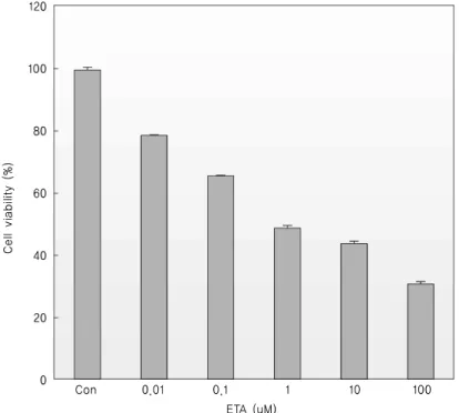
Fig. 1. Dose-dependent inhibitory effect on the viability of K-562 cells. Cells (1 × 10⁶ cells/well in 6-well plates) were treated with each different concentrations of ETA for 18 hrs and then, cell numbers were measured by trypan blue staining. Trypan blue-positive cells were considered as nonviable and their percentage was estimated by bright-field microscopy.
5. Cell cycle analysis
Cells were harvested in PBS-EDTA solution, fixed in cold 70% ethanol, and stored at -20°C. Fixed cells were subsequently washed, treated with 100 μg/mL RNase A, and stained with 50 g/mL propidium iodide. the analysis of DNA contents was performed in a FACScan flow cyto- meter (Becton Dickinson) with a minimum of 1 × 10⁴ events collected for analysis using Becton Dickinson Cell Quest software. Cells were sorted based on the expression of green fluorescent protein and DNA contents were analyzed in these cells.
III. RESULTS
1. ETA on cell viability assay
To confirm the viability of cells, the above mentioned trypan blue assay was performed with K-562 cells. ETA treatment on K-562 cells decreased the viability of the cells in a dose-dependent manner (Fig. 1) and in a
time-dependent manner (data not shown). Therefore, ETA had a significant inhibitory effect on the viability of K-562 cells.
2. The cytotoxic effect of ETA
MTT assays were performed to evaluate the cytotoxic effects of ETA. As shown in Fig. 2, K-562 cells were incubated with increasing concentrations of ETA. The results obtained revealed cytotoxic effects in a does-dependent manner.
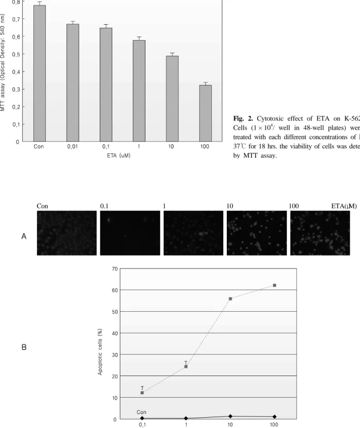
3. Apoptosis induced by ETA
To confirm the morphological changes of K-562 cells by ETA, DAPI staining assay was performed. Fig. 3 shows the morphology of apoptotic cells determined by DAPI assay with K-562 cells. Morphological changes were observed in the sample treated with 10 and 100 μM of ETA. Fluorescence increased in a does-dependent manner. The percentages of apoptotic cells in K-562 cells
Con 0.1 1 10 100 ETA(μM)
Fig. 3. Morphological change by ETA in K-562 cells. For DAPI staining assay, 1 × 10⁶ cells were plated in 2 mL growth medium in the presence or absence of various concentrations (1~100 μM) of ETA in 6-well plates and cultured at 37℃ in 5% CO2.
Fig. 2. Cytotoxic effect of ETA on K-562 cells.
Cells (1 × 10⁴/ well in 48-well plates) were each treated with each different concentrations of ETA at 37℃ for 18 hrs. the viability of cells was determined by MTT assay.
A
B
Fig. 4. ETA regulated Sub-G₁phase arrest of K-562 cells. Cells were plated in 2 mL growth medium in the presence or absence of various concentrations of ETA in 6-well plates and cultured at 37℃ in 5% CO2 for 18 hrs. Each panel represents the flow-cytometric measurements of DNA contents shown after PI staining.
Fig. 5. Measurement of Annexin-V binding of K-562 cells. For Annexin-V Binding Assay, 1 × 10⁴ cells were plated in 2 mL growth medium in the presence or absence of various concentrations of ETA in 6-well plates and cultured at 37℃ in 5% CO2 for 18 hrs.
To confirm the apoptosis of the cells by ETA, Annexin-V flow cytometric experiments were performed as described by the manufacturer.
treated with ETA were increased continuously.
4. ETA controlled sub-G1 arrest
To determine ETA controlled sub-G₁arrest in K-562 cells, flow cytometric cell cycle analyses were performed
following the PI staining of nuclei. Fig. 4 shows the results of a representative experiment in which K-562 cells were incubated for 18 hrs with various concentration of ETA.
5. Detection of Annexin-V binding from K-562 cells
To confirm the apoptosis of K-562 cells by ETA, Annexin- V flow cytometric experiments were performed.
The change in location of phosphatidyl-serine in the cell membrane during apoptosis can be detected with Annexin V. Co-staining with Annexin V and PI allowed differ- entiation of viable cells (Annexin V-negative, PI-negative) from early apoptotic cells (Annexin V-positive, PI-nega- tive) and late apoptotic cells (Annexin V-positive, PI- positive). K-562 cells treated with various concentrations of ETA were incubated for 18 hrs. As shown in Fig. 5, the percentages of both early (bottom right quadrant) and late (top right quadrant) apoptotic cells increased in a dose-dependent manner.
IV. DISCUSSION
A number of micro-organisms not only kill cells with virulent factors that interact with key molecules of the cell death pathway, but also cause cancer (Weinrauch et al, 1999; Blaser et al, 1995). Nonetheless, these bacteria were interestingly used in the treatment of cancer nearly 150 years ago. It has been reported that mammalian cancers were reduced when they were infected with microbial pathogens (Coley et al, 1991; Alexandroff et al, 1999).
Because most tumors contain large and poorly vas- cularized areas, hypoxia is pathophysiologic chara- cteristics of most solid tumors (Helmlinger et al, 1997;
Brown et al, 1998). Recently, considering this point, anaerobic bacteria, devoid of its toxic genes, were used in treatment of cancer, resulting in significant regression of subcutaneous tumors (Dang et al, 2001). However, there are still difficulties produced by live bacteria, which need to be overcome before the safe treatment of human cancer.
Numerous studies have focused on the targeted induction of apoptosis in order to control the cancer cells. Bacteria
can induce apoptosis by a variety of different mechanisms including the inhibition of several survival pathways active in the mammalian cells (Alexandroff et al, 1999), the secretion of protein synthesis inhibitors and the molecules activating the endogenous death machinery in the infected cells (Helmlinger et al, 1997). P. aeruginosa frequently causes pneumonia (Brown et al, 1998), sep- ticemia, and other acute infections (Dang et al, 2001) in immunocompromised patients. This gram- negative bacte- rium produces several extracellular products such as proteases, hemolysins, ETA, exoenzyme S, elastase and pyocyanin (Blaser et al, 1995). It has been reported that P. aeruginosa induced apoptosis in human respiratory epithelial cells (Coley et al, 1991) and endothelial cells (Alexandroff et al, 1999). ETA is a highly active anticancer drug that triggers apoptosis in a wide spectrum of cancer cells (Ofir et al, 2002; Huisman et al, 2002).
This is the first study to provide experimental evidence demonstrating that a dose-dependent cytotoxic effect by ETA in K-562 cells exists. ETA caused preferentially apoptotic cell death on K-562 cells. In this study, ETA produces a significant dose-dependent decrease in cell proliferation and induced an apoptotic- type cell death.
Apoptosis, as described by Wyllie et al. (1980), involves the condensation of chromatin, the restructuring of the cytoplasm, the blebbing of cytoplasmic membranes, and finally the fragmentation of the cells into apoptotic bodies that are phagocytosed by neighbouring cells, and features that distinguish the process from necrotic cell death. Apoptosis is initially characterized by morpho- logical changes of dying cells. Morphological change (DNA fragmentation) by various concentrations of ETA was observed. The primary mechanism of the action of ETA is attributed to its ability to bind to microtubules and prevent their assembly, causing cells to arrest in the Sub-G1 phase and then progress to apoptosis. This study demonstrated that ETA induced Sub-G₁arrest in K-562 cells in a dose-dependent manner. In addition, our report confirmed that the apoptosis of K-562 cells by ETA
increased in a dose-dependent manner.
The results presented in our study indicated that ETA is capable of killing K-562 cells through apoptosis me- chanism. In conclusion, ETA may provide a therapeutic choice in the clinical application for the treatment of cancer cells.
References
1. Alexandroff AB, Jackson AM, O'Donnell MA, James K. BCG immunotherapy of bladder cancer 20 years on. Lancet 353:1689-1694, 1999.
2. Blaser MJ, Perez-Perez GI, Kleanthous H, Cover TL, Peek RM, Chyou PH, Stemmermann GN, Nomura A.
Infection with Helicobacter pylori strains possessing cagA is associated with an increased risk of deve- loping adenocarcinoma of the stomach. Cancer Res 55:2111-2115, 1995.
3. Brown JM, Giaccia AJ. The unique physiology of solid tumors : opportunities (and problems) for cancer therapy. Cancer Res 58:1408-1416, 1998.
4. Christopher EJ, Swiatoniowski A, Issekutz AC, Lin TJ. Pseudomonas aeruginosa exotoxin A induces human mast cell apoptosis by a caspase-8 and -3- dependent Mechanism. J Biol Chem 279:37201-37207, 2004.
5. Coley WB. The treatment of malignant tumors by repeated inoculations of erysipelas with a report of ten original cases. Clin Orthop Relat Res 262:3-11, 1991.
6. Dang LH, Bettegowda C, Huso DL, Kinzler KW, Vogelstein B. Combination bacteriolytic therapy for
the treatment of experimental tumors. Proc Natl Acad Sci USA 98:15155-15160, 2001.
7. Helmlinger G, Yuan F, Dellian M, Jain RK. Inter- stitial pH and pO2 gradients in solid tumors in vivo:
high-resolution measurements reveal a lack of corr- elation. Nat Med 3:177-182, 1997.
8. Huisman C, Ferreira CG, Broker LE, Rodriguez JA, Smit EF, Postmus PE, Kruyt FA, Giaccone G.
Paclitaxel triggers cell death primarily via caspase- independent routes in the non-small cell lung cancer cell line NCI-H460. Clin Cancer Res 8:596–606, 2002.
9. Kaufmann SH. Induction of endonucleolytic DNA cleavage in human acute myelogenous leukemia cells by etoposide, camptothecin, and other cytotoxic anti- cancer drugs : a cautionary note. Cancer Res 49 : 5870-5879, 1989.
10. Meyn RE, Stephens LC, Hunter NR. Silas L. Apop- tosis in murine tumors treated with chemotherapy agents. Anticancer Drugs 6:443-450, 1995.
11. Nagata S. Apoptosis by death factor. Cell 88:355-365, 1997.
12. Ofir R, Seidman R, Rabinski T, Krup M, Yavelsky V, Weinstein Y, Wolfson M. Taxol-induced apoptosis in human SKOV3 ovarian and MCF7 breast carcinoma cells is caspase-3 and caspase-9 independent. Cell Death Differ 9:636-642, 2002.
13. Weinrauch Y, Zychlinsky A. The induction of apop- tosis by bacterial pathogens. Ann Rev Microbiol 53:
155-187, 1999.
14. Wyllie AH, Kerr JFR, Currie AR. Cell death : the significance of apoptosis. Int Rev Cytol 68:251-306, 1980.
국문 초록
인간 백혈병 세포에서 Psuedomonas aeruginosa exotoxin A에 대한 세포독성과 세포자멸사 효과
약 100년 전에 박테리아가 암을 억제한다는 보고를 바탕으로 다양한 미생물이 항암효과를 가지는 백신 개발 에 이용되거나 또는 미생물의 세포 밖 독소 단백질을 찾아내고 있다. Psuedomonas aeruginosa exotoxin A(ETA) 는 암세포에서 세포성장을 억제하고 세포 죽음을 유발하는 것으로 알려져 있다. 하지만 ETA가 세포 자멸사를 유도하는 정확한 기전은 아직 알려져 있지 않다. 따라서 본 연구에서는 세포자멸사의 유도를 확인하기 위해 K562 cell을 이용하여 세포의 형태학적 변화, 세포독성, Annexin-V binding assay 그리고 세포주기를 분석하였으 며, 그 결과로 ETA는 K-562세포에서의 세포증식과 성장을 억제하였고, 세포자멸사 기작을 통한 K-562 암세포의 사멸을 일으켰음을 관찰하였다. 또한 flow cytometric analysis에서는 ETA가 세포주기 중 특히 sub-G1 기를 정지 시키는 것으로 나타났다. 본 연구는 ETA가 인간 백혈병 K-562 암세포의 세포성장을 억제하고 sub-G1 기를 정 지시킴으로서 세포자멸사를 유도하고 있음을 확인하였다.