Endoplasmic recticulum stress와 관련된 유전자기능과 전사조절인자의 in silico 분석
김태민․여지영․박찬선1․이문수1․정명호*
부산대학교 한의학전문대학원,1한국생명공학연구원
Received July 8, 2009 /Accepted July 28, 2009
In Silico Analysis of Gene Function and Transcriptional Regulators Associated with Endoplasmic Recticulum (ER) Stress. Tae-Min Kim, Jiyoung Yeo, Chan Sun Park1, Moon Soo Rhee1 and Myeong Ho Jung*. School of Oriental Medicine, Pusan National University, Yangsan 626-810, Gyeongnam, Korea,
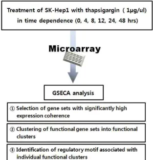
1Korea Research Institute of Bioscience and Biotechnology, Yusong, Taejon 305-600, Korea - It has been postu- lated that endoplasmic (ER) stress is involved in the development of several diseases. However, the detailed molecular mechanisms have not been fully understood. Therefore, we characterized a genetic network of genes induced by ER stress using cDNA microarray and gene set expression coherence analysis (GSECA), and identified gene function as well as several transcription regulators associated with ER stress. We analyzed time-dependent gene expression profiles in thapsigargin-treated Sk-Hep1 using an oligonucleotide expression chip, and then selected functional gene sets with significantly high expression coherence which was processed into functional clusters according to the expression similarities. The functions related to sugar binding, lysosome, ribosomal protein, ER lumen, and ER to golgi transport increased, whereas the functions with mRNA processing, DNA replication, DNA repair, cell cycle, electron transport chain and helicase activity decreased. Furthermore, functional clus- ters were investigated for the enrichment of regulatory motifs using GSECA, and several transcrip- tional regulators associated with regulation of ER-induced gene expression were found.
Key words : Endoplasmic recticulum (ER) stress, gene expression microarray, gene set expression coherence analysis (GSECA), gene function, transcription regulator
*Corresponding author
*Tel:+82-51-510-8468, Fax:+82-51-510-8437
*E-mail : [email protected]
서 론
Endoplasmic reticulum (ER)는 단백질 합성 및 새로 만들어 진 단백질의 합성후변형 뿐만 아니라 세포내 칼슘이온의 저장 등에도 중요한 역할을 담당한다. 대부분의 분비단백 혹은 막 결합단백은 ER에서 정상적인 구조를 획득하여 분비경로를 따 라 이동하게 되나, 그렇지 못한 단백질(misfolded or unfolded protein)의 경우 ER의 분해기능에 의해서 제거된다. 그런데 ER stress는 이러한 ER의 단백질처리능력과 부하간의 불균형 로 인해 일어나는 현상으로, 초기단계에서는 chaperon의 발현 증가를 통한 단백질의 합성 후 변형 능력증가, 단백질합성억 제, 그리고 비정상적 단백질제거 등의 보호반응(Unfolded Protein Response, UPR)으로 세포가 대처하나, 적응한계를 넘 어서는 과도한 스트레스의 경우, 세포사멸(apoptosis)을 포함 한 여러 가지의 병리적인 현상들을 유발한다[8].
지금까지 과도한 ER stress는 프리온병, 알츠하이머병, 파킨 슨병 등의 신경질환의 원인으로 보고되어졌으나, 최근에는 ER stress가 제2형 당뇨병의 발병에도 중요한 병인으로서 관 여함이 보고되어졌다[9]. 당뇨병의 경우, 여러 조건에서 유발 되는 ER stress가 c-jun N-terminal kinase (JNK)의 활성화를
통해 간이나 지방조직에서 인슐린 저항성(insulin resistance) 을 유도하고, 인슐린을 분비하는 췌장조직에서 β-cell의 세포 사멸을 유발함으로써 당뇨병을 일으키는 것으로 알려져 있다 [5]. 특히, 비만유도가 된 동물모델과 고열량식을 섭취한 동물 의 간과 지방조직에서 ER stress가 증가하고, 이에 따라 in- sulin receptor substrate-1 (IRS-1)의 tyrosine 인산화와 Akt의 serine 인산화가 감소하여 인슐린저항성이 유도됨을 밝혀, 비 만에 의한 ER stress 증가가 당뇨병의 원인이 됨을 증명하였다 [6]. 한편, 이러한 ER stress을 감소시키는 chemical chaperon 들을 처리하면 당뇨쥐에서 당뇨상태가 개선됨이 또한 보고되 었다[7].
ER stress와 질병과의 관련성에도 불구하고 ER stress에 의 한 기능적인 변화 및 핵심적인 조절인자는 극히 일부만이 밝 혀진 상태이다. ER stress에 의한 유전자발현의 조절은 전사적 수준(transcriptional level)에서 이루어져, ATF6, ATF4, CHOP, XBP-1 등의 몇몇 전사조절인자가 관여하는 것으로 알 려져 있으나[12], 고등동물의 전사조절이 다수의 전사조절인 자 및 조절특이염기서열의 복잡한 조합으로 이루어진다는 점 을 고려할 때 훨씬 많은 수의 전사조절인자가 상호작용하여 ER stress 반응을 조절할 것으로 여겨진다. 그러므로 ER stress 와 관련된 핵심적인 전사조절인자를 찾아 이들의 작용기전을 밝히면, 질병발병에서의 역할규명 및 분자적 중재 방법 등을 개발할 수 있을 것이다.
따라서 본 연구에서는 ER stres와 관련된 기능적 변화와 핵심적인 조절인자를 규명하기 위해, 인슐린 작용조직인 간세 포에서 ER stress를 시간별로 유도한 후, expression micro- array로 유전자의 발현을 분석하였고, 이 expression profiles 을 GSECA의 방법으로 분석하였다.
재료 및 방법 세포배양
실험에 사용한 세포주는 간암세포인 SK-Hep1으로 ATCC 로부터 분양받았으며, 15% fetal bovine serum (FBS), 2 mM glutamine, 100 IU/ml penicillin, 100 μg/ml streptomycin등 이 포함된 DMEM 성장배지를 사용하여 5% CO2, 37oC의 조건 에서 배양하였다. ER stress 유도를 위해 SK-Hep1에 thapsi- gargin을 1 μg/ml 농도로 4, 8, 12, 24, 48 시간 처리하였다.
RNA 분리 및 Affymetrix GeneChip
Thapsigargin을 시간별로 처리한 cell들과 처리하지 않은 cell들로부터 total RNA를 TRizol reagent (GibcoBRL, Invitrogen Corporation, USA)으로 분리하였으며, 이를 이용 하여 double stranded cDNA 합성하였다. Biotin-labelling은 Affymetrix Labelling Assay kit (Affymetrix Inc, USA)을 이용 하여 cDNA로부터 cRNA을 합성하면서 labelling하였으며, factionation된 biotin-labelled cRNA는 Human U133-Plus 2.0 (Affymetrix Inc) chip과 18 시간 동안 hybridization을 행하였 다. Hybridization image는 GeneChip operating Software (Affymetrix Inc)으로 scan하였으며, Affymetrix Inc에서 제공 하는 Biconductor package로 expression level을 정량하였다.
Functional gene sets 작성
Normalization값을 gene ontology (GO) 데이터베이스를 이용하여 유전자를 기능적으로 분류하였고, 통계프로그램인 Hypergeometric distribution (GSEA) [3]를 이용하여 유의한 유전자군의 기능을 밝혔다. 유전자들을 기능별로 분류하기 위 해 public gene database인 GO, KEGG, GenMAPP들을 바탕 으로 유전자들을 grouping하였다.
Functional gene sets의 expression coherence을 이 용한 functional clustering
각 functional gene set에 대해 GSECA [8]을 이용하여 각 유전자들의 상관성을 결정하였다. 모든 가능한 유전자쌍들에 대해 Pearson correlation coefficient (PCC)을 계산하여 그 평 균값을 각 functional gene set에 대해 expression coherence로 사용하였다. 매우 높은 expression coherence를 가진 func- tional gene set을 위해 gene set에 속해있는 평균 expression값 을 계산하여 비슷한 expression pattern을 나타내는 functional
gene set을 hierachical clustering으로 나타내었다.
Functional cluster들의 전사조절인자 확인
Regulatory motif gene set을 이용하여 각각의 functional cluster의 putative transcriptional regulator을 GSEA 방법[3]
을 변형하여 분석하였다. 전반적인 방법은 GSEA방법과 유사 하나, GSECA [2]에서는 gene ordering parameter로서 single to noise ratioo (SNR) 보다는 PCC를 사용하였다.
결 과
ER stress을 유도한 SK-Hep1에서 gene expression microarray
SK-Hep1에 ER stress를 유도하기 위해 ER stress 유도물질 인 thapsigargin을 시간별(0, 4, 8, 12, 24, 48 시간)로 처리하였으 며, ER stress marker protein인 peIF를 측정함으로써 ER stress 가 유도되었음을 확인하였다(data not shown). Expression mi- croarray은 thapsigargin을 처리한 각각의 세포로부터 total RNA을 분리한 후 Affymetrix의 Human U133-Plus 2.0 chip을 사용하여 유전자들의 발현을 분석하였다.
ER stress 에 의한 유전자의 기능적 변화
ER stress에 의한 유전자들의 기능변화를 GSECA의 방법[8]
으로 분석하였다. 매우 유의적으로 expression coherence을 나 타내는 gene sets을 functional annotation으로 우선적으로 선 별한 후, 이러한 functional gene sets을 다시 발현이 비슷한 것끼리 묶고 일반적인 k-means clustering algorithm을 이용 하여 약 12종류의 clusters을 분류하였다(Fig. 2). 이들 중 발현
Fig. 1. Study procedure.
Fig. 2. Functional clustering of ER stress-related functional gene sets. Hierachical clustering was used to measure the dis- tances between functional gene sets and those with sim- ilar expression patterns were grouped into individual functional clusters by k-means clustering algorithm. The expression level of a functional gene set is the mean ex- pression value of the genes belonging to the gene sets.
이 ER stress 유도에 따라 증가하는 cluster들과 감소하는 clus- ter들에 대해 유전자들의 기능들을 분석하였다(Table 1). Table 1에서 보는 바와 같이 ER stress 유도에 따라 lysosome, sugar binding, ER lumen에 관련된 기능이 증가하는 반면, mRNA processing, mitosis, DNA replication, DNA repair, cell cycle, electron transport chain, response to DNA damage, helicase activity, small nucleolar ribonucleoprotein complex, DNA-dependent ATPase activity, RNA transcription 과 관련 된 기능은 감소하였다.
ER stress에 관련된 전사조절인자
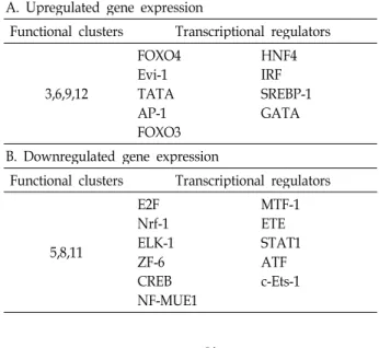
각각의 functional clusters에 속해있는 유전자들의 발현을 조절하는 조절인자를 확인하기 위해, 각 유전자들의 promoter 에 존재하는 putative transcription factor motif을 이용하여 GSECA의 방법[2]으로 분석하였다(Table 2). Cluster 3, 5, 7 과 cluster 5, 8, 7, 9의 유전자들의 전사조절인자를 분석한 결과, ER stress 유도에 따라 발현이 증가하는 cluster들(Cluster 3, 5, 7)에 대해서는 FOXO4, AP-1, FOXO3, HNF4, IRF-1, GATA 등의 전사인자들이 공통적으로 존재하였으며, ER stress유도 에 따라 발현이 감소하는 유전자들의 cluster들(Cluster 3, 5, 7)에서는 E2F, Nrf-1, Elk-1, YY1, CREB, MTF-1, STAT-1, ATF 등의 전사조절인자들이 공통적으로 존재하였다.
Table 1. Annotated function of representative functional clus- ters
A. Upregulated gene expression
Cluster Annonated function
3, 6, 9, 12
lysosome sugar binding
endoplasmic reticulum lumen ER to golgi transport B. Downregulated gene expression
Cluster Annonated function
5, 8, 11
mRNA processing mitosis
DNA replication cell division cell_cycle DNA repair
small nucleolar ribonucleoprotein complex DNA-dependent ATPase activity
electron transport chain helicase activity
response to DNA damage stimulus
transcription from RNA polymerase II promoter
Table 2. Lists of transcriptional regulators significantly en- riched in functional clusters
A. Upregulated gene expression
Functional clusters Transcriptional regulators 3,6,9,12
FOXO4 Evi-1 TATA AP-1FOXO3
HNF4 IRFSREBP-1 GATA
B. Downregulated gene expression
Functional clusters Transcriptional regulators
5,8,11
E2F Nrf-1 ELK-1 ZF-6 CREBNF-MUE1
MTF-1 ETE STAT1 ATF c-Ets-1
고 찰
ER은 단백질 합성 및 새로 만들어진 단백질의 합성 후 변형 이 일어나는 세포소기관으로 세포내 칼슘이온의 저장에도 중 요한 역할을 담당한다. 그러나유전자의 손상에 의한 돌연변 이, 포도당 결핍과 같은 대사이상이나 감염, 약물 등의 스트레 스에 의해 소포체내에 비정상적인 단백질이 증가하면 ER stress 반응기전이 활성화된다. 이러한 ER stress 반응의 초기
단계에서는 단백질합성억제, 비정상적 단백질제거 등의 적응 반응을 통해 보상될 수 있으나, 적응한계를 넘어서는 과도한 ER stress의 경우 궁극적으로 세포는 세포사멸(apoptosis)을 일으키는 등 질병을 유발하게 된다.
ER stress 반응에 관여하는 전사조절인자로는 ATF6, ATF4, CHOP, XBP-1등이 알려져 있으나[1], 다른 전사조절인자들에 대해서는 구체적으로 규명되지 않은 상태이다. 최근에는 ER stress가 당뇨병의 핵심 병태생리로 작용한다는 것이 알려지 고 있으나, 구체적으로 ER stress 반응중 어떠한 조절인자가 작용하며, 어떠한 기전으로 당뇨병에서 나타나는 인슐린 분비 장애나 인슐린 저항성 등을 유발하는 지는 아직 구체적으로 알려진 바가 없다.
그런데 ER stress 유도에 따른 표적유전자규명과 유전자네 트워크구성은 gene expression microarray와 유전자의 pro- moter 영역에 해당하는 염기서열을 분석하는 생물정보학적방 법을 이용하면 가능하다. 특히, 고등동물의 전사적조절이 다 수의 전사조절인자와 결합염기서열간의 다양한 결합패턴에 의해서 결정된다는 점을 고려할 때, 훨씬 많은 수의 전사인자 가 ER stress에 관여할 것으로 예측되어진다. 결국 전사조절인 자들의 상위 및 하위레벨에 해당하는 또 다른 전사인자와 이 들과 상호작용하는 유전자들이 밝혀져야 할 것이다. 따라서, 본 연구에서는 ER stress 반응을 유도하는 조건하에서 당뇨관 련조직인 간세포에서 유전자발현의 profiling을 확보하고, 이 를 이용하여 ER stress에 의한 유전자들의 기능적 변화와 핵심 전사조절전사인자를 in silico로 분석하였다.
Thapisigargin을 시간별로 처리한 Sk-Hep1 세포에서 total RNA를 분리하고 expression microarray을 행하여 유전자 발 현 profiling을 확보하였다. 매우 유의적으로 expression co- herence을 나타내는 gene set을 functional annotation으로 우 선적으로 선별한 후, 이러한 functional gene set을 다시 발현 이 비슷한 것끼리 묶어 약 12 종류의 cluster로 분류하였다.
이들의 유전자들 기능을 GSECA 의 방법[2]으로 분석하였을 때, lysosome, sugar binding, ER lumen (Cluster 6)에 관련된 기능이 증가하는 반면, mRNA processing, mitosis, nuclear mRNA splicing, DNA replication, DNA repair, cell cycle, electron transport chain, response to DNA damage, helicase activity, helicase activity, electron transport chain, response to DNA damage 등의 기능은 감소하였다. 이러한 기능의 변 화는, ER stress가 유도되면 ER에 관련된 기능(ER lumen, ER to Golgi transporter)이 증가하여, ER stress에 대응하여 세포 를 방어하는 기능들이 증가하나, 새로운 단백질의 생성(DNA replication, mRNA processing)이나, ATP 생성(Electron transport chain), DNA 수선(Response to DNA damage, DNA repair)과 세포분열(Mitosis) 등의 기능은 감소하는 것으 로 나타났다. 따라서 ER stress가 유도되면, 1) ER에 주어지는 과도한 부하를 감소시키는 기능들이 증가되어지나 2) ER
stress가 점차 더 증가하면 mRNA transcription, ATP 생성이 나 DNA repair 더 나아가 세포분열의 기능이 감소하여 세포 의 활성이 감소됨을 알 수 있었다.
ER stress에 의한 유전자의 조절에 관련된 전사조절인자들 을 역시 생물정보학적 방법(GSECA)으로 분석하였을 때, FOXO4, AP-1, FOXO3, HNF4, IRF-1, GATA 등의 전사인자들 이 ER stress에 의해 발현이 증가하는 유전자들의 promoter에 공통적으로 존재하였으며, E2F, Nrf-1, Elk-1, YY1, CREB, MTF-1, STAT-1, ATF 등의 전사인자들이 발현이 감소하는 유 전자들의 promoter에서 공통적으로 존재하여, 이들의 전사인 자들이 ER stress에 의한 유전자의 발현조절에 중요한 역할을 하는 전사조절인자임을 알 수 있었다.
이들 전사조절인자 중 HNF4, Foxo, AP-1 등은 gluconeo- genesis에 관여하는 phosphoenolpyruvate carboxylase kin- ase (PEPCK)의 발현을 증가시키는 대표적인 전사인자[10, 4, 11]로, ER stress가 HNF4, Foxo, AP-1 등의 전사조절인자의 활성증가를 통해 PEPCK의 발현을 증가시켜 당뇨병을 유발할 것으로 판단되었다. 또한 미토콘드리아 biogenesis에 매우 중 요한 전사인자인 Nrf-1이 ER stress에 의해 발현이 감소되는 유전자들의 promoter에서 발견되어 졌는데, 이는 ER stress에 의해 전자전달계의 기능이 감소하는 데에 관여할 것으로 생각 되어졌다. 즉, ER stress에 의해 Nrf-1의 활성이 감소됨으로써 전자전달계에 관여하는 유전자들의 발현이 감소되어, 전자전 달계의 기능이 감소할 뿐만 아니라 미토콘드리아의 기능도 감소할 것으로 판단되었다.
요 약
ER stress에 관련된 유전자의 기능변화와 전사조절인자 분 석하기 위해 ER stress를 유도한 간세포에서 expression micro- array로 유전자 발현을 확보한 후 GSECA로 분석하였다. ER stress가 유도되면, ER에 주어지는 과도한 부하를 감소시키는 기능들이 증가하는 반면, ER stress가 더 증가함에 따라 ATP 생성이나 DNA repair, 더 나아가 세포분열의 기능이 감소하는 등 세포가 damage을 받음을 알 수 있었다. ER stress에 관련된 전사조절인자로는 FOXO4, AP-1, FOXO3, HNF4, IRF-1, GATA 등의 전사조절인자들이 ER stress에 의해 발현이 증가 하는 유전자들의 promoter에 공통적으로 존재하였으며, E2F, Nrf-1, Elk-1, YY1, CREB, MTF-1, STAT-1, ATF 등의 전사인자 들이 발현이 감소하는 유전자들의 promoter에서 공통적으로 존재하여, 이들의 전사인자들이 ER stress에 의한 유전자의 발 현조절에 중요한 역할을 하는 전사조절인자임을 알 수 있었다.
감사의 글
이 논문은 부산대학교 자유과제 학술연구비(2년)에 의하여
연구되었음.
References
1. Iwakoshi, N. N., A. H. Lee, and L. H. Glimcher. 2003. The X-box binding protein-1 transcription factor is required for plasma cell differentiation and the unfolded protein response. Immunol. Rev. 194, 29-38.
2. Kim, T. M., Y. J. Chung, M. G. Rhyu, and M. H. Jung. 2007.
Inferring biological functions and associated transcriptional regulators using gene set expression coherence analysis.
BMC Bioinformatics 17, 453-563.
3. Kim, T. M. and M. H. Jung. 2006. Identification of transcrip- tional regulators using binding site enrichment analysis.In Silico Biol. 6, 531-544.
4. Mounier, C. and B. I. Posner. 2006. Transcriptional regu- lation by insulin: from the receptor to the gene. Can. J.
Physiol. Pharmacol. 84, 713-724.
5. Oyadomari, S., E. Araki, and M. Mori. 2002. Endoplasmic reticulum stress-mediated apoptosis in pancreatic beta-cells.
Apoptosis 7, 335-345.
6. Ozcan, U., Q. Cao, E. Yilmaz, A. Lee, N. Iwakoshi, E.
Ozdelen, G. Tuncaman, C. Gorgun, and G. Hotamisligil.
2004. Endoplasmic reticulum stress links obesity, insulin ac-
tion, and type 2 diabetes. Science306, 457-461.
7. Ozcan, U., E. Yilmaz, L. Ozca, M. Furuhashi, E. Vailancourt, R. Smith, C. Gorgun, and G. Hotamisligil. 2006. Chemical chaperones in a mouse model of type 2 diabetes.Science 313, 1137-1140.
8. Ron, D. and P. Walter. 2007. Signal integration in the endo- plasmic reticulum unfolded protein response.Nat. Rev. Mol.
Cell. Biol. 8, 519-529.
9. Tiotra, P. and C. Tsigos. 2006. Stress, the endopalsmic retic- ulum, and insulin resistance. Ann. N. Y. Acad. Sci. 1083, 63-76.
10. Wang, J. C., P. E. Strömstedt, T. Sugiyama, and D. K.
Granner. 1999. The phosphoenolpyruvate carboxykinase gene glucocorticoid response unit: identification of the func- tional domains of accessory factors HNF3 beta (hepatic nu- clear factor-3 beta) and HNF4 and the necessity of proper alignment of their cognate binding sites.Mol. Endocrinol. 13, 604-618.
11. Yeagley, D., J. M. Agati, and P. G. Quinn. 1998. A tripartite array of transcription factor binding sites mediates cAMP induction of phosphoenolpyruvate carboxykinase gene transcription and its inhibition by insulin.J. Biol. Chem. 273, 18743-18750.
12. Zhao, L. and S. Ackerman. 2006. Endoplasmic reticulum stress in health and disease.Curr. Opin. Cell Biol.18, 444-452.