Regulation of Endoplasmic Reticulum Stress Response by the Immobilization Stress
Kisang Kwon
1†, Young-Sook Kwon
3†, Seung-Whan Kim
2, Dong-Woon Kim
1and O-Yu Kwon
1*
1
Departments of Anatomy, School of Medicine, Chungnam National University, Taejon 301-747, Korea
2
Department of Emergency Medicine, Chungnam National University Hospital, Taejon 301-721, Korea
3
Department of Nursing, Joongbu University, Chungnam 312-702, Korea
Received July 3, 2012 /Revised July 11, 2012 /Accepted August 8, 2012Many kind of cell stresses induce gene expression of unfolded protein response (UPR)-associated factors. This study demonstrated that up- and down-regulation of gene expression of endoplasmic re- ticulum (ER) stress chaperones and ER stress sensors was induced by immobilization stress in the rat organs (adrenal gland, liver, lung, muscle). However, no statistically significant regulation was de- tected in the others (heart, spleen, thymus, kidney, testis). The results are the first to show that im- mobilization stress induces UPR associated gene expression, will help to explain immobilization stress-associated ER stress.
Key words : Immobilization stress, endoplasmic reticulum (ER) stress, ER chaperone, ER stress sensor
*Corresponding author
*Tel:+82-42-580-8206, Fax:+82-42-586-4800
*E-mail : [email protected]
†These authors contributed equally to this work.
서 론
조직을 이루고 있는 각각의 세포는 외부의 급격한 환경변화 에 적응하여 정상적인 생리 상태를 유지하기 위한 세포내 자 체 조절능력을 가지고 있다. 세포의 정상적인 생리 상태를 교 란시키는 것을 총칭하여 세포스트레스(cell stress)라고 한다.
특히, 세포소기관인 소포체(endoplasmic reticulum, ER)에 크 게 영향을 미치는 세포스트레스를 소포체스트레스(ER stress) 라고 한다[8]. 분비단백질은 ER내에서 posttranslational mod- ification을 거쳐 정상적으로 세포외로 분비되어야 하지만 다 양한 ER stress에 의해서 소포체내강(ER lumen)에 축적된다.
변이단백질의 발현과 비정상적인 환경, 정상 분비단백질의 과 잉생산이 대표적인 원인으로 생각할 수 있다. 이 같은 ER stress에 세포가 대응하는 가장 기본적인 응답을 소포체스트 레스응답(unfolded protein response, UPR)로 총칭하며 특정 단백질의 전사유도와 세포의 총 단백질 번역을 억제하는 sys- tem이다[2]. 이때에 ER lumen에서 UPR에 관여하는 단백질들 을 소포체샤페론(ER chaperone)이라고 하다. 이들은 ER lu- men에서 정상적으로 단백질이 folding & assembly할 수 있는 환경을 제공해주는 일에 중추적인 역할을 한다. 즉, ER chap- erone은 새롭게 합성된 폴리펩다이드의 folding, 단백질의 세 포내 이동 혹은 세포외 분비, 비정상적인 구조를 가진 단백질 의 분해 등, 단백질이 처음 만들어져서 성숙하여 분비 및 분해 될 때까지의 전 과정에 걸쳐서 관여하는 단백질이다. 이처럼
ER chaperone은 세포내의 여러 가지 단백질이 정상적인 기능 을 가질 수 있도록 종합적인 품질관리기능을 수행하고 있다 (ER quality control, ERQC) [9]. ERQC기능에 의해서 불량한 분비단백질이 과도하게 ER lumen에 축적되면 질병으로 나타 나는데 이를 포괄적으로 ER storage disease (ERSD)라고하며, congenital hypothyroidism, albinism tyrosinase deficiency, polyglutamine diseases와 같은 질환이 알려져 있다[3]. ER stress는 다양한 inducer에 의해서 유도된다. 대표적인 것으로 는 tunicamycin 처리에 의해서 당단백질의 glycosylation이 방 해 받았을 때, A23187 혹은 thapsigagin에 의해서 ER lumen의 Ca
2+depletion이 일어날 때, ER lumen에 과도한 malfolded 단백질이 축적될 때, DTT와 같은 환원제에 의해서 단백질의 3차구조에 이상이 생길 때, virus 감염 혹은 cancer와 같은 병 리적인 상태가 될 때이다. 이때에 관여하는 ER signal path- way에 직접 작용하는 3종류의 ER stress sensor (IRE1, PERK, ATF6)가 알려져 있다[11]. 세포가 ER stress를 받으면 ER lu- men에서 BiP과 결합하고 있는 IRE1이 monomer에서 인산화 된 dimer가 되어, 세포질의 XBP-1 mRNA의 splicing이 일어 나 XBP-1단백질이 생산되어 chaperone 생합성을 촉진한다.
PERK 역시 인산화된 dimer가 되면서 핵전사인자인 eIF2α의 인산화를 유도하여 세포 전체의 단백질합성을 저해함으로써 세포들 보호한다. 그리고 ATF6은 ER stress를 받으면 세포질 쪽의 단편이 떨어져 나와서 ERSE (ER stress element)와 결합 한다. 이같이 세포는 ER stress를 받을 경우에 자신을 보호할 수 있는 기전을 가지고 있다. 이처럼 다양한 자극에 의해서 ER stress가 유도되어 세포가 적응을하는것이 알려져있다[10].
설치류를 포함하는 동물은 자가 방어기전의 하나로 타 동물
로 부터 멀리 도망가려는 습성을 가지고 있다. 실험적으로도
설치류를 작은 상자에 가두면 스트레스를 받아서 신경발생이
- Note -
저해되어 우울증(depression)과 같은 현상을 보이지만, 항우 울제(antidepressants)를 투여하면 반대효과가 나타난다. 이처 럼 동물을 움직이지 못하게 하는 스트레스를 부동스트레스 (immobilization stress)라고 한다. 지금까지 알려진 대표적인 부동스트레스 관련연구는 다음과 같다. 부동스트레스는 cata- cholamine의 생합성 조절, peroxiredoxin I & II의 발현 조절, leptin signaling 조절, acetylcholinesterase 활성조절, 지방-단 백질-DNA의 oxidative damage 원인에 깊이 관여한다[1,4-7].
그러나 아직까지 부동스트레스에 의한 ER stress유도에 관련 된 보고는 없다. 본 연구는 처음으로 부동스트레스가 세포생 존에 중요한 기전중의 하나인 ER chaperone 및 ER signaling 의 발현을 조절하는지를 알아보았다.
재료 및 방법
실험동물인 Sprague Dawley계 흰쥐(수컷, 4주령)는 다물 사이언스(대전시)에서 구입하여 사용하였다. 각각 3마리씩 대 조군과 부동스트레스를 가한 군으로 나누어 실험하였다. 부동 스트레스 방법은 Kvetnansky & Mikulaj 실험방법을 사용하 였다[4]. 할로탄을 사용하여 가볍게 흡입마취 시킨 후 평평한 아클릴 판에 흰쥐의 네발에 시판하는 청테이프를 이용하여 등이 아래를 향하도록 하여 움직이지 못하게 붙였다. 그리고 난 후 6시간동안 방치하여 부동스트레스를 주었다.
조직으로부터 total RNA을 얻기 위하여 cold PBS로 충분히 세정한 다음에 사용한다. 1.5 ml tube에 RNA isolation re- agent (TRI-REAGENT)를 500 μl와 잘게 절단된 조직을 넣고 초음파분쇄기로 2-3분 동안 충격을 준다. 여기에 100 μl의 chloroform을 첨가하여 충분히 섞어준 다음 13,000 rpm, 4℃
에서 15분 동안 원심 분리하였다. 약 500 μl의 상등액을 취하여 새로운 tube로 옮기고 동량의 isopropanol 넣고 상온에서 10 분 정도 처리한 후 13,000 rpm으로 10분 동안 원심분리하고 tube의 바닥에 얻어진 pellet에 75% ethanol을 초기 RNA iso- lation reagent 양과 동일한 500 μl 넣고 12,000 rpm으로 5분 동안 원심 분리하여 최종적으로 total RNA를 얻었다. DEPC가 처리된 증류수에 녹여 UV spectrophotometer로 정량하였다.
RT-PCR (reverse transcription polymerase chain reaction)은 RNA (3 μg)를 80℃에서 3분 가열하여 denaturation 시킨 후 바로 얼음에 담가둔다. 10x buffer 3 μl, dNTP 4 μl, 1 μl의 oligo-dT (300 ng), 10,000 U의 역전사효소와 RNase inhibitor 를 첨가하고 총 30 μl가 되게 한 후 42℃에서 1시간 30분 간 반응시켜 cDNA를 합성한다. 반응이 끝난 후 94℃에서 2분 반응시켜 역전사효소를 inactivation시킨 다음 최종 100 μl로 맞춘다. 그 다음 단계로 cDNA를 증폭하기 위해서 PCR을 수 행하였다. PCR 반응액 20 μl에 각각의 forward 와 reverse pri- mer를 넣어서 94℃ 5분, 94℃ 30분, 57℃ 40초, 72℃ 40초로 27회 반복하여 전기영동으로 확인하였다. 사용된 forward 와
reverse primer는 아래와 같다. F (5'-AGTGGTGGCCACT AATGGAG-3’) and R (5'-TCTTTTGTCAGGGGTCGTTC-3’) for Bip; F (5'-TGTGGATGGCACGGTAGAAG-3’) and R (5'-GGTGCCCAGGTTTTTAACCA-3’) for GRp94; F (5'- GCATCATGCCATCTCTGCTA-3’) and R (5'-GGCATCTTC ATCCCAGTCAT-3’) for CNAX; F (5'-GGACTGGGACGAA GAGATGG-3’) and R (5'-CCTCTGCTCCTCATCCTGCT-3’) for Calr; F (5'-AAACTCCTCCCAGCGTTTCA-3’) and R (5'-TAGGCCTCCAAGGACTGGAA-3’) for EDEM; F (5'-CA GAGTTCTGCCACCGCTTC-3'), and R (5'-TCCTCGAGATC GTCATCATC-3') for PDI; F (5'-AGTGGTGGCCACTAAT GGAG-3’) and R (5'-TCTTTTGTCAGGGGTCGTTC-3’) for ERp29; F (5'-TGATTGGACACCTCCACCTG-3'), and R (5'-GGTCACCGACTCCCTGAAAG-3') for ERp72; F (5'-TGT GCTGTCAAACCCTGCCATT-3’) and R (5'-ATTGATGCTT GCGTGTAGGCCA-3’) for Ero1; F (5'-CTAGGCCTGGAGG CCAGGTT-3’) and R (5'-ACCCTGGAGTATGCGGGTTT-3’) for ATF6; F (5'-CAGAGTTCTGCCACCGCTTC-3'), and R (5'-TCCTCGAGATCGTCATCATC-3') for PDI; and R (5'-CC ACCCTGGACGGAAGTTTG-3’) for IRE1; F (5'-GGTCTG GTTCCTTGGTTTCA-3’) and R (5'-TTCGCTGGCTGTGTA ACTTG-3’) for PERK; F(5'-TGAGTCTCTGCCTTTCGCCTTT- 3'), and R (5'-TCAGCAAGCTGTGCCACTTT-3') for Chop;
F (5'-CGTTCAGACAGAGGCCAGTTC-3'), and R (5'-CGA GGACCACCATCATCC-3') for Rpn1; F (5’-AGCCATGTAC GTAGCCATCC-3’) and R (5’-CTCTCAGCTGTGGTGGTG AA-3’) for β-actin
Western blotting은 Protein electrophoresis kit (ATTO Co., Japan)를 사용하여 12% SDS-PAGE에 준비된 sample을 전기 영동 하였다. 전기영동이 끝난 후 transfer kit (Bio-RAD)를 사용하여 gel의 protein을 PVDF cell membrane에 transfer buffer (20 mM Tris-HCl, 150 mM glycine, 20% methanol, pH 8.3)를 사용하여 transfer하였다. Transfer가 끝난 후 mem- brane을 3.75% skim milk in PBST (PBS, 0.05% Tween 20)를 사용하여 상온에서 1시간 동안 blocking하였다. Blocking이 끝 난 후 1차 항체를 반응 시킬 때에는 5% skim milk in PBST에 1:1,000의 비율로 희석하여 4℃에서 2시간 동안 반응시켰다.
1차 항체 반응이 끝난 후 membrane을 PBST를 10분씩 5회
상온에서 shaker를 사용하여 세척하였다. 세척이 끝난 후 2차
항체를 반응 시킬 때에는 PBST에 1:2,000의 비율로 희석하여
상온에서 1시간 동안 반응 시켰으며, 반응이 끝난 후 mem-
brane을 PBST를 10분씩 5회 상온에서 shaker를 사용하여 세
척하였다. 세척이 끝난 후 West save (Lab Frontier, Korea)을
사용하여 발색반응을 유도한 후 X-ray film에 감광하여 결과
를 분석하였다.
A
B
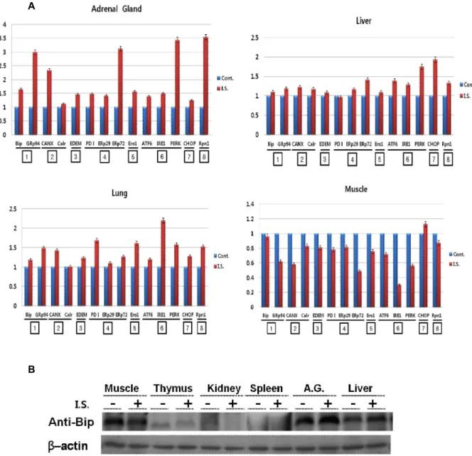
Fig. 1. Effect of the immobilization stress on the unfolded protein response (UPR)-associated gene expression in the each organ.
Adult Sprague Dawley rats were housed under controlled conditions on a 12 h light/12 h dark cycle and provided with food and water at libitum. Rats were handled daily for 7 days before experiments to minimize the stress of handling.
Rat were immobilized by strapping for 6 h according to Kvetnansky & Mikulaj methord [4]. Control rats walked freely.
Each gene expression was estimated by RT-PCR(A) and tested by Western blotting to estimate those translational level(B).
Vertical axis of Fig. 1A indicates relative gene (mRNA) expression. Detailed preparation of each sample was described in the
Material & Method
. Rabbit anti-Bip antibody and anti-actin antibody were obtained from Santa Cruz Biotechnology (Santa Cruz, CA, USA). The bar graph shows the averages of the quantified data from 3 independent experiments. I.S.(immobilization stress) & A.G. (adrenal gland).
결과 및 고찰
부동스트레스를 받은 rat를 희생시켜서 RT-PCR을 하기 위 한 total RNA와 SDS-PAGE를 하기 위한 단백질을 각각 조직 (heart, spleen, thymus, kidney, testis, adrenal gland, liver, lung, muscle)에서 분리하였다. 부동스트레스가 각 장기에서 ER chaperones과 ER signaling 관련 유전자들의 발현조절에
관여하는지를 알기 위하여 RT-PCR로 그들의 발현을 알아보
았다. Bip (immunoglobulin heavy chain-binding protein)과
GRp94 (glucose regulated protein 94 kDa)는 ER lumen에서
미숙 혹은 불완전한 단백질의 folding & assembly를 돕는
general chaperones, calnexin과 calreticulin은 ER lumen에서
당단백질의 folding & assembly를 돕는 lectin chaperones,
ER-associated glycoprotein degradation에 관여하는 EDEM
(ER degradation-enhancing alpha-mannosidase-like 1), ER lumen에서 잘못 형성된 단백질의 -S-S-를 인식하여 고쳐주는 PDI (protein disulfide isomerase)와 유사한 기능의 ERp29, ERp72 그리고 전자공여단백질인 Ero1 (ER oxidoreductin 1) 을 포함하는 redox chaperones, ER stress signal의 sensor 역 할을 하는 ATF6 (activating transcription factor 6), IRE1 (inositol requiring ER-to-nucleus signal kinase 1), PERK [double-stranded RNA-activated kinase (PKR)-like ER kin- ase] 그리고 전사수준에서 UPR을 apoptosis 혹은 in- flammation을 결정하는 전사인자인 CHOP (CCAAT/en- hancer binding protein homologous protein). Control로 ER membrane에 있는 Rpn1 (ribophorin 1)을 사용하였다.
Heart, spleen, thymus, kidney, testis에서는 어떤 유전자도 통계적으로 유의성 있는 발현의 차이가 관찰되지 않았다, 그 러나 adrenal gland, liver, lung, muscle에서는 유의성 있는 유전자의 발현차이가 관찰되었다(Fig. 1A). 전체적인 발현양 상은 adrenal gland, liver, lung에서는 발현이 상승하는 유전 자들이 관찰된 반면에 muscle에서는 전체적으로 발현이 억제 되었다. 가장 심한 유전자발현의 차이를 보인 것은 adrenal gland였다. Adrenal gland에서는 ER lumen의 Bip (약 1.5배) 과 GRp94 (약 3배)는 동시에 상승 발현하였지만 calreticulin은 변화가 없고 calnexin (약 2.5배)은 상승 발현하였다. Redox chaperone중에서는 ERp72 (약 3배)만 높은 발현을 보였다. 그 리고 ER stress sensor중에서는 PERK(약3.5배) 발현이 상승하 였지만 나머지는 큰 변화를 보이지 않았다. 일반적인 스트레스 는 대뇌피질에서 인식하여 시상하부(hypothalamus)로 전달되 며 시상하부는 뇌하수체(pituitary)로 corticotropin-releasing hormone (CRH)을 방출한다. 이로 인해 혈장으로 분비된 cor- ticotropin이 부신피질(adrenal cortex)의 corticotropin re- ceptors를 자극하여 혈액내로 cortisol을 분비한다. 시상하부의 cortisol receptor는 CRH 생성을 감소시켜 항상성을 유지한다.
이와 같이 일반적인 스트레스를 가장 잘 전달하고 조절하는 기관이 adrenal gland이다. 이런 관점에서 부동스트레스 역시 타 종류의 스트레스와 동일하게 작용 및 응답하기 때문에 가 장 강하게 ER stress와 관련 유전자의 변화를 유도하는 것으로 추정할 수 있다. Liver의 CHOP의 상승발현(약 2배)이 주목된 다, liver에서의 경우는 부동스트레스가 apoptosis 혹은 in- flammation을 선택하는 전사인자 CHOP의 발현만이 높아진 것은 이들의 선택에 밀접한관계가 있을 것으로 추정된다.
Lung에서는 IRE1의 발현이 2배 이상 상승하였으며 나머지는 특이적으로 주목할 만한 발현변화는 없었다. Muscle의 경우는 전체적으로 발현 감소하였다. Control에 비교하여 1/2 정도까 지 발현이 감소된 것은 ERp29, IRE1이다, 그중에서 IRE1이 가장 강하게 발현이 억제되었다. 근육에서 이처럼 모든 유전 자발현이 감소되는 것은 동물에게 부동스트레스로 말미암아 서 새로운 단백질합성이 일어나지 않기 때문에 ER lumen에서
단백질생합성에 관련하는 모든 유전자들의 활성이 떨어졌기 때문으로 추정된다. 부동스트레스는 ER stress 관련유전자의 변화는 각각 차이가 많지만, Rpn1의 발현이 약 3.5배 상승한 것으로 보아서 ER lumen에 UPR과 관련 있는 변화가 심하게 일어나 비정상적인 단백질축적에 의해서 ER 자체가 많이 커 진 것으로 추측할 수 있다. 위의 결과는 유전자 전사수준에서 조절되는 것을 확인인한 결과이다. Fig. 1B에서는 이들의 변화 가 단백질 번역수준에서도 적용되는지를 알기위하여 ER lu- men의 대표적인 chaperone인 Bip의 변화를 관찰하였다. 전사 수준의 양상과 동일하게 muscle에서는 부동스트레스에 의해 서 발현이 억제되고 adrenal gland, liver에서는 발현이 상승하 였다. 나머지는 발현의 변동이 관찰되지 않았다.
결론적으로 부동스트레스는 세포의 ER stress와 관련된 유 전자발현의 변화를 유도한다. 즉 Heart, spleen, thymus, kid- ney, testis에서는 유전자발현 변화가 없었지만 adrenal gland, liver, lung에서는 유의할만한 상승변화가 있었다. 그러나 muscle에서는 다른 것들과 대조적으로 발현이 감소되었다. 이 결과는 부동스트레스도 다른 종류의 cell stress와 같이 세포수 준에서 UPR을 조절할 수 있다는 최초의 보고이다.
감사의 글
This work was supported by the National Research Foundation of Korea (NRF) grant funded by the Ministry of Education, Science and Technology of Korea (MEST) (No.2012-0001830).
References
1. Das, A., Kapoor, K., Sayeepriyadarshini, A. T., Dikshit, M., Palit, G. and Nath, C. 2000. Immobilization stress-induced changes in brain acetylcholinesterase activity and cognitive function in mice.
Pharmacol. Res.
42, 213-217.2. Hetz, C. 2012. The unfolded protein response: controlling cell fate decisions under ER stress and beyond.
Nat. Rev.
Mol. Cell Biol.
13. 89-102.3. Kim, P. S. and Arvan, P. 1998. Endocrinopathies in the fam- ily of endoplasmic reticulum (ER) storage diseases: dis- orders of protein trafficking and the role of ER molecular chaperones.
Endocr. Rev.
19. 173-202.4. Kvetnansky, R. and Mikulaj, L. 1970. Adrenal and urinary catacholamines in rats during adaptation to repeated im- mobilization stress.
Endocrinology
87, 738-743.5. Larco, D. O., Cruthirds, D. F., Weiser, M. J., Handa, R. J.
and Wu, T. J. 2012. The effect of chronic immobilization stress on leptin signaling in the ovariectomized (OVX) rat.
Endocrine
[Epub ahead of print].6. Liu, J., Wang, X., Shigenaga, M. K., Yeo, H. C., Mori, A.
and Ames, B. N. 1996. Immobilization stress causes oxida- tive damage to lipid, protein, and DNA in the brain of rats.
초록:부동스트레스에 의한 소포체스트레스반응 조절 권기상
1․권영숙
2․김승환
3․김동운
1․권오유
1*
(
1충남의대 해부학교실,
2중부대학교 간호학과,
3충남대병원 응급의학과)
많은 종류의 세포스트레스는 unfolded protein response (UPR)관련인자의 유전자발현을 조절한다. 본연구결 과 부동스트레스(immobilization stress)는 세포의 소포체스트레스(ER stress)와 관련된 유전자발현의 변화를 유도 한다; Heart, spleen, thymus, kidney, testis에서는 유전자발현 변화가 없었지만 adrenal gland, liver, lung에서는 유의할만한 상승변화가 있었다. 그러나 muscle에서는 다른 것들과 대조적으로 발현이 감소되었다. 이 결과는 부 동스트레스도 다른 종류의 세포스트레스와 같이 세포수준에서 UPR을 조절할 수 있다는 최초의 보고이다.
FASEB J.
10, 1532-1538.7. Paek, N. H., Kwak, S. S., Lee, D. S. and Lee, Y. H. 2006.
Characterization of peroxiredoxins in the gray matter in the spinal cord after acute immobilization stress.
J. Korean Soc.
Traumatol.
19. 105-112.8. Parmar, V. M. and Schröder, M. 2012. Sensing endoplasmic reticulum stress.
Adv. Exp. Med. Biol.
738. 153-168.9. Saijo, Y. 2010. ER quality control of immune receptors and
regulators in plants.
Cell Microbiol.
12. 716-724.10. Shore, G. C., Papa, F. R. and Oakes, S. A. 2011. Signaling cell death from the endoplasmic reticulum stress response.
Curr. Opin. Cell Biol.
23. 143-149.11. Walter, P. and Ron, D. 2011. The unfolded protein response:
from stress pathway to homeostatic regulation.
Science
334.1081-1086.