SREBP-1c Ablation Protects Against ER Stress-induced Hepatic Steatosis by Preventing Impaired Fatty Acid Oxidation
Young-Seung Lee
1, Timothy F. Osborne
2, Young-Kyo Seo
3and Tae-Il Jeon
1*
1Department of Animal Science, Chonnam National University, Gwangju 61186, Korea
2Institute for Fundamental Biomedical Research, Department of Medicine and Biological Chemistry, Johns Hopkins University School of Medicine, St. Petersburg, FL 33701, USA
3Aging Research Center, Korea Research Institute of Bioscience and Biotechnology, Daejeon 34141, Korea Received July 9, 2021 /Revised July 28, 2021 /Accepted July 30, 2021
Hepatic endoplasmic reticulum (ER) stress contributes to the development of steatosis and insulin resistance. The components of unfolded protein response (UPR) regulate lipid metabolism. Recent studies have reported an association between ER stress and aberrant cellular lipid control; moreover, research has confirmed the involvement of sterol regulatory element-binding proteins (SREBPs)—the central regulators of lipid metabolism—in the process. However, the exact role of SREBPs in control- ling lipid metabolism during ER stress and its contribution to fatty liver disease remain unknown.
Here, we show that SREBP-1c deficiency protects against ER stress-induced hepatic steatosis in mice by regulating UPR, inflammation, and fatty acid oxidation. SREBP-1c directly regulated inositol-requir- ing kinase 1α (IRE1α) expression and mediated ER stress-induced tumor necrosis factor-α activation, leading to a reduction in expression of peroxisome proliferator-activated receptor γ coactivator 1-α and subsequent impairment of fatty acid oxidation. However, the genetic ablation of SREBP-1c prevented these events, alleviating hepatic inflammation and steatosis. Although the mechanism by which SREBP-1c deficiency prevents ER stress-induced inflammatory signaling remains to be elucidated, al- teration of the IRE1α signal in SREBP-1c-depleted Kupffer cells might be involved in the signaling.
Overall, the results suggest that SREBP-1c plays a crucial role in the regulation of UPR and in- flammation in ER stress-induced hepatic steatosis.
Key words : ER stress, fatty acid oxidation, hepatic steatosis, SREBP-1c, unfolded protein response
*Corresponding author
*Tel : +82-62-530-2127, Fax : +82-62-530-2129
*E-mail : [email protected]
This is an Open-Access article distributed under the terms of the Creative Commons Attribution Non-Commercial License (http://creativecommons.org/licenses/by-nc/3.0) which permits unrestricted non-commercial use, distribution, and reproduction in any medium, provided the original work is properly cited.
Introduction
Non-alcoholic fatty liver disease (NAFLD) is the most prevalent chronic liver disorder, affecting approximately 25% of the global population. NAFLD encompasses a wide histological variety of hepatic steatosis, steatohepatitis (NASH), fibrosis, and cirrhosis [33]. Although its pathogenesis and progression are complex and have not been fully elucidated, triglyceride (TG) accumulation is likely the first step of NAFLD pathophysiology, and it results from increased lipo- genesis, imbalanced fat import/export flux, and/or de- creased fat oxidation [23].
Sterol regulatory element binding proteins (SREBPs), which belong to the basic helix-loop-helix leucine zipper
(bHLH-LZ), regulate the expression of genes necessary for maintaining cellular lipid homeostasis [20]. There are three mammalian SREBP isoforms; SREBP-1a and 1c are encoded by a single gene owing to differential promoter usage and alternative splicing, whereas SREBP-2 is encoded by a differ- ent gene. Although there is some functional overlap among the isoforms, SREBP-1 primarily regulates fatty acid and TG synthesis whereas SREBP-2 is mainly involved in cholesterol biosynthesis [21]. SREBPs are synthesized as inactive pre- cursors bound to endoplasmic reticulum (ER) membrane. In response to low sterol levels, SREBP cleavage-activating pro- tein (SCAP) is dissociated from insulin-induced gene (INSIG) and then SCAP-SREBP complex translocates to the Golgi ap- paratus, where SREBPs are proteolytically processed to yield the transcriptionally active form. This process is also acti- vated by insulin signaling and ER stress [10].
ER stress results from the disruption of ER homeostasis,
such as unfolded or misfolded protein accumulation in its
lumen, and is associated with aberrant cellular lipid accumu-
lation, which is common in patients with obesity and NAFLD
[3]. Because SREBPs activate lipid biosynthesis genes and
are maintained as precursors in the ER membrane, it was reasonable to hypothesize that an enhanced SREBP process- ing due to ER stress would explain the mechanism of lipid overload in NAFLD. The first experimental observation that led to such mechanism was the processing of the ER mem- brane-bound precursor form of activating transcription fac- tor 6 (ATF6), an ER stress-related transcription factor that requires identical Golgi-located proteases involved in SREBP maturation [32]. Additionally, ER retention of ATF6- caused by its interaction with the ER stress-related chaperone 78 kDa glucose-regulated protein (GRP78) [27] and overex- pression of GRP78 in the livers- decreased steatosis and in- hibited SREBP-1 processing in the liver and primary hep- atocytes of ob/ob mice treated with both insulin and ER stress inducers [12]. These results suggest a more intimate relationship between ER stress and SREBPs than what was previously recognized in homocysteine-induced hepatic steatosis [30].
The unfolded protein response (UPR) signaling induced by ER stress is mediated by three ER membrane-bound pro- teins: protein kinase RNA (PKR)-like ER kinase (PERK), in- ositol-requiring kinase 1α (IRE1α), and ATF6 [25]. IRE1α acti- vation promotes the splicing of X box-binding protein 1 (XBP1) mRNA and subsequent transcription of molecular chaperones and genes involved in ER-associated degrada- tion (ERAD) [16]. IRE1α also appears to mediate the cellular signaling pathways involved in inflammation, insulin action, and apoptosis via c-Jun-NH
2-terminal kinase (JNK), p38 mi- togen-activated protein kinase, and nuclear factor-kappa B (NF-κB) [2]. Recent studies demonstrated that IRE1α phos- phorylation and XBP1 splicing were enhanced in the liver of mice with high-fat diet-induced insulin resistance [19] and that a liver-specific knockout of XBP-1 resulted in down- regulated expression of a subset of lipogenic genes and re- duced plasma lipid levels because of a decrease in hepatic TG secretion [14]. These studies suggest a close association between ER stress response and hepatic lipid control.
In this study, SREBP-1c directly activates the expression of IRE1α and lipogenic genes in hepatocytes; a decrease in fatty acid oxidation (FAO) by ER stress was reversed in SREBP-1c knockout mice, thereby contributing to protection against hepatic steatosis. These findings suggest a link be- tween the IRE1α-XBP1 pathway, inflammation, and lipid metabolism in an SREBP-1c-dependent manner.
Materials and Methods
Animals
C57BL/6J and SREBP-1c knockout mice on a B6:129S6 mixed background were purchased from Jackson Laborato- ries (stock number: 000664 and 004365, respectively). All mice were maintained on a chow diet for one week and 12-hr light/dark cycle for acclimatization. Then, 10-week-old male SREBP-1c knockout (1cKO) and wild-type (WT) litter- mates were injected intraperitoneally with 1 mg/kg body weight of tunicamycin (Tu; Sigma-Aldrich) or vehicle in 150 mM dextrose and sacrificed at 8:00 AM (end of the dark cycle) by CO
2asphyxiation at 8, 24, and 72 hr after injection.
For fasting/refeeding experiments, WT mice were fasted for 24 hr with or without refeeding with a regular chow diet for 12 hr before sacrifice. All animal experiments were per- formed in accordance to the accepted standards for animal protection welfare and with permission from the Institutio- nal of Animal care and Use Committees of the Chonnam National University (YB-2014-41).
Cell lines, adenoviral infection, and transfection AML12 and HeLa cells were purchased from the Amer- ican Type Culture Collection. AML12 cells were cultured in DMEM/F-12 media containing 1% ITS, 10% FBS, 10 ng/μl dexamethasone, and 1% antibiotics at 5% CO
2and 37℃.
HeLa cells were maintained in DMEM supplemented with 10% FBS and 1% antibiotics. The AML12 cells were treated with 0.5 mM palmitic acid (PA) complexed with BSA at a 5:1 molar ratio. The PA-BSA conjugates were prepared as described previously [1]. The HeLa cells were transfected for 48 hr with small RNA, including siRNA targeting human SREBP-1 or negative control (Dharmacon), using Lipofect- amine RNAiMAX (Invitrogen). The cells were also infected with Ad-hSREBP-1c (I. Shechter, Uniformed Services Univer- sity, Bethesda, Maryland, USA) or Ad-GFP for 48 hr. After adenoviral infection or siRNA transfection, the cells were incubated with 1 μM thapsigargin (Tg; Sigma-Aldrich) at an indicated time.
Primary hepatocyte isolation
To isolate the primary hepatocytes from WT and 1cKO
mice, the mice were anesthetized with isoflurane (Hana
Pharm. Co., South Korea) and perfused with Earle’s bal-
anced salt solution (Invitrogen) through a cannulated portal
vein for 3 min, followed by Liberase
TM(Roche Diagnostics),
Table 1. Primer sequences used in this study
Genes RT-qPCR primers
Forward Reverse
Chop Grp78 Gadd34
Ire1α Srebp-1c
Fasn Gpat Dgat2 Pgc-1α Mcad Atp5b Tnf-α Il-1β Xbp1 L32 IRE1α GAPDH
CTGCCTTTCACCTTGGAGAC GAAAGGATGGTTAATGATGCTGAG
TTTTGGCAACCAGAACCG ACACCGACCACCGTATCTCA TGGATTGCACATTTGAAGACAT
GCTGCGGAAACTTCAGGAAAT ACAGTTGGCACAATAGACGTTT GCGCTACTTCCGAGACTACTT AACCACACCCACAGGATCAGA
AGGTTTCAAGATCGCAATGG GGTTCATCCTGCCAGAGACTA
CGAGTGACAAGCCTGTAGCC ATGAGAGCATCCAGCTTCAA TTACGGGAGAAAACTCACGGC
ACATTTGCCCTGAATGTGGT GCCACCCTGCAAGAGTATGT
GAAGGTGAAGGTCGGAGTC
CGTTTCCTGGGGATGAGATA GTCTTCAATGTCCGCATCCTG GGAGATAGAAGTTGTGGGCG CTCAGGATAATGGTAGCCATGTC
GCCAGAGAAGCAGAAGAG AGAGACGTGTCACTCCTGGACTT
CCTTCCATTTCAGTGTTGCAGA GGGCCTTATGCCAGGAAACT TCTTCGCTTTATTGCTCCATGA
CTCCTTGGTGCTCCACTAGC AATCCCTCATCGAACTGGACG
AGCTGCTCCTCCACTTGGT TGAAGGAAAAGAAGGTGCTC GGGTCCAACTTGTCCAGAATGC
ATCCTCTTGCCCTGACCTT ATGTTGAGGGAGTGGAGGTG GAAGATGGTGATGGGATTTC
Genes ChIP primers
Forward Reverse
Ire1α Acc2
CTTCTAGCGCCCAGGATAGG GCAGGTAAGTAAGTGTGCTG
ATAAGTGCCTTGTGTCCCGG GCCACCAGTTCCATTCTCAG
a collagenase solution, for the next 3 min. The digested livers
were minced gently, and the hepatocytes were collected and washed twice with a buffer solution by centrifugation at 50×
g for 1 min. The cells were further purified with a Percoll
®buffer (GE Healthcare) by centrifugation at 100× g for 10 min. The primary hepatocytes were allowed to attach to col- lagen-coated plates for 6 hr in Williams’ Medium E (Thermo Fisher Scientific) with GlutaMAX (Life Technologies), anti- biotics, and 10% FBS. The cells were treated with 10 μg/ml Tu at an indicated time or with recombinant mouse TNF-α concentration (R&D System) for 24 hr.
Chromatin immunoprecipitation (ChIP) assay Chromatin preparations for ChIP assays with mouse liv- ers were performed as described previously [9]. For gene- specific ChIP, quantitative polymerase chain reaction (qPCR) analysis of SREBP-1 bound to specific gene promoters was performed with a standard curve method, and enrichment was normalized relative to the IgG control. The qPCR oligo- nucleotide pairs are shown in Table 1.
RNA analysis
The total RNA from the cultured cells and liver tissues was isolated using TRIzol™ (Invitrogen) and QIAGEN
RNAeasy isolation kits (QIAGEN), and cDNA was synthe- sized using an iScript™ cDNA Synthesis Kit (Bio-Rad).
qPCR was performed using SYBR Green Master Mix (Bio- Rad) with an iQ5 Real-Time PCR Detection System (Bio- Rad). The primer sequences used in this study are shown in Table 1. The mRNA levels were normalized for expression to mouse ribosomal protein L32 and human glyceraldehyde 3-phosphate dehydrogenase (GAPDH) as the control and were calculated using the comparative threshold cycle meth- od. Reverse transcription (RT)-PCR analysis of the Xbp-1 mRNA splicing was performed as described previously [16].
Immunoblotting
The hepatocytes were lysed in ice-cold RIPA buffer (50
mM Tris, 150 mM NaCl, 0.1% SDS, 0.5% Na deoxycholate,
and 1% Triton X) with protease and phosphatase inhibitors
(Thermo Fisher Scientific). The lysates were incubated on ice
for 20 min, further sonicated, and centrifuged at 27,000× g
for 20 min at 4℃. The supernatants were collected and stor-
ed at -80℃. Protein concentration was measured using a
BCA Kit (Thermo Fisher Scientific). Equal amounts of pro-
tein samples were separated by SDS-PAGE gel and were
then transferred to nitrocellulose membranes (Pall Corpora-
tion). The membranes were blocked in 5% skim milk for 1
hr and incubated with anti-SREBP-1 (2A4, Santa Cruz Bio- technology Inc.), anti-FASN, anti-IRE-1α, anti-p-PERK, anti- PERK, anti-GRP78, anti-CHOP, anti-sXBP1 (Cell Signaling Technology), and anti-β-actin (Sigma-Aldrich) as primary antibodies. After incubation with horseradish peroxidase- conjugated secondary antibodies, blots were developed us- ing a SuperSignal West Femto Maximum Sensitivity Sub strate (Thermo Fisher Scientific).
Histology and immunohistochemistry
Mouse livers were fixed with 4% paraformaldehyde, em- bedded in paraffin wax, and sectioned sagittally (4-µm thick). The slices were stained with hematoxylin and eosin dye to determine the morphological changes. The sections were dewaxed in xylene and rehydrated, and antigen re- trieval was performed by heating the sections with 10 mM citrate buffer at pH 6. The sections were blocked for 30 min in 1% BSA, 0.02% Triton X-100, and 10% normal goat serum, reacted with anti-F4/80 (1:400, sc-377009C-7, Santa Cruz Biotechnology Inc.) overnight at 4℃, and incubated with Alexa Fluor 488-conjugated goat anti-mouse secondary anti- bodies (Invitrogen). The sections were mounted by Vecta- shield with DAPI (Vector laboratories). Images from serial sections were acquired using a confocal laser scanning mi- croscope (A1R VAAS, Nikon) with an NIS-Elements AR software. F4/80-positive cells were counted using an Image J software.
Liver lipid measurement
TG and cholesterol levels were measured using a modi- fied method from a published study [28]. The liver samples were homogenized in PBS. Total lipids were extracted using a chloroform-methanol (2:1) solution for 12 hr at 4℃ and were dried with nitrogen gas. The lipid pellets were dis- solved in ethanol with 25% Triton X-100. TG and cholesterol levels were measured using commercially available kits (Thermo Fisher Scientific).
Fatty acid oxidation assays
FAO assay was performed as described previously [4].
The liver homogenates were incubated in [
3H]-PA (60μ Ci/
mM) bound to 2% BSA and were then transferred to a tube containing cold 10% trichloroacetic acid. The tubes were cen- trifuged at 8,500× g for 10 min at 4℃. The supernatant was immediately removed, mixed with 6N NaOH, and applied to an ion-exchange resin (DOWEX 1; Sigma-Aldrich). The
eluate was collected, measured by liquid scintillation ana- lyzer (PerkinElmer), and normalized to the amount of total protein.
Statistical analysis
The data are presented as mean ± SEM. A two-tailed, un- paired Student’s t-test was used for the pairwise comparison of treatments. One-way or two-way ANOVA was used to compare three or more groups, followed by Tukey’s multi- ple comparison test, as shown in the figures. The analyses were performed using the statistical software package Prism 6.0 (GraphPad Software). The differences were considered significant at p<0.05.
Results
SREBP-1c regulates IRE1α expression during ER stress
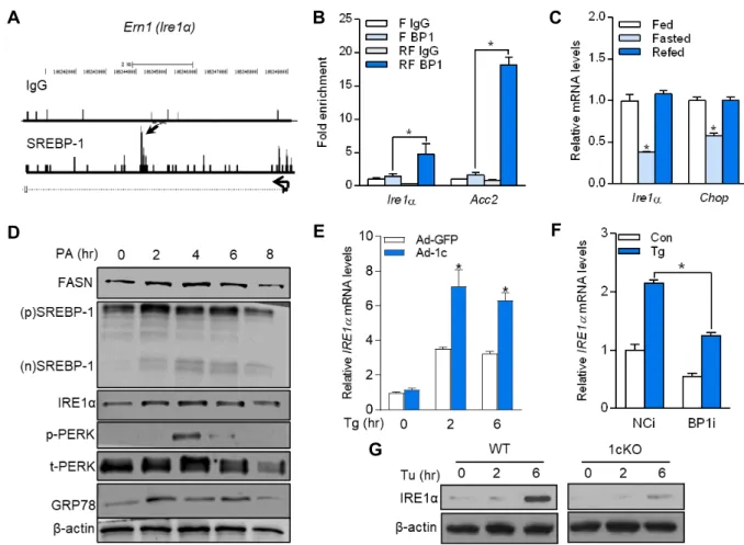
Using our previous ChIP sequencing dataset for SREBP-1 [26], SREBP-1 binding peaks were found in the promoter region of an ER stress sensor Ire1α (Fig. 1A). To confirm the direct binding of SREBP-1 to Ire1α promoter, ChIP analysis was performed with chromatin isolated from the livers of mice after fasting–refeeding transition. As shown in Fig.
1B, SREBP-1 binding to the Ire1α promoter region was en- riched by 3.4-fold in the chromatin from refed mice com- pared with that in the chromatin from fasted mice. The mRNA expression level of Ire1α and C/EBP homologous protein (Chop), an ER stress marker, was also significantly increased in the refed condition where the SREBP-1c ex- pression level was drastically increased (Fig. 1C) [5]. Exoge- nous saturated fatty acids, such as PA, potently induced the ER stress and lipogenesis [15]. PA treatment elicited a time-dependent increase in the cleavage of the nuclear form of SREBP-1, which was detected after 2 hr of PA treatment, along with its downstream target gene fatty acid synthase (FASN) in the mouse AML12 hepatocyte cell line (Fig. 1D).
ER stress sensors such as IRE1α, phospho-PERK, and GRP78 were also regulated in parallel with protein levels of SREBP- 1 and FASN (Fig. 1D). To determine if IRE1α regulation dur- ing ER stress is SREBP-1-dependent, HeLa cells were in- fected with an adenovirus expressing a nuclear SREBP-1c;
then, Tg, an ER stress inducer, was added to the cells. As
shown in Fig. 1E, the Tg-induced mRNA expression of IRE1α
was increased when SREBP-1c was overexpressed; however,
SREBP-1c silencing by siRNA completely blunted ER stress-
A B C
D E F
G
Fig. 1. SREBP-1c activates IRE1α transcription. (A) Sequence reads from SREBP-1c ChIP-Seq experiments were mapped onto the mouse genome in the University of California Santa Cruz (UCSC) Genome Browser. The SREBP-1c binding site at the Ire1
α
gene locus is pointed by arrows. (B) ChIP assay for SREBP-1 (BP1) binding in liver chromatin from fasted (F) or refed (RF) mice. Acetyl-CoA carboxylase 2 (Acc2) was used as a positive control. (C) Relative mRNA levels of Ire1α
and Chop in the liver of fed, fasted, or refed mice. (D) AML12 cells were treated with 0.5 mM palmitic acid (PA)-BSA conjugates at an indicated time. Immunoblots for precursor (p) and nuclear (n) SREBP-1 and ER stress markers (E and F). Ire1α
mRNA levels in HeLa cells infected with recombinant adenovirus expressing SREBP-1c (Ad-1c) or transfected with siRNA targeting SREBP-1c (BP1i), followed by incubation with 1 μM thapsigargin (Tg). (G) Primary hepatocytes isolated from WT and 1cKO mice were treated with 10 μg/ml tunicamycin (Tu) at an indicated time. IRE1α was analyzed by immunoblotting. Data are mean ± SEM. *p<0.05 versus F BP1 (B) or Fed (C) by one-way ANOVA; * p<0.05 by Student’s t-test (E and F).mediated IRE1α induction (Fig. 1F). The induction of mouse IRE1α proteins by Tu, an ER stress inducer, was also sig- nificantly reduced in 1cKO primary hepatocytes (Fig. 1G).
These findings suggest that SREBP-1c directly regulates IRE1 α in both mouse and human liver cells.
SREBP-1c deficiency protects against ER stress- induced hepatic steatosis
To further investigate the role of SREBP-1c in ER stress-ac- tivated UPR in vivo, Tu was intraperitoneally injected into WT and 1cKO mice. Consistent with our in vitro results, 72 hr after Tu injection, ER stress-related genes such as Chop,
Grp78, growth arrest and DNA damage-inducible protein 34(Gadd34), and Ire1α were significantly upregulated in the liv-
er of WT mice; however, the expression of these genes was
downregulated in 1cKO mice (Fig. 2A). IRE1α is a bifunc-
tional transmembrane kinase with both protein kinase and
endoribonuclease activities to process the splicing (activa-
tion) of the XBP1 mRNA [2]. The splicing of the Xbp1 (sXbp1)
mRNA persisted in WT mice at 72 hr after Tu injection,
whereas 1cKO mice only had unspliced Xbp1 (uXbp1) at 72
hr (Fig. 2B). Further analysis revealed that the protein levels
of IRE1, sXBP1, and CHOP were also significantly decreased
in the liver of 1cKO mice at all time points (Fig. 1C). ER
stress or UPR induction contributes to the development of
hepatic steatosis by disrupting lipid homeostasis [13]. Similar
A B
C
D E
F
Fig. 2. SREBP-1c deficiency protects against ER stress-induced hepatic steatosis. (A) Relative mRNA expression of ER stress markers in the liver of WT and 1cKO mice 72 hr after treatment with 1 mg/kg body weight of Tu. (B) Xbp1 splicing was analyzed by RT-PCR. (C) Immunoblots for liver ER stress markers. (D) H&E staining of the liver of mice. Scale bars, 100 μm. (E) Liver triglyceride and total cholesterol levels. (F) Hepatic mRNA levels of genes encoding SREBP-1c and lipogenic enzymes.
Data are mean ± SEM. # p<0.05 versus WT-Vehicle (Veh) and * p<0.05 versus WT-Tu by one-way ANOVA (A); * p<0.05 versus WT by two-way ANOVA (E).
to the findings of a previous study [24], Tu significantly in- creased the hepatic lipid accumulation in WT mice based on the H&E staining and TG content analysis, whereas 1cKO mice exhibited a significant reduction in hepatic steatosis and lipid accumulation (Fig. 2D, Fig. 2E). However, although hepatic steatosis can be induced by an increased SREBP-1c-
mediated lipogenesis, most lipogenic genes including SREBP-
1c were significantly decreased by Tu in both WT and 1cKO
mice at all time points (Fig. 2F). These results suggest that
SREBP-1c deficiency protects against ER stress-induced hep-
atic steatosis though a lipogenesis-independent mechanism.
A B
C
D E
Fig. 3. SREBP-1c deficiency suppresses ER stress-induced inflammation and fatty acid oxidation dysregulation. (A) Relative mRNA expression of genes involved in fatty acid oxidation in the liver of WT and 1cKO mice 72 hr after Tu injection. (B) Rate of fatty acid oxidation. (C) Immunohistochemistry for a macrophage marker F4/80 (green). Scale bars, 100 μm. The number of F4/80-positive cells was counted using Image J. (D) Relative mRNA levels of TNF-α and IL-1β. (E) mRNA levels of Pgc-1
α
from WT and 1cKO primary hepatocytes treated with TNF-α in a dose-dependent manner. Data are mean ± SEM.# p<0.05 versus WT-Veh and * p<0.05 versus WT-Tu by one-way ANOVA (A, B, and C); * p<0.05 versus WT by two-way ANOVA (D).
SREBP-1c deficiency prevents ER stress-impaired fatty acid oxidation
Hepatic steatosis can also be stimulated by reducing FAO and disturbing lipoprotein and VLDL secretion [10]. Not- ably, the mRNA expression of genes involved in mitochon- drial FAO, including peroxisome proliferator-activated re- ceptor γ coactivator 1-α (Pgc-1α), medium-chain acyl-CoA dehydrogenase (MCAD), and ATP synthase (Atp5b), was sig-
nificantly downregulated by Tu in the liver of WT mice;
however, the downregulation was blunted in the liver of
1cKO mice (Fig. 3A). Moreover, Tu-induced ER stress drasti-
cally suppressed the rate of FAO in WT mice but 1cKO pre-
vented the impairment of mitochondrial FAO (Fig. 3B). The
inflammatory cytokine tumor necrosis factor-α (TNF-α) re-
duced the expression level of Pgc-1α in human cardiac cells
and in a mouse model, resulting in metabolic dysregulation
observed in heart failure [22]. Because IRE1α regulates in- flammatory cytokine production via ribonuclease and kinase activities [18, 29], the involvement of inflammatory cytokines in Tu-impaired mitochondrial FAO was examined. As shown in Fig. 3C, Tu markedly enhanced the recruitment of F4/80
+Kupffer cells (liver macrophages) in the liver of WT mice, whereas it was not observed in the liver of 1cKO mice. Tu-induced TNF-α and IL-1β mRNA levels were also completely blunted in the liver of 1cKO mice (Fig. 3D).
Furthermore, we confirmed that the TNF-α treatment mark- edly suppressed Pgc-1α expression in the primary hep- atocytes of WT and 1cKO mice (Fig. 3E), suggesting that the protective effect of SREBP-1c deficiency on FAO alteration is mediated by a reduction in Tu-induced inflammatory responses.
Discussion
Because the SREBP pathway is affected by regulatory in- puts that are not solely associated with fundamental aspects of lipid metabolism, it is likely that SREBPs regulate genes associated with additional physiological processes. In fact, there are several studies that support this hypothesis. In some studies, functional SREBP binding sites were localized in the regulatory regions of specific genes involved in apop- tosis and cell cycle control, such as caspase 2 [17] and p21/
WAF [8], respectively. Our previous study also supports the roles of SREBP-1 in processes other than lipid metabolism [26]. The ChIP-seq approach was used with SREBP-1 anti- bodies and hepatic chromatin from fasted/refed mice, which are conditions known to increase the abundance of nuclear SREBP-1c. When the genes associated with SREBP-1 binding peaks were categorized by gene ontology (GO), enriched clusters involved in carbohydrate and lipid metabolism were identified as expected. However, several other GO categories exhibited a high degree of enrichment, including protein me- tabolism and trafficking, apoptosis, cell structure, and pro- liferation/differentiation. In this study, to identify new SREBP-1c target genes not related to lipid metabolism, we used the abovementioned ChIP-seq dataset and found that the promoter site in Ire1α, one of the three principal ER stress transmembrane sensors, was occupied by SREBP-1. ER stress-induced IRE1α expression was also downregulated in SREBP-1c-deficient mouse and human hepatocytes. We fur- ther showed that, in the liver, SREBP-1c deficiency com- pletely blocked Tu-induced lipid accumulation.
To study the role of SREBP-1c in ER stress-induced hep- atic steatosis, the expression of lipogenic genes was initially analyzed; however, differences in the mRNA level of lipo- genic genes between WT and 1cKO mice were not identified.
Consistent with our observation, other studies have shown a reduction in the expression levels of lipogenic genes, in- cluding SREBP-1c, in Tu-challenged mouse liver [11, 24]. In addition to the attenuation of lipogenesis, the expression lev- el of FAO-related genes, including Pgc-1α, and the rate of FAO were also decreased in Tu-challenged WT mouse liver;
however, these changes were reversed in SREBP-1c-deficient mouse liver. A recent study demonstrated that the activation of Kupffer cells in response to pro-inflammatory stimuli in- duces lipid metabolism changes in hepatocytes; these changes include TG accumulation and reduced FAO, which are mediated by Kupffer cell-derived TNF-α [7]. Consistent with this previous study, Pgc-1α expression was also sig- nificantly downregulated by TNF-α treatment in the primary hepatocytes of mice, suggesting TNF-α as a potential media- tor of the detrimental effects of ER stress on FAO in hepato- cytes. However, TNF-α signaling in hepatocytes was not af- fected by SREBP-1c ablation because no difference between genotypes was observed in Pgc-1α expression in response to TNF-α. Upon ER stress, the activated IRE1α activates JNK and NF-κB, which are implicated in the transcriptional acti- vation of inflammatory cytokines such as IL-1β and TNF-α, which are released by Kupffer cells (liver macrophages) [6, 31]. Notably, ER stress-induced inflammatory responses were significantly decreased in the liver of 1cKO mice, as indicated by macrophage infiltration and expression of in- flammatory cytokines, such as IL-1β and TNF-α.
In summary, we showed that SREBP-1c deficiency sup- pressed ER stress-induced IRE1α expression and pro-in- flammatory cytokine production, thus preventing the dysre- gulation of mitochondrial FAO and hepatic steatosis.
Although SREBP-1c deficiency plays an important role in the regulation of UPR, inflammation, and lipid oxidation to protect against ER stress-induced hepatic steatosis, further studies are required to address the crosstalk between SREBP- 1c and UPR in ER stress-induced activation of Kupffer cells into their pro-inflammatory phenotype.
Acknowledgment
This study was supported by NRF-2014R1A1A2057994.
The Conflict of Interest Statement
The authors declare that they have no conflicts of interest with the contents of this article.
References
1. Chen, J., Fontes, G., Saxena, G., Poitout, V. and Shalev, A.
2010. Lack of TXNIP protects against mitochondria-medi- ated apoptosis but not against fatty acid-induced ER stress- mediated beta-cell death. Diabetes 59, 440-447.
2. Chen, Y. and Brandizzi, F. 2013. IRE1: ER stress sensor and cell fate executor. Trends Cell Biol. 23, 547-555.
3. Cnop, M., Foufelle, F. and Velloso, L. A. 2012. Endoplasmic reticulum stress, obesity and diabetes. Trends Mol. Med. 18, 59-68.
4. Djouadi, F., Bonnefont, J. P., Munnich, A. and Bastin, J. 2003.
Characterization of fatty acid oxidation in human muscle mitochondria and myoblasts. Mol. Genet. Metab. 78, 112-118.
5. Horton, J. D., Bashmakov, Y., Shimomura, I. and Shimano, H. 1998. Regulation of sterol regulatory element binding proteins in livers of fasted and refed mice. Proc. Natl. Acad.
Sci. USA. 95, 5987-5992.
6. Hu, P., Han, Z., Couvillon, A. D., Kaufman, R. J. and Exton, J. H. 2006. Autocrine tumor necrosis factor alpha links endo- plasmic reticulum stress to the membrane death receptor pathway through IRE1alpha-mediated NF-kappaB activa- tion and down-regulation of TRAF2 expression. Mol. Cell.
Biol. 26, 3071-3084.
7. Huang, W., Metlakunta, A., Dedousis, N., Zhang, P., Sipula, I., Dube, J. J., Scott, D. K. and O'Doherty, R. M. 2010. Deple- tion of liver Kupffer cells prevents the development of di- et-induced hepatic steatosis and insulin resistance. Diabetes 59, 347-357.
8. Inoue, N., Shimano, H., Nakakuki, M., Matsuzaka, T., Naka- gawa, Y., Yamamoto, T., Sato, R., Takahashi, A., Sone, H., Yahagi, N., Suzuki, H., Toyoshima, H. and Yamada, N.
2005. Lipid synthetic transcription factor SREBP-1a activates p21WAF1/CIP1, a universal cyclin-dependent kinase inhibitor.
Mol. Cell. Biol. 25, 8938-8947.
9. Jeon, T. I., Esquejo, R. M., Roqueta-Rivera, M., Phelan, P.
E., Moon, Y. A., Govindarajan, S. S., Esau, C. C. and Osborne, T. F. 2013. An SREBP-responsive microRNA operon contrib- utes to a regulatory loop for intracellular lipid homeostasis.
Cell Metab. 18, 51-61.
10. Jeon, T. I. and Osborne, T. F. 2012. SREBPs: metabolic in- tegrators in physiology and metabolism. Trends Endocrinol.
Metab. 23, 65-72.
11. Jo, H., Choe, S. S., Shin, K. C., Jang, H., Lee, J. H., Seong, J. K., Back, S. H. and Kim, J. B. 2013. Endoplasmic reticulum stress induces hepatic steatosis via increased expression of the hepatic very low-density lipoprotein receptor. Hepatol- ogy 57, 1366-1377.
12. Kammoun, H. L., Chabanon, H., Hainault, I., Luquet, S., Magnan, C., Koike, T., Ferre, P. and Foufelle, F. 2009. GRP78
expression inhibits insulin and ER stress-induced SREBP-1c activation and reduces hepatic steatosis in mice. J. Clin.
Invest. 119, 1201-1215.
13. Lebeaupin, C., Vallee, D., Hazari, Y., Hetz, C., Chevet, E.
and Bailly-Maitre, B. 2018. Endoplasmic reticulum stress sig- nalling and the pathogenesis of non-alcoholic fatty liver disease. J. Hepatol. 69, 927-947.
14. Lee, A. H., Scapa, E. F., Cohen, D. E. and Glimcher, L. H.
2008. Regulation of hepatic lipogenesis by the transcription factor XBP1. Science 320, 1492-1496.
15. Li, H., Min, Q., Ouyang, C., Lee, J., He, C., Zou, M. H. and Xie, Z. 2014. AMPK activation prevents excess nutrient-in- duced hepatic lipid accumulation by inhibiting mTORC1 signaling and endoplasmic reticulum stress response. Bio- chim. Biophys. Acta 1842, 1844-1854.
16. Lin, J. H., Li, H., Yasumura, D., Cohen, H. R., Zhang, C., Panning, B., Shokat, K. M., Lavail, M. M. and Walter, P.
2007. IRE1 signaling affects cell fate during the unfolded protein response. Science 318, 944-949.
17. Logette, E., Le Jossic-Corcos, C., Masson, D., Solier, S., Sequeira-Legrand, A., Dugail, I., Lemaire-Ewing, S., Desoche, L., Solary, E. and Corcos, L. 2005. Caspase-2, a novel lipid sensor under the control of sterol regulatory element bind- ing protein 2. Mol. Cell. Biol. 25, 9621-9631.
18. Martinon, F., Chen, X., Lee, A. H. and Glimcher, L. H. 2010.
TLR activation of the transcription factor XBP1 regulates in- nate immune responses in macrophages. Nat. Immunol. 11, 411-418.
19. Ning, J., Hong, T., Ward, A., Pi, J., Liu, Z., Liu, H. Y. and Cao, W. 2011. Constitutive role for IRE1alpha-XBP1 signal- ing pathway in the insulin-mediated hepatic lipogenic pro- gram. Endocrinology 152, 2247-2255.
20. Osborne, T. F. 2000. Sterol regulatory element-binding pro- teins (SREBPs): key regulators of nutritional homeostasis and insulin action. J. Biol. Chem. 275, 32379-32382.
21. Osborne, T. F. and Espenshade, P. J. 2009. Evolutionary con- servation and adaptation in the mechanism that regulates SREBP action: what a long, strange tRIP it's been. Genes Dev.
23, 2578-2591.
22. Palomer, X., Alvarez-Guardia, D., Rodriguez-Calvo, R., Coll, T., Laguna, J. C., Davidson, M. M., Chan, T. O., Feldman, A. M. and Vazquez-Carrera, M. 2009. TNF-alpha reduces PGC-1alpha expression through NF-kappaB and p38 MAPK leading to increased glucose oxidation in a human cardiac cell model. Cardiovasc. Res. 81, 703-712.
23. Postic, C. and Girard, J. 2008. Contribution of de novo fatty acid synthesis to hepatic steatosis and insulin resistance: les- sons from genetically engineered mice. J. Clin. Invest. 118, 829-838.
24. Rutkowski, D. T., Wu, J., Back, S. H., Callaghan, M. U., Ferris, S. P., Iqbal, J., Clark, R., Miao, H., Hassler, J. R., Fornek, J., Katze, M. G., Hussain, M. M., Song, B., Swathir- ajan, J., Wang, J., Yau, G. D. and Kaufman, R. J. 2008. UPR pathways combine to prevent hepatic steatosis caused by ER stress-mediated suppression of transcriptional master regulators. Dev. Cell 15, 829-840.
초록:지방산 산화 장애 제어를 통한 SREBP-1c 결핍의 소포체 스트레스 유발 비알콜성지방간 보호작용
이영승
1․티모씨 에프 오스본
2․서영교
3․전태일
1*
(1전남대학교 농업생명과학대학 동물자원학부, 2존스 홉킨스 의과대학, 3한국생명공학연구원 노화제어전문연구단)
간 소포체(ER) 스트레스는 비알콜성지방간과 인슐린 저항성의 발달에 기여하고, unfolded protein response (UPR)의 구성요소는 지질 대사를 조절한다. 최근 연구에 따르면 ER 스트레스와 비정상적인 세포 지질 대사 사이 의 연관성이 보고되었으며, 이 과정에서 지질 대사의 중심 조절자인 sterol regulatory element binding proteins (SREBPs)의 관련성이 확인되었다. 그러나 ER 스트레스 동안 지질 대사를 조절하는 SREBP의 정확한 역할과 비알 콜성지방간에 대한 기여는 아직 밝혀지지 않았다. 본 연구에서 SREBP-1c 결핍은 UPR, 염증 및 지방산 산화 조절 을 통해 ER 스트레스에 의해 유도된 비알콜성지방간으로부터 생쥐를 보호한다는 것을 보여준다. SREBP-1c는 in- ositol requiring kinase 1α (IRE1α) 발현을 직접적으로 조절하고 ER 스트레스에 의해 유도된 tumor necrosis fac- tor-α의 활성화를 매개하여 peroxisome proliferator-activated receptor γ coactivator 1-α (PGC1α)의 감소와 그에 따른 지방산 산화의 장애를 유발한다. 그러나, SREBP-1c의 유전적 결핍은 이러한 현상을 보호하여 간 염증과 지방 축적을 완화시킨다. SREBP-1c 결핍이 ER 스트레스에 의해 유도된 염증 신호를 방지하는 메커니즘은 아직 밝혀지 지 않았지만, SREBP-1c가 결핍된 Kupffer 세포에서 IRE1α 신호의 변화가 염증 신호에 관여할 수 있을 것으로 생 각된다. 본 연구결과는 SREBP-1c가 ER 스트레스에 의해 유도된 비알콜성지방간에서 UPR 및 염증의 조절에 중요 한 역할을 함을 시사한다.
25. Schroder, M. and Kaufman, R. J. 2005. The mammalian un- folded protein response. Annu. Rev. Biochem. 74, 739-789.
26. Seo, Y. K., Chong, H. K., Infante, A. M., Im, S. S., Xie, X.
and Osborne, T. F. 2009. Genome-wide analysis of SREBP-1 binding in mouse liver chromatin reveals a preference for promoter proximal binding to a new motif. Proc. Natl. Acad.
Sci. USA. 106, 13765-13769.
27. Shen, J., Chen, X., Hendershot, L. and Prywes, R. 2002. ER stress regulation of ATF6 localization by dissociation of BiP/GRP78 binding and unmasking of Golgi localization signals. Dev. Cell 3, 99-111.
28. Truong, X. T., Nguyen, T. T. P., Kang, M. J., Jung, C. H., Lee, S., Moon, C., Moon, J. H. and Jeon, T. I. 2019. Pear extract and malaxinic acid reverse obesity, adipose tissue inflammation, and hepatosteatosis in mice. Mol. Nutr. Food Res. 63, e1801347.
29. Urano, F., Wang, X., Bertolotti, A., Zhang, Y., Chung, P., Harding, H. P. and Ron, D. 2000. Coupling of stress in the ER to activation of JNK protein kinases by transmembrane
protein kinase IRE1. Science 287, 664-666.
30. Werstuck, G. H., Lentz, S. R., Dayal, S., Hossain, G. S., Sood, S. K., Shi, Y. Y., Zhou, J., Maeda, N., Krisans, S. K., Malinow, M. R. and Austin, R. C. 2001. Homocysteine-induced endo- plasmic reticulum stress causes dysregulation of the choles- terol and triglyceride biosynthetic pathways. J. Clin. Invest.
107, 1263-1273.
31. Yang, Q., Kim, Y. S., Lin, Y., Lewis, J., Neckers, L. and Liu, Z. G. 2006. Tumour necrosis factor receptor 1 mediates en- doplasmic reticulum stress-induced activation of the MAP kinase JNK. EMBO Rep. 7, 622-627.
32. Ye, J., Rawson, R. B., Komuro, R., Chen, X., Dave, U. P., Prywes, R., Brown, M. S. and Goldstein, J. L. 2000. ER stress induces cleavage of membrane-bound ATF6 by the same proteases that process SREBPs. Mol. Cell 6, 1355-1364.
33. Younossi, Z. M., Koenig, A. B., Abdelatif, D., Fazel, Y., Henry, L. and Wymer, M. 2016. Global epidemiology of nonalcoholic fatty liver disease—meta analytic assessment of prevalence, incidence, and outcomes. Hepatology 64, 73-84.