CASE REPORT
pISSN 1225-7737/eISSN 2234-8042 http://dx.doi.org/10.12701/yujm.2013.30.2.120 YUJM 2013;30(2):120-3120
YUJM VOLUME 30, NUMBER 2, DECEMBER 2013에르고노빈 심초음파로 확진된 승모판 폐쇄부전을 유발한 혈관연축 1예
차정준, 경찬희, 조장호, 김용훈, 김혜원, 이성주1, 임세중, 최의영 연세대학교 의과대학 내과학교실, 1충주의료원 심장내과
Severe Mitral Regurgitation Due to Coronary Vasospasm, Confirmed by Ergonovine Echocardiography
Jung-Joon Cha, Chan Hee Kyung, Jang Ho Cho, Yong Hoon Kim, Haewon Kim, Sung-Joo Lee
1, Se-Joong Rim, Eui-Young Choi
Department of Internal Medicine, Yonsei University College of Medicine, Seoul,
1
Division of Cardiology, Department of Internal Medicine, Chungju Medical Center, Chungju, Korea
The common causes of organic mitral regurgitation (MR) include mitral valve prolapse (MVP) syndrome, rheumatic heart disease, and endocarditis. MR also occurs secondary to dilated cardiomyopathy and coro- nary artery disease. In acute severe MR, the hemodynamic overload often cannot be tolerated, and mitral valve repair or replacement must be performed immediately. We report herein a case of severe MR due to coronary vasospasm that was confirmed via ergonovine echocardiography in a 70-year-old man. He was scheduled to undergo mitral valve surgery, but it did not push through and he was put on medical therapy.
Key Words: Mitral valve insufficiency, Coronary vasospasm, Ergonovine, Echocardiography, Stress
Received: June 2, 2013, Revised: July 3, 2013, Accepted: July 9, 2013
Corresponding Author: Eui-Young Choi, Division of Car- diology, Gangnam Severance Hospital, Yonsei University College of Medicine, 211, Eonjuro, Gangnam-gu, Seoul 135-720, Korea
Tel: 82-10-2521-8022, Fax: 82-2-2019-3452 E-mail: [email protected]
서 론
승모판 폐쇄부전은 승모판 탈출증, 류마티스성 판막질환, 심내막염 등에 의한 일차적인 원인과 관동맥질환 및 확장성 심근병증 등에 동반되는 이차적인 원인으로 분류할 수 있다.
이와 같은 분류에 상관없이, 중증 승모판 폐쇄부전은 좌심방 과 좌심실의 혈류량 증가를 야기하며, 이로 인해 전부하의 증가와 심박출량의 증가가 이루어지게 된다. 그러나 보완적 인 편심성 심비대가 없다면 대동맥으로의 심박출량이 감소 되게 되고 폐부종이 유발된다. 이와 같이 혈역학적으로 불안
정한 중증의 승모판 폐쇄부전 환자에게는 승모판 수술이 권 장된다.1 저자들은 약물로 조절되지 않는 정상 관동맥의 승 모판 폐쇄부전에 대하여 수술적 치료를 계획하고 있던 환자 에게서 에르고노빈 심초음파(ergonovine stress echocardio- graphy)를 통해 관동맥 혈관연축(coronary vasospasm)으로 인한 일시적인 중증의 승모판 폐쇄부전이 확인된 것을 경험 하였기에 문헌고찰과 함께 보고하고자 한다.
증 례
환 자: 남자, 70세
주 소: 운동시 발생하는 호흡곤란과 두근거림, 간헐적인 흉통
현병력: 환자는 특이 내과적 과거력이 없는 분으로 운동시 발생하는 호흡곤란, 두근거림 및 간헐적인 흉통을 주소로 시행한 경흉부심초음파 검사상 중증의 승모판 폐쇄부전이 관찰되어 베타차단제와 이뇨제를 복용하며 경과를 관찰하였
Vasospasm, Confirmed by Ergonovine Test
YUJM VOLUME 30, NUMBER 2, DECEMBER 2013
121
Fig. 2. Transthoracic echocardiography shows mild mitral regur- gitation in asymptomatic status.
Fig. 1. Increased haziness in both hilar areas which implies pul-
monary congestion. Fig. 3. Improvement of haziness in both hilar areas at the time
patient’s symptom disappeared.
으나, 증상 악화가 반복되어 수술적 치료 계획하에 수술전 평가로 관동맥 조영 검사를 시행하였고, 정상 관동맥이 관찰 되어 승모판 치환술 혹은 교정술을 하기 위해 본원으로 전원 되었다.
가족력: 특이사항 없음
진찰 소견: 신체검진에서 혈압 128/76 mm Hg, 맥박 67회/
분, 호흡 18회/분, 체온 36.5℃로 측정되었다. 심음 청진상 심첨부에서 범수축기 잡음과 수포음이 청진되었고, 환자는 주로 운동시에 호흡곤란 및 두근거림을 호소하였다.
검사 소견: 입원 당시 말초 혈액검사에서 백혈구 6,670 mm3, 혈색소 12.6 g/dL, 혈소판 154,000/mm3였다. 혈청 생 화학 검사에서 BUN/Creatinine 19.5/0.91 (mg/dL), Na/K/Cl/
tCO2 138/4.3/102/23 (mmol/L)였고, 혈당 95 mg/dL, AST/
ALT 21/ 15 IU/L, protein/albumin 6.0/4.0 (g/dL), Ca/P 8.7/3.4 (mg/dL), CK 73 U/L, CK-MB 1.10 mcg/L, Troponin T 0.016 mcg/L였다. 흉부 단순촬영상 양폐문의 음영 증가되어 있는 폐부종 소견이 관찰되었고(Fig. 1), 심전도 검사상 이상 소견 은 없었다.
치료 및 경과: 본원 내원 후 환자 증상이 호전 양상을 보여 다시 시행한 경흉부심초음파상 좌심실 박출률은 62%였고 좌심방 용적지수는 32.69 mm3/m2였으며, 좌심실은 LVEDD/
LVESD 42/29 (mm)로 정상 크기였다. 경도의 승모판 폐쇄부 전이 관찰되었으나, 국소벽 운동장애는 관찰되지 않았고, 흉 부 단순촬영에서도 입원 당시 관찰되었던 폐부종 소견은 호 전되었다(Fig. 2, 3). 승모판의 정밀한 진단을 위해 경식도 심초음파 검사에서 승모판의 류마티스성 판막질환과 승모판 탈출증은 관찰되지 않았다(Fig. 4). 흉통과 동반되어 호흡곤
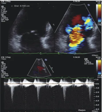
란이 발생하였으며, 관동맥 조영술상 정상 소견이라는 것에 착안하여 관동맥 연축 등에 인한 이차성 승모판 폐쇄부전 원인 감별을 위해 에르고노빈 심초음파를 시행하였고, 검사 중 50 μg의 ergonovine을 투여 시 흉통과 동반되어 심전도상 Lead II, III, aVF 의 ST분절 하강(Fig. 5)과 전유두근을 포함하 는 심저부 후측벽 심근의 무운동이 관찰되었고, 이로 인한 승모판 후엽의 고정으로 중증의 승모판 폐쇄부전이 관찰되
Jung-Joon Cha et al.
122
YUJM VOLUME 30, NUMBER 2, DECEMBER 2013Fig. 6. Baseline transthoracic echocardiography shows mild mitral regurgitation with good leaflets coaptation (6A). However, during ergonovine challenge test (50 μg), severe mitral regurgitation devel- oped due to incomplete mitral leaflets coaptation (white arro- whead), which is associated with severe hypokinesis of left vent- ricular posterior segment containing papillary muscular base (arrow) (6B).
Fig. 4. Transesophageal echocardiography does not show any fin- dings of primary mitral valvular disease such as rheumatic invol- vement or leaflets prolapse.
Fig. 5. During ergonovine challenge test (50 μg), ST depression was noted at lead II, III, aVF.
었다(Fig. 6). 환자는 호흡곤란을 호소하였고, 측정한 혈압은 77/43 mm Hg으로 감소하였으며, 심전도상 부정맥은 관찰 되지 않았으나, 맥박수는 125회/분으로 증가하였으며, 청진 상 양폐야의 수포음이 청진되어 폐부종 발생이 의심되었다.
니트로글리세린(nitroglycerin) 0.5 mg 정주 후 관찰되던 국소 벽운동장애는 소실되었고, 승모판 폐쇄부전은 호전되었으며 활력징후는 안정 상태로 회복되었다. 환자는 관동맥 혈관연 축에 대하여 칼슘통로차단제 및 니트로글리세린을 처방하였 고 증상없이 퇴원하였다.
고 찰
관동맥 혈관연축은 급성관동맥 허혈의 중요한 기전으로 심혈관질환의 다양한 임상적 징후의 발생에 기여한다. 또한 관동맥 혈관연축은 운동성협심증 유무와 심근경색 과거력의 유무 및 동맥경화의 유무에 관계없이 발생할 수 있다.2 관동 맥 혈관연축은 흉통, 실신 등의 원인이 되며,3 일부 경우에는 고도의 방실차단, 급성심근경색, 심실빈맥 및 심실세동 등으 로 급성 심장사가 발생한다.4 동양인과 백인의 인종에 따른 관동맥 조영술 형태와 관동맥 혈관연축의 유병률에는 차이 점이 있으며, 관동맥 혈관연축의 유병률은 백인에 비해 동양 인에서 높다.5 또한 한 연구에서는 최근 심근경색이 있었던 동양인과 백인의 혈관운동의 반응 차이에서 동양인이 백인 에 비해 3배 정도 혈관연축의 발생률이 높고, 아세틸콜린
Vasospasm, Confirmed by Ergonovine Test
YUJM VOLUME 30, NUMBER 2, DECEMBER 2013
123
부하검사에서도 더 큰 혈관연축 반응을 보였다.6
본 증례는 증상이 있는 중증의 승모판 폐쇄부전에 대한 수술적 치료를 고려하여 전원 되었으나, 환자의 승모판 역류 정도가 감소되었으며, 약물 복용 중에도 증상의 호전 및 악화가 반복되며, 승모판 폐쇄부전증을 일으킬 만한 일차적 승모판 이상과 좌심실의 구조적 혹은 기능적 변화도 찾지 못하였다.
이에 관동맥 조영술상 협착이 없던 점을 감안하여 관동맥 혈관연축으로 인한 허혈성 승모판 폐쇄부전을 의심해 시행한 에르고노빈 심초음파에서 국소벽 심근장애가 동반된 심한 승모판 폐쇄부전증과 폐부종을 관찰함으로써, 관동맥 혈관 연축과 관련되어 발생한 허혈성 승모판 폐쇄부전증을 강력히 의심할 수 있었다.
고정된 관동맥 협착 소견이 없는 환자에서 관동맥 혈관연 축의 진단은 관동맥 조영술 중 시행하는 약물 유발검사였다.7 그러나 최근 미국내 의료기관에서는 일상적인 관동맥 조영 술의 과정에서 유발 검사 빈도를 줄이고 있는데, 이는 관동맥 내 에르고노빈 정주의 안전성에 대한 고려와 고도의 기술이 요구되어지는 검사로 인한 주치의의 본의 아닌 선택이 유발 검사 빈도의 감소에 기여하는 것으로 보인다.8
에르고노빈 심초음파는 관동맥 혈관연축의 유발 검사 중 의 하나로 의심되는 환자에게 시행하는 것에 대해 관동맥 조영술에 비하여 안전성과 비용-효율성이 있다.2 또한 관동 맥 조영술 중 일상적으로 사용되는 니트로글리세린에 의하 여 혈관 긴장도의 소실이 발생하여 유발 검사를 시행하지 못하거나 시행해도 음성 결과가 관찰되는 경우가 있는데,9 이러한 경우에도 에르고노빈 심초음파는 관동맥 혈관연축의 진단에 유용한 비침습적인 검사로 알려져 있으며,10 관동맥 혈관연축으로 인한 허혈성 승모판 폐쇄부전증이 의심된다면 가장 유용한 검사라고 할 수 있다.
본 증례와 같이 원인불명의 호흡곤란 및 일시적으로 심한 승모판 폐쇄부전증이 발생한 환자에서는 관동맥질환 여부 확인을 위한 검사와 관동맥 혈관연축으로 인한 허혈성 승모 판 폐쇄부전증의 발생 확인을 위한 에르고노빈 심초음파는 고려해 봐야 할 검사가 될 것이다.
참고문헌