• 검색 결과가 없습니다.

비정질 칼슘 포스페이트 나노 입자의 합성과 특성

N/A
N/A
Protected

Academic year: 2021

Share "비정질 칼슘 포스페이트 나노 입자의 합성과 특성"

Copied!
6
0
0

로드 중.... (전체 텍스트 보기)

전체 글

(1)

Article

https://doi.org/10.14478/ace.2018.1089

1)

1. Introduction

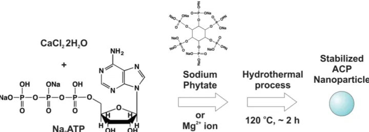
Calcium phosphate (CaP) is one of the most important biomaterials with outstanding biocompatibility[1] because their mineralized form known as hydroxyapatite (HAP) is one of the main inorganic con- stituents of vertebrate and human bones and teeth[2-6]. Synthetic HAP has been developed for biomedical applications such as tissue en- gineering and regenerative repair of bones and tooth[7]. Among vari- ous forms of CaP including HAP, amorphous calcium phosphate

† Corresponding Author: Pusan National University,

School of Chemical, Biomolecular, and Environmental Engineering, 2, Busandaehak-ro 63beon-gil, Geumjeong-gu, Busan 46241, Korea Tel: +82-51-510-2397 e-mail: sungwook.chung@pusan.ac.kr

pISSN: 1225-0112 eISSN: 2288-4505 @ 2018 The Korean Society of Industrial and Engineering Chemistry. All rights reserved.

(ACP) is one of the particular phases that formed first from a super- saturated aqueous solution of calcium (II) cations (Ca

2+

) and phosphate (III) (PO

43-

) anions with almost no long-range and atomic scale order- ing that typical crystalline CaPs, such as HAP and tricalcium phos- phate (TCP) have. Deciphering the role of ACP during the formation of bone minerals is critical to understand the process of bone for- mation[6]. Moreover, ACP has a superior osteoconductivity and bio- degradability than TCP and HAP and can promote cell proliferation and promotion via enhanced alkaline phosphatase enzyme activities[8], which makes ACP a promising candidate as one of the novel bio- materials potentially employed in the area of regenerative medicine.

The structural unit of ACP has been proposed to be a spherical clus- ter known as “Posner’s cluster”. This neutral cluster composed of cal- cium and phosphate ions has an empirical formula of Ca

9

(PO

4

)

6

with an approximate diameter of 0.9~1.4 nm[6,9]. The resulting ACP has

비정질 칼슘 포스페이트 나노 입자의 합성과 특성

한지훈⋅정성욱*,†

부산대학교 화학공학⋅고분자공학과 , *부산대학교 화공생명⋅환경공학부 (2018년 8월 23일 접수, 2018년 8월 27일 심사, 2018년 9월 17일 채택)

Synthesis and Characterization of Amorphous Calcium Phosphate Nanoparticles

Ji-Hoon Han and Sungwook Chung

*,

Department of Polymer Science and Chemical Engineering, Pusan National University, 2, Busandaehak-ro 63beon-gil, Geumjeong-gu, Busan 46241, Korea

*

School of Chemical, Biomolecular, and Environmental Engineering, Pusan National University, 2, Busandaehak-ro 63beon-gil, Geumjeong-gu, Busan 46241, Korea

(Received August 23, 2018; Revised August 27, 2018; Accepted September 17, 2018)

초 록

본 연구에서는 비정질 칼슘 포스페이트(ACP) 나노 입자의 합성과 특성 분석을 진행하였다. 염화칼슘(calcium chloride (CaCl

2

))과 아데노신 인산나트륨(disodium adenosine triphosphate (Na

2

ATP)) 그리고 피트산 나트륨(sodium phytate) 첨가 제를 열수 반응을 통해 상대적으로 단분산된 100 nm 크기 이하의 ACP 나노 입자를 성공적으로 합성하였고 나노 입자 의 화학적 조성과 구조를 재료 분석을 통해 확인하였다. 피트산 나트륨 첨가제의 사용을 통해 얻은 ACP 나노 입자는 비정질성을 유지하고 결정성 하이드록시아파타이트(HAP)로의 전환을 방지하는 안정성이 향상되었음을 발견하였다.

본 연구를 통해 발견된 향상된 안정성을 가지는 ACP 나노 입자는 재생 의학 분야에서의 생체 적합 물질로의 응용에 중요한 잠재적 용도가 있을 것이라 사료된다.

Abstract

The synthesis and characterization of amorphous calcium phosphate (ACP) nanoparticles were reported in this work. We show that relatively monodisperse ACP nanoparticles with a size of sub-100 nm can be prepared by a hydrothermal reaction of calcium chloride (CaCl

2

) and disodium adenosine triphosphate (Na

2

ATP) in the presence of sodium phytate as an additive.

Their compositions and structures were confirmed using a series of material characterization techniques. Our results exhibit that ACP nanoparticles synthesized using sodium phytate enhanced the stability of maintaining their amorphous nature and prevented from a conversion to crystalline hydroxyapatite (HAP). ACP nanoparticles with the improved stability have potential uses in biomaterial applications in regenerative medicine.

Keywords: amorphous calcium phosphate, nanoparticles, hydroxyapatite, hydrothermal reaction

(2)

been considered to be made up of particles containing a number of Posner’s clusters with water molecules in the intervening spaces, and it has been proposed as first nuclei to be involved in the formation of polymorphic crystalline CaPs including HAP and dicalcium phosphate dihydrate[3,4,6,10-23]. Once formed in aqueous environments, this partly hydrated and unstructured ACP is relatively unstable and there- fore can be rapidly transformed to crystalline HAP through a series of processes such as dissolution, nucleation, and crystal growth unless it is stabilized in some manner that the organisms likewise exploit to control the stability of ACP during biomineralization[15,21,22,24]. For examples, many trace amounts of chemical components discovered in the structure of bone and tooth minerals were confirmed to affect the stability and the transformation of ACP to HAP[25-27]. Moreover, it has been found that the stability and lifetime of metastable ACP in aqueous solutions depend on various factors such as the number ratio of calcium and phosphate ion (Ca/P ratio)[10], pH[12], ionic strength[13], and additives[4,14,15,19-23,25-27]. One of the ways to stabilize ACP is to incorporate Mg and adenosine triphosphate (ATP) with bulk ACP in aqueous solutions, and the presence of Mg and ATP inhibits the conversion of ACP to HAP[21]. Unfortunately, this method does not always guarantee nanostructured ACP with well-defined sizes and shapes.

Herein, we report a hydrothermal method to synthesize ACP nano- particles using hydrated calcium chloride (CaCl

2

⋅2H

2

O) as a source of calcium ions and disodium adenosine triphosphate (Na

2

ATP) as a source of phosphate ions in the presence of sodium phytate as an ef- fective additive for ACP stabilization. We show that as-synthesized ACP nanoparticles from our hydrothermal method have reasonably uni- form sizes that are strongly affected by the presence of sodium phytate in the reaction mixture. We demonstrate that as-synthesized ACP nano- particles in the presence of sodium phytate are highly stabilized, i.e.

do not undergo the transformation to crystalline HAP, for the extended period of time while pristine ACP nanoparticles made without sodium phytate do not preserve their amorphous state after a short induction period.

2. Materials and Methods

Our ACP nanoparticles were synthesized via a hydrothermal reaction based on the scheme shown in Figure 1. An aqueous solution of 0.11 g sodium salt of ATP (Na

2

ATP) dissolved in 10 mL triply distilled H

2

O was dropwise added (i.e., ~10 mL per 10 mins) to the aqueous solution of 0.15 g CaCl

2

⋅2H

2

O in 30 mL triply distilled H

2

O at room temperature while vigorously stirring at 150 rpm. Additives such as 0.08 g MgCl

2

or 0.05 g sodium phytate were added to the aqueous mixture, and the pH of the solution was adjusted to ~5.0. The result- ing aqueous solution was transferred to a 100 mL Teflon-lined stain- less steel autoclave and heated to ~120 ℃ for 2 h. Once the reaction was complete, the autoclave was cooled to room temperature. The white precipitates were then filtered through a mixed cellulose ester membrane with a pore diameter of 0.45 µm, washed with an excess of deionized H

2

O and dried in a vacuum oven at room temperature to give ~7 and ~67 mg of ACP nanoparticles in the presence of MgCl

2

and sodium phytate, respectively. The final product was stored in the desiccator at ~4 ℃.

X-ray powder diffraction (XRD) data of the as-synthesized ACP nanoparticles and HAP nanorods were recorded by using a Philips X’Pert-MPD Diffractometer with a monochromatized source of Cu K

α1

radiation (λ = 0.15405 nm) at 1.6 kW power (40 kV, 30 mA). Fourier transform infrared spectroscopy (FT-IR) was performed by using a Spectrum GX FT-IR spectrometer with both KBr pellet and attenuated total reflection (ATR) techniques. The morphologies of ACP nano- particles and HAP nanorods were investigated by field emission scan- ning electron microscopy (FESEM) (Zeiss Supra 40 FESEM) with an accelerating voltage ranged from 5 to 10 kV. Energy dispersive X-ray spectroscopy (EDS) was performed using Zeiss Supra 40 FESEM equipped with an Oxford X-ray energy dispersive spectrometer for ele- mental analysis. Transmission electron microscopy (TEM) of ACP nanoparticles and HAP nanorods was performed on a Hitachi H-7600 microscope operating at 80 kV. The stability of as-synthesized ACP nanoparticles was also investigated by XRD, FT-IR, and FESEM.

Figure 1. Schematic illustration of the preparation of amorphous calcium phosphate (ACP) nanoparticles via a hydrothermal method.

(3)

3. Results and Discussion

Figure 2 shows FESEM micrographs of ACP nanoparticles and HAP nanorods. Based on FESEM measurements, as-synthesized ACP nano- particles synthesized with or without the additives such as MgCl

2

and sodium phytate via our hydrothermal reaction possessed a similar spherical shape and morphology. Figure 3 shows FESEM micrographs of ACP nanoparticles synthesized with and without MgCl

2

additive, which also exhibited an analogous morphology. Their corresponding EDS measurements in Figure 3 indicated that they were primarily com- posed of C, O, P, and, Ca. Regarding the samples formed in the pres- ence of Mg

2+

ions from MgCl

2

, they revealed the signature of Mg.

Based on the FESEM measurements in Figure 2, the average size of as-synthesized ACP nanoparticles produced in the presence of Mg

2+

ions was determined to be 480 ± 50 nm. However, the average size of as-synthesized ACP nanoparticles formed in the presence of sodium phytate was measured to be 91 ± 7 nm. Therefore, our results sug- gested that the average size was significantly decreased and their par- ticle size distribution was improved by the incorporation of sodium phytate in the aqueous reaction mixtures.

Figure 4 shows XRD patterns of as-synthesized ACP nanoparticles and HAP nanorods. The XRD pattern of HAP nanorods (green curve) showed a group of diffraction peaks that were successfully indexed to a single crystalline phase of HAP with a hexagonal symmetry (JCPDS

No. 09-0432). The diffraction peaks located at 2θ = ~10.8, 22.9, 25.9, 28.9, 31.8, 32.2, 32.9, 34.1, 35.5°, and 39.8° corresponded to the Figure 2. FESEM images of as-synthesized ACP nanoparticles. (A) ACP nanoparticles synthesized in the presence of MgCl

2

. Inset is a higher magnification FESEM image. (B) ACP nanoparticles synthesized in the presence of sodium phytate. (C) Crystalline hydroxyapatite (HAP) nanorods that were transformed from ACP nanoparticles synthesized with no additives after a short induction period. Inset is a higher magnification FESEM image.

Figure 3. FESEM images and their corresponding EDS spectra of as-synthesized ACP nanoparticles. (A) ACP nanoparticles synthesized with no additives. (B) ACP nanoparticles synthesized in the presence of MgCl

2

.

Figure 4. X-ray powder diffraction (XRD) patterns of ACP

nanoparticles and HAP nanorods. Blue curve (ACP

1

) exhibits the

XRD pattern of the ACP nanoparticles synthesized in the presence of

MgCl

2

(blue curve). Red curve (ACP

2

) shows the XRD pattern of the

ACP particles synthesized in the presence of sodium phytate. Green

curve (HAP) corresponding to the XRD pattern of HAP nanorods

shows their characteristic peaks (denoted with asterisk marks) that are

assigned based on JCPDS No. 09-0432 (For interpretation of the

references to color in this figure legend, the reader is referred to the

web version of this article).

(4)

(100), (111), (002), (210), (211), (112), (300), (202), (301), and (310) planes of hexagonal HAP, respectively[28]. However, the XRD pat- terns of the as-synthesized ACP nanoparticles formed in the presence of Mg

2+

ions (red curve) and sodium phytate (blue curve) were consid- erably broadened with a diffraction peak centered around 28~30°. Our results implied that the as-synthesized nanoparticles indeed had ACP phase confirmed by the existence of the broad peak at 2θ = ~30°[18]

and the overall broad shape of the patterns were clearly distinguished from XRD patterns of HAP nanorods.

A series of FT-IR spectra of the as-synthesized CaP nanoparticles are shown in Figure 5. The FT-IR spectrum of Na

2

ATP starting materi- al (black curve) exhibited the following IR bands ~1,259, ~1,099,

~1,030, ~960, and ~606 cm

-1

, among which exist in between 1,000 and 1,300 cm

-1

were due to the molecular vibrations of -OPO

32-

groups attached as a triphosphate to the sugar ribose of the Na

2

ATP[29,30].

Note that the characteristic IR bands at ~1,096, ~1,030, ~604, and

~563 cm

-1

in the green curve were assigned likely due to the presence of the phosphate (PO

43-

) groups of HAP nanorods (green curve). These values were consistent with the reported ones of ~1,093 and ~1,030 cm

-1

matched with PO

43-

bend ν

3

vibrations and ~606 and ~561 cm

-1

with PO

43-

bend ν

4

vibrations of crystalline HAP, respectively[28].

However, the FT-IR spectrum of ACP nanoparticles formed in the presence of Mg

2+

ions (red curve) exhibited the following IR bands

~1,105, ~933, and ~566 cm

-1

. As for the case of ACP nanoparticles formed in the presence of sodium phytate, their spectrum (blue curve) showed the following IR bands ~1,126, ~1,105, ~992, and ~540 cm

-1

. It is worth to mention that their specific IR bands such as ~1,105 and ~566 cm

-1

(red curve) and ~1,105 and ~540 cm

-1

(blue curve) were slightly shifted from ones within the ranges of 1,055~1,128 cm

-1

and 540~570 cm

-1

which were previously assigned to PO

43-

bend ν

3

and ν

4

vibrations observed from the amorphous state of bulk calcium phosphate, respectively[18,31]. Although not clearly visible due to their relatively weak intensity, there might be a few specific IR bands in the spectrum (blue curve) that were probably due to the existence of cal- cium phytate complexes[32]. Overall, comparisons of the red, blue, green, and black curves in Figure 5 revealed that the IR characteristics of the as-synthesized nanoparticles were reasonably reminiscent of ACP as a major component rather than neither Na

2

ATP nor sodium phytate nor HAP nanorods. However, the discrepancies of character- istic phosphate IR bands between as-synthesized ACP nanoparticles and bulk ACP were probably due to structurally distinct and chemi- cally dissimilar surroundings of the phosphate groups bound within the nanoparticles. In addition, the striking resemblance between the red and blue curves in Figure 5 implied that the nanoparticles of significantly different sizes still might provide the phosphate groups of ACP nano- particles with considerably similar chemical environments with no long-range order.

It has been reported that the metastable ACP can be stabilized in aqueous solution by the use of Mg

2+

ions to prevent the transformation from ACP to HAP[20,21]. Preliminary control experiments were con- ducted to verify the stability of as-synthesized ACP nanoparticles formed in the presence of sodium phytate and MgCl

2

versus bulk ACP.

Three samples of bulk ACP formed by CaCl

2

and NaH

2

PO

4

in the presence of no additive, as-synthesized ACP nanoparticles formed in the presence of Mg

2+

ions from MgCl

2

, and as-synthesized ACP nano- particles formed in the presence of sodium phytate were stored in an aqueous PBS buffer (pH = 7.4) at room temperature. After a period of a week or so, they were taken out of the solution, filtered, washed with an excess of triply distilled H

2

O, and dried in a vacuum oven at room temperature. Only the sample of bulk ACP was indeed trans- formed to crystalline HAP, and the remaining samples maintained their amorphous nature, which was confirmed by FT-IR and XRD measurements. Therefore, our results suggested that the stability of ACP nanoparticles was indeed enhanced by the addition of sodium phytate analogous to the case of Mg

2+

ions.

Why the usage of sodium phytate as an additive during the hydro- thermal synthesis of ACP nanoparticles improved their stability is un- certain, but the potential explanation is that sodium phytate may partic- ipate in the reaction via its dissociation and release of phytate ions as organic phosphates and the presence of phytate ions may play a sig- nificant role as surfactants or ligands in stabilizing the surface of ACP nanoparticles by lowering their surface energy, therefore inhibiting the transformation of ACP to HAP. In-depth surface analysis of ACP nanoparticles would be required to shed light on the fundamental as- pect of the stabilization mechanism.

4. Conclusions

We successfully prepared amorphous calcium phosphate (ACP) nanoparticles using CaCl

2

and Na

2

ATP via the described hydrothermal method. By incorporating the additives such as MgCl

2

and sodium phytate in the aqueous mixture during the hydrothermal reaction, we Figure 5. FT-IR spectra of as-synthesized ACP nanoparticles and HAP

nanorods. Black curve corresponds to FT-IR of Na

2

ATP. Red (ACP

1

)

and blue (ACP

2

) curves correspond to the spectra from the

as-synthesized ACP nanoparticles in the presence of MgCl

2

and

sodium phytate, respectively. Green (HAP) curve corresponds to HAP

nanorods that were transformed from ACP nanoparticles synthesized

with no additive. The positions of characteristic ν

3

and ν

4

vibrations

of PO

43-

groups were embodied with the arrows in each spectrum (For

interpretation of the references to color in this figure legend, the

reader is referred to the web version of this article).

(5)

were able to produce large ACP nanoparticles with ~500 nm diameter in the presence of Mg

2+

ions from MgCl

2

and small with ~100 nm in the presence of sodium phytate with fairly narrow size distributions.

The structure, morphology, and composition of the ACP nanoparticles were thoroughly characterized using FESEM and EDS measurements.

Their amorphous structure was confirmed by FT-IR and XRD measurements. When no additive was added during the hydrothermal synthesis, as-synthesized ACP nanoparticles were not stable and even- tually transformed to crystalline HAP nanostructures within a day or so. However, the presence of MgCl

2

or sodium phytate resulted in en- hancing the stability of ACP nanoparticles by maintaining their amor- phous nature for a relatively extended period of a week or longer. The differences in the stability of the ACP nanoparticles synthesized with or without the additives such as MgCl

2

and sodium phytate were likely in qualitative agreement with expectations based on their potential role of lowering of surface energy via passivation of the surface of ACP nanoparticles. Additional studies of characterizing the surfaces of stabi- lized ACP nanoparticles would be required to uncover the mechanistic role of sodium phytate as a stabilizing ligand in the near future. In summary, our results provide a facile means of producing ACP nano- particles of relatively narrow size distributions via a hydrothermal method and improving their stability of preserving the amorphous phase by using sodium phytate.

Acknowledgements

This work was supported by a 2-Year Research Grant of Pusan National University.

References

1. C. Qi, J. Lin, L. H. Fu, and P. Huang, Calcium-based biomaterials for diagnosis, treatment, and theranostics, Chem. Soc. Rev., 47, 357-403 (2018).

2. C. Combes, S. Cazalbou, and C. Rey, Apatite biominerals, Minerals, 6, 1-25 (2016).

3. W. J. Jin, S. Q. Jiang, H. H. Pan, and R. K. Tang, Amorphous phase mediated crystallization: Fundamentals of biomineralization, Crystals, 8, 1-24 (2018).

4. H. R. Wang et al., Oriented and ordered biomimetic remineraliza- tion of the surface of demineralized dental enamel using HAP@ACP nanoparticles guided by glycine, Sci. Rep., 7, 40701-40713 (2017).

5. E. Beniash, R. A. Metzler, R. S. K. Lam, and P. U. P. A. Gilbert, Transient amorphous calcium phosphate in forming enamel, J.

Struct. Biol., 166, 133-143 (2009).

6. A. Dey et al., The role of prenucleation clusters in surface-induced calcium phosphate crystallization, Nat. Mater., 9, 1010-1014 (2010).

7. H. Zhou and J. Lee, Nanoscale hydroxyapatite particles for bone tissue engineering, Acta Biomater., 7, 2769-2781 (2011).

8. M. Nagano, T. Nakamura, T. Kokubo, M. Tanahashi, and M.

Ogawa, Differences of bone bonding ability and degradation be- havior in vivo between amorphous calcium phosphate and highly

crystalline hydroxyapatite coating, Biomaterials, 17, 1771-1777 (1996).

9. A. L. Boskey, Amorphous calcium phosphate: The contention of bone, J. Dent. Res., 76, 1433-1436 (1997).

10. S. Kim, H. S. Ryu, H. Shin, H. S. Jung, and K. S. Hong, In situ observation of hydroxyapatite nanocrystal formation from amor- phous calcium phosphate in calcium-rich solutions, Mater. Chem.

Phys., 91, 500-506 (2005).

11. C. G. Wang et al., Crystallization at Multiple Sites inside Particles of Amorphous Calcium Phosphate, Cryst. Growth Des., 9, 2620-2626 (2009).

12. A. L. Boskey and A. S. Posner, Conversion of amorphous calcium phosphate to microcrystalline hydroxyapatite. A pH-dependent, sol- ution-mediated, solid-solid conversion, J. Phys. Chem., 77, 2313-2317 (1973).

13. G. H. Nancollas and B. Tomazic, Growth of calcium-phosphate on hydroxyapatite crystals - Effect of supersaturation and ionic me- dium, J. Phys. Chem., 78, 2218-2225 (1974).

14. H. C. Margolis, S. Y. Kwak, and H. Yamazaki, Role of minerali- zation inhibitors in the regulation of hard tissue biomineralization:

Relevance to initial enamel formation and maturation, Front.

Physiol., 5:339 (2014).

15. S. Q. Jiang, W. Jin, Y.-N. Wang, H. Pan, Z. Sun, and R. Tang, Effect of the aggregation state of amorphous calcium phosphate on hydroxyapatite nucleation kinetics, RSC Adv., 7, 25497-25503 (2017).

16. Z. Zyman, D. Rokhmistrov, and V. Glushko, Structural changes in precipitates and cell model for the conversion of amorphous cal- cium phosphate to hydroxyapatite during the initial stage of precip- itation, J. Cryst. Growth, 353, 5-11 (2012).

17. H. Füredi-Milhofer, L. Brečević, and B. Purgarić, Crystal growth and phase transformation in the precipitation of calcium phos- phates, Faraday Discuss. Chem. Soc., 61, 184-193 (1976).

18. S. Q. Jiang, H. H. Pan, Y. Chen, X. R. Xu, and R. K. Tang, Amorphous calcium phosphate phase-mediated crystal nucleation kinetics and pathway, Faraday Discuss., 179, 451-461 (2015).

19. R. Wuthier and E. Eanes, Effect of phospholipids on the trans- formation of amorphous calcium phosphate to hydroxyapatite in vitro, Calcif. Tissue Res., 19, 197-210 (1975).

20. R. Z. LeGeros et al., Amorphous calcium phosphates (ACP):

Formation and stability, Key Eng. Mater., 284, 7-10 (2005).

21. N. C. Blumenthal, F. Betts, and A. S. Posner, Stabilization of amorphous calcium-phosphate by Mg and ATP, Calcif. Tissue Res., 23, 245-250 (1977).

22. Y. Chen, W. J. Gu, H. H. Pan, S. Q. Jiang, and R. K. Tang, Stabilizing amorphous calcium phosphate phase by citrate adsorp- tion, Cryst. Eng. Comm., 16, 1864-1867 (2014).

23. Z. Amjad, Inhibition of the amorphous calcium phosphate phase transformation reaction by polymeric and non-polymeric inhibitors, Phosphorus Res. Bull., 7, 45-54 (1997).

24. C. Qi, Y.-J. Zhu, X.-Y. Zhao, B.-Q. Lu, Q.-L. Tang, J. Zhao, and F. Chen, Highly stable amorphous calcium phosphate porous nano- spheres: Microwave-assisted rapid synthesis using ATP as phos- phorus source and stabilizer, and their application in anticancer drug delivery, Chemistry, 19, 981-987 (2013).

25. Y. Tanizawa and T. Suzuki, Effects of silicate ions on the for-

mation and transformation of calcium phosphates in neutral aque-

ous solutions, J. Chem. Soc. Faraday Trans., 91, 3499-3503

(6)

(1995).

26. P. Bar-Yosef Ofir, R. Govrin-Lippman, N. Garti, and H.

Füredi-Milhofer, The influence of polyelectrolytes on the formation and phase transformation of amorphous calcium phosphate, Cryst.

Growth Des., 4, 177-183 (2004).

27. M. J. Root, Inhibition of the amorphous calcium phosphate phase transformation reaction by polyphosphates and metal ions, Calcif.

Tissue Int., 47, 112-116 (1990).

28. C. Qi, Q. L. Tang, Y. J. Zhu, X. Y. Zhao, and F. Chen, Microwave-assisted hydrothermal rapid synthesis of hydroxyapatite nanowires using adenosine 5’-triphosphate disodium salt as phos- phorus source, Mater. Lett., 85, 71-73 (2012).

29. F. Syberg, Y. Suveyzdis, C. Koetting, K. Gerwert, and E.

Hofmann, Time-resolved Fourier transform infrared spectroscopy of the nucleotide-binding domain from the ATP-binding cassette transporter MsbA, J. Biol. Chem., 287, 23923-23931 (2012).

30. M. Liu, M. Krasteva, and A. Barth, Interaction of phosphate groups of ATP and aspartyl phosphate with the Sarcoplasmic Reticulum Ca

2+

-ATPase: A FTIR study, Biophys. J., 89, 4352-4363 (2005).

31. Z. F. Zhou et al., Calcium phosphate-phosphorylated adenosine hy- brid microspheres for anti-osteosarcoma drug delivery and osteo- genic differentiation, Biomaterials, 121, 1-14 (2017).

32. Z. He, C. W. Honeycutt, T. Zhang, and P. M. Bertsch, Preparation and FT-IR characterization of metal phytate compounds, J.

Environ. Qual., 35, 1319-1328 (2006).

수치

Figure 1. Schematic illustration of the preparation of amorphous calcium phosphate (ACP) nanoparticles via a hydrothermal method.
Figure 4 shows XRD patterns of as-synthesized ACP nanoparticles  and HAP nanorods. The XRD pattern of HAP nanorods (green curve)  showed a group of diffraction peaks that were successfully indexed to  a single crystalline phase of HAP with a hexagonal symm

참조

관련 문서

*단어 사이의 공통성과

상기 신입생 장학금 외에도 본교는 신입생장학금-재학생장학금-해외연수장학금-대학원진학장학금에 이르는 전주기 장학제도를 운영하고 있으며, 다양한 교외장학금

약국은 당초 수집 목적과 합리적으로 관련된 범위에서 정보주체에게 불이익이 발생하는지 여부, 암호화 등 안전성 확보에 필요한 조치를 하였는지 여부 등을

1 John Owen, Justification by Faith Alone, in The Works of John Owen, ed. John Bolt, trans. Scott Clark, "Do This and Live: Christ's Active Obedience as the

멘토링 과정에서는 많은 어려움이 발생하기 때문에 초지일관 자신 감과참을성을 가지고 끈질기게 멘토링에 참여하는 것이 필요하다. 계획된 멘토링에 진지하고 헌신적으로

이렇게 작동을 하게 하려면 각 예제 폼들을 자식 윈 도우로 설정해야 하는데 그 방법으로는 다음과 같이 자식으로 생성될 폼 들의 속성을

[r]

그러므로 ㉥ ‘김 선생님’은 현재의 담화 상황에 참여하지 않는 인물을 지칭하는 표현이라는 설명은 적절하다.. 그러므로 ㉤이 아버지가 지금까지 은주와 나눈 대화의 화제