DOI: 10.4196/kjpp.2009.13.6.475
475
ABBREVIATIONS: TLR2, toll-like receptor2; PXR, pregnane X receptor; RIF, rifampicin.
Received November 9, 2009, Revised November 13, 2009, Accepted December 3, 2009
Corresponding to: Seong-Beom Lee, Institute of Hansen’s Disease, Department of Pathology, College of Medicine, The Catholic University of Korea, 505, Banpo-dong, Seocho-gu, Seoul 137-701, Korea. (Tel) 82-2-590-7313, (Fax) 82-2-595-2241, (E-mail) [email protected]
*Seong-Beom Lee and Gue Tae Chae are co-corresponding authors.
Rifampicin Inhibits the LPS-induced Expression of Toll-like Receptor 2 via the Suppression of NF-κB DNA-binding Activity in RAW 264.7 Cells
Seong Keun Kim1,2, Young Mi Kim1,2, Chung Eun Yeum1, Song-Hyo Jin1, Gue Tae Chae1,2,*, and Seong-Beom Lee1,2,*
1Institute of Hansen’s Disease, 2Department of Pathology, College of Medicine, The Catholic University of Korea, Seoul 137-701, Korea
Rifampicin is a macrocyclic antibiotic which is used extensively for treatment against Mycobacterium tuberculosis and other mycobacterial infections. Recently, a number of studies have focused on the immune-regulatory effects of rifampicin. Therefore, we hypothesized that rifampicin may influence the TLR2 expression in LPS-activated RAW 264.7 cells. In this study, we determined that rifampicin suppresses LPS-induced TLR2 mRNA expression. The down-regulation of TLR2 expression coincided with decreased production of TNF-α. Since NF-κB is a major transcription factor that regulates genes for TLR2 and TNF-α, we examined the effect of rifampicin on the LPS-induced NF-κB activation.
Rifampicin inhibited NF-κB DNA-binding activity in LPS-activated RAW 264.7 cells, while it did not affect IKKα/β activity. However, rifampicin slightly inhibited the nuclear translocation of NF-κB p65.
In addition, rifampicin increased physical interaction between pregnane X receptor, a receptor for rifampicin, and NF-κB p65, suggesting pregnane X receptor interferes with NF-κB binding to DNA.
Taken together, our results demonstrate that rifampicin inhibits LPS-induced TLR2 expression, at least in part, via the suppression of NF-κB DNA-binding activity in RAW 264.7 cells. Thus, the present results suggest that the rifampicin-mediated inhibition of TLR2 via the suppression of NF-κB DNA- binding activity may be a novel mechanism of the immune-suppressive effects of rifampicin.
Key Words: Rifampicin, TLR2, LPS, Pregnane X receptor, NF-κB
INTRODUCTION
The toll-like receptor 2 (TLR2) is essential for the devel- opment of innate immunity against microbial pathogens.
TLR2-deficient mice are susceptible to infections by Myco- bacterium tuberculosis, Staphylococcus aureus, and Toxo- plasma gondii (Mun et al., 2003). TLR2, a pattern recog- nition receptor, has been implicated in the production of iNOS and numerous proinflammatory cytokines, including TNF-α, IL-1β, IL-6, and IL-12 in response to microbial products (Aravalli et al., 2005). Its expression is strongly induced by proinflammatory cytokines and microbial prod- ucts, including lipopolysaccharide (LPS) and is dependent on NF-κB and STAT5 activity in murine macrophages (Haehnel et al., 2002).
Rifampicin has been widely used in the treatment of vari- ous mycobacterial infections. It has also been reported to possess immune-suppressive effects. A previous study of a mouse model showed that rifampicin prolonged the survival
time of heart allografts by suppressing humoral and cel- lular immunity (Bellahsene and Forsgren, 1980). It has also exerted therapeutic effects in psoriasis, a T-cell mediated disease, in clinical practice (Tsankov and Angelova, 2003).
Although the exact mechanism underlying immune-suppre- ssion by rifampicin remains unclear, the inhibition of the NF-κB pathway by rifampicin appears to play a role in this immune-suppression since rifampicin has been shown to in- hibit the expression of NF-κB regulated genes by suppress- ing NF-κB activity (Pahlevan et al., 2002; Mlambo and Sigola, 2003) and NF-κB is an important transcriptional regulator of immune and inflammatory responses (Ghosh et al., 1998).
Rifampicin is a potent ligand for the human pregnane X receptor (PXR). PXR is a member of nuclear receptor family and expressed at high levels in the liver and intestine (Moreau et al., 2008). PXR functions as a transcription factor for various genes, encoding xenobiotics metabolizing en- zymes and transporters, and is also implicated in lipid me- tabolism, where it inhibits lipid catabolism, increases fatty acid uptake and lipogenesis in the liver (Moreau et al., 2008).
In addition, the activation of PXR shows anti-inflammatory effects. It has been reported that repression of the NF-κB signaling pathway by PXR may be a possible mechanism
to explain the anti-inflammatory effect of rifampicin in in- flammatory bowel diseases (Zhou et al., 2006). However, precisely how PXR suppresses NF-κB signaling pathway is currently unclear.
PXR has been found to suppress gene expression by di- rectly interfering with other transcription factors. Previous studies showed that PXR binds directly to transcription fac- tors such as FoxA2 and HNF4α, resulting in the suppression of their regulated genes (Li and Chiang, 2005; Nakamura et al., 2007). Furthermore, NF-κB activity can be suppressed through physical interactions between NF-κB p65 and nuclear receptors, including the glucocorticoid (GC) re- ceptor (Ray and Prefontaine, 1994), the estrogen receptor (Ray et al., 1994; Stein and Yang, 1995), the progesterone receptor (Kalkhoven et al., 1996) and the androgen receptor (Palvimo et al., 1996) in response to a corresponding ligand.
Thus, we hypothesized that rifampicin may suppress NF-κB activity through physical interactions between PXR and NF-κB p65, and that rifampicin may inhibit the LPS-in- duced TLR2 expression by suppressing NF-κB activity in RAW 264.7 cells, a murine macrophage cell line. However, the major focus of previous studies has been the effects of rifampicin and PXR on the NF-κB pathway in intestinal inflammation (Shah et al., 2007) and hepatic metabolism (Gu et al., 2006). Little information is available concerning their effects on the NF-κB pathway in the regulation of macrophage function, an important modulator of immune and inflammatory responses.
The objective of the current study was to assess the mech- anisms underlying the rifampicin-suppressed expression of TLR2 in LPS-activated RAW 264.7 cells, a murine macro- phage cell line. We initially attempted to assess the in- hibitory effect of rifampicin on the expression of TLR2. We then examined the issue of whether PXR is expressed in RAW 264.7 cells and, if expressed, whether rifampicin is able to active mouse PXR. The cellular mechanism by which rifampicin inhibits the expression of TLR2 was examined, with emphasis on the rifampicin-mediated inhibition of the NF-κB pathway. In addition, we also assessed whether the presence of rifampicin increased the degree of physical as- sociation between PXR and NF-κB subunits.
METHODS Reagents and antibodies
Rifampicin, BAY 11-7085, LPS, and actinomycin D1 were obtained from Sigma-Aldrich Co. Ltd (St. Louis, MO).
Rifampicin and BAY 11-7085 were dissolved in dimethyl sulfoxide (DMSO). The final vehicle concentration was ad- justed to 0.1% (v/v) and the control medium contained the same quantity of vehicle. Antibodies against phospho-IKK α/β, IKKβ, IκBα, and NF-κB p65 were obtained from Cell Signaling Technology (Beverly, MA). Antibodies against PXR and GAPDH, and horseradish peroxidase and Cy3- conjugated secondary antibodies were obtained from Santa Cruz Biotechnology (Santa Cruz, CA).
Cell culture
The RAW 264.7 murine macrophage cell line was acquired from ATCC (Manassas, VA) and grown in Dulbecco’s Modified Eagle’s medium (GIBCO BRL, Grand Island, NY) supple- mented with 20 mM HEPES (Fisher Scientific, Atlanta,
GA), 10% FBS (GIBCO BRL), 100 U/ml of penicillin, and 100 μg/ml of streptomycin (Bio Whittaker Inc., Walkersville, MD). The RAW 264.7 cells were plated onto 6-well plates at 2×10⁵ cells/well or onto 96-well plates at 4×10⁴ cells/well for 24 hours before treatment. The cells were pretreated with various concentration of rifampicin for 1 hour and then treated with LPS (5 ng/ml).
Immunoblot analysis
At designated times, the treated cells were removed from the incubator and placed on ice. The cells were then washed 3 times with ice-cold PBS. The cells were then lysed for 30 minutes with RIPA lysis buffer (50 mM Tris-HCl [pH 7.4], 1% Triton X-100, 150 mM NaCl, 0.1% SDS, 0.5% so- dium deoxycholate, 100 mM phenylmethylsulfonyl fluoride, 1 μg/ml of leupeptin, 1 mM Na3VO4, and 1× CompleteTM Protease Inhibitor Cocktail [Santa Cruz Biotechnology]).
Equal amounts of protein were loaded onto 10∼15% SDS- PAGE gels, electrophoresed, and transferred onto PVDF membranes (Millipore, Bedford, MA). The membranes were blocked in Tris-Buffered Saline with 0.05% Tween 20 (TBST) supplemented with 5% powdered milk or 5% BSA, and then incubated with primary antibodies against the designated proteins. The blots were then washed with TBST and incubated with a horseradish peroxidase-con- jugated secondary antibody in TBST plus 5% powdered milk. The bound antibodies were detected with Super Signal Ultra chemiluminescence reagents (Pierce Biotechnology, Inc., Rockford, IL).
Reverse transcriptase (RT)-PCR and quantitative real time-RT-PCR
The expression of PXR mRNA was determined by RT- PCR and those of TLR2, TNF-α, and CYP27A1 mRNA were determined by quantitative real time-RT-PCR. Briefly, to- tal RNA was extracted using a High Pure RNA isolation kit (Roche Diagnostics, Mannheim, Germany) and converted to cDNA using an Advantage RT-for-PCR kit (Clontech, Hampshire, UK), according to the manufacturer’s instruc- tions. To confirm PXR mRNA expression, PCR was per- formed at 94oC for 3 min followed by 35 cycles of amplifica- tion (94oC for 1 min, 58.5oC for 1 min, and 72oC for 30 sec) with the following primer set, forward primer (5’-GTC TTC AAA TCT GCC GTG TA-3’) and reverse primer (5’-CCT TGA AGT GGG AGA AAG TT-3’). The PCR products were then separated on a 2% polyacrylamide gel. To quantify TLR2, TNF-α, and CYP27A1 mRNA, quantitative real time RT-PCR was performed with a iQTM SYBRⓇ Supermix kit (BIO-RAD Laboratories, Hercules, CA) in a Peltier Thermal Cycler-200 system (MJ Research, Berlin, Germany). Real time PCR was performed in triplicate at 95°C for 3 min followed by 35 cycles of amplification (94oC for 30 sec, 58oC for 30 sec, and 72oC for 30 sec). The relative amounts of mRNA for TLR2, TNF-α and CYP27A1 were determined by subtracting the cycle threshold (Ct) values for these genes from the Ct values for the GAPDH or 36B4. The fol- lowing primers for TLR2, TNF-α, CYP27A1, GAPDH, and 36B4 were used: For TLR2, forward primer (5’-GGA ACT GTC GGA GGT AGA GTT CG-3’) and reverse primer (5’-TTT CTA CTT TAC CCA GCT CGC TCA-3’). For TNF-α, forward primer (5’-GCC TCT TCT CAT TCC TGC TT-3’) and reverse primer (5’-CAC TTG GTG GTT TGC TAC GA-3’). For CYP27A1, forward primer (5’-TGC CTG GGT
CGG AGG AT-3’) and reverse primer (5’-GAG CCA GGG CAA TCT CAT ACT T-3’). For GAPDH, forward primer (5’-GGG AAG CTC ACT GGC ATG G-3’) and reverse primer (5’-CTT CTT GAT GTC ATC ATA CTT GGC AG-3’). For 36B4, forward primer (5’-CCA GGA AGG CCT TGA CCT TT-3’) and reverse primer (5’-CTG ATC ATC CAG CAG GTG TT-3’).
TLR2 mRNA stability assay
For TLR2 mRNA stability assay, RAW 264.7 cells were pre-treated with rifampicin (50 μg/ml) and then treated with LPS (5 ng/ml) for 2 hours before adding actinomycin D1 (5 μg/ml). At designated times, the level of TLR2 mRNA was determined by quantitative real time-RT-PCR.
Electrophoretic mobility shift assay (EMSA)
The NF-κB DNA-binding activity was determined by EMSA. After treatment, nuclear and cytoplasmic extracts were prepared using the NE-PERⓇ Nuclear and Cytoplasmic Extraction Reagents (Pierce Biotechnology, Inc.). EMSA probes were created by biotinylating the 3’ end of the single- stranded oligonucleotides using a biotin 3’ end DNA labeling kit (Pierce Biotechnology, Inc.) according to the manufac- turer’s protocol. The biotinylated oligonucleotides were an- nealed by boiling for 1 min and then allowing them to slow- ly cool to room temperature. The consensus nucleotide se- quence for NF-κB was 5’-AGA GAT TGC CTG ACG TCA GAC AGC TAG-3’. The EMSA binding reaction was per- formed by utilizing a LightShift chemiluminescent EMSA kit (Pierce Biotechnology, Inc.). A nuclear extract was in- cubated in 1× binding reaction buffer containing 50 mM KCl, 10 mM EDTA, 25 ng/ml poly dI-dC, 5 mM MgCl2, and the biotinylated probe. After a 20 min incubation at room temperature, the reaction mixture was electrophoresed on a non-denaturing 6% polyacrylamide gel and then trans- ferred to a nylon membrane. The transferred mixture was UV-cross-linked to the membrane and detected by chem- iluminescent reagents (Pierce Biotechnology, Inc.). For the competition assay, a 200-fold excess of unlabeled probe was added together with the labeled probe. For the supershift assay, 1 μg of antibody against NF-κB p65 was added to- gether with the nuclear extract.
Immunostaining
The expression pattern of PXR in RAW 264.7 cells was examined by immunostaining with an anti-PXR antibody.
The cells were fixed in 2% paraformaldehyde in PBS and were then permeabilized by treatment with 5% Triton X-100 in PBS. The fixed cells were rinsed with PBS and incubated in blocking solution (0.1 M NH4Cl, 0.2% gelatin, and 0.3% Triton X-100 in PBS) for 20 min. The cells were then incubated overnight with an anti-PXR antibody (Santa Cruz Biotechnology) in an incubation solution (0.2% gelatin, 0.3% Triton X-100, and 3% goat serum in PBS) at 4oC. After washing with PBS, cells were incubated with the corre- sponding Cy3-conjugated secondary IgG at room temper- ature for 2 h. Nuclei were counterstained for 15 min with 10 μM Hoechst 33342 (Sigma-Aldrich Co. Ltd). The neg- ative control was processed without the presence of the pri- mary antibody. Immunofluorescence was visualized by in- verted fluorescence microscopy (IX71/IX51, Olympus Corpo-
ration, Tokyo, Japan).
Co-immunoprecipitation
A co-immunoprecipitation assay was conducted to exam- ine the physical association between PXR and NF-κB subunits. Briefly, the cells were collected and lysed in RIPA buffer and the protein concentration was determined by a BCA assay. After centrifuging the cell lysate for 5 min at 15,000 g, the clear lysate was immunoprecipitated with an irrelevant rabbit IgG as a negative control and antibody against NF-κB p65, together with protein A SepharoseTM CL-4B (Amersham Biosciences, Uppsla, Sweden) overnight at 4oC on a rotator. The immunoprecipitates were washed three times with washing buffer (20 mM Tris-HCl [pH 7.5], 10% glycerol, 2% nonidet, 1 mM EDTA, 1 mM EGTA, 150 mM NaCl, and 2 mM Na3VO4), resuspended in 1× SDS- PAGE sample buffer (60 mM Tris-HCl [pH 6.8], 25% glycerol, 2% SDS, 14.4 mM 2-mercaptoethanol, and 0.1% bromophe- nol blue), and heated for 5 min at 100oC. After a spin-down, equal amounts of protein from the supernatants were elec- trophoresed on 10% SDS-PAGE gels, transferred onto PVDF membranes (Millipore, Bedford, MA), and immunoblotted with an antibody against PXR and NF-κB p65.
Statistical analysis
All results are expressed as the means±SD of data from at least three separate experiments. Statistical significance was determined via the Student’s t-test for two points. p<
0.01 was considered to be statistically significant.
RESULTS
Rifampicin inhibits the LPS-induced expression of TLR2 mRNA, and the expression of TLR2 mRNA is dependent on the NF-κB pathway
We initially assessed the inhibitory effects of rifampicin on the LPS-induced expression of TLR2 in RAW 264.7 cells.
As shown in Fig. 1A, pre-treatment with rifampicin in- hibited the LPS-induced expression of TLR2 at the mRNA level, in a concentration-dependent manner. We then as- sessed whether the decreased mRNA level of TLR2 by ri- fampicin is due to a reduced TLR2 mRNA stability. However, there was no significant difference in TLR2 mRNA stability between rifampicin-treated and -untreated cells after LPS treatment (Fig. 1B). These results suggest that rifampicin inhibits transcription of the TLR2 gene in LPS-activated RAW cells.
Next, we assessed whether the LPS-induced expression of TLR2 is dependent on the NF-κB pathway. Consistent with the results of a previous report (Haehnel et al., 2002), BAY 11-7085, an inhibitor of NF-κB, inhibited the expre- ssion of TLR2 mRNA in LPS-activated RAW cells (Fig. 1C).
In addition, we also assessed whether rifampicin inhibits expression of TNF-α mRNA, a NF-κB regulated gene, in LPS-activated RAW 264.7 cells. Rifampicin, in a concen- tration-dependent manner, inhibited the LPS-induced ex- pression of TNF-α mRNA (Fig. 1D). These results suggest that rifampicin inhibits the LPS-induced TLR2 expression by suppressing NF-κB activity.
Fig. 1. Rifampicin inhibits the LPS- induced expression of TLR2 mRNA, and the expression of TLR2 mRNA is dependent on the NF-κB pathway.
(A, D) RAW 264.7 cells were pre- treated with rifampicin at the indi- cated concentrations or with DMSO for 1 h and then stimulated with LPS (5 ng/ml) for 4 hours. mRNA levels of TLR2 (A) and TNF-α (D) were determined by quantitative real time RT-PCR. (B) For TLR2 mRNA stability assay, RAW 264.7 cells were pre-treated with rifampicin (50 μg/ml) and then treated with LPS (5 ng/ml) for 2 hours before adding actinomycin D1 (5 μg/ml). At designated times, the level of TLR2 mRNA was deter- mined by quantitative real time-RT- PCR. (C) RAW 264.7 cells were pre- treated with BAY 11-7085, an inhi- bitor of NF-κB, at the indicated concentrations or with DMSO for 1 h and then stimulated with LPS (5 ng/ml) for 4 hours. mRNA levels of TLR2 were determined by quantita- tive real time RT-PCR. The results are shown as the means±SD of data from at least three separate experi- ments, each performed with triplicate samples. *p<0.01 versus non-treated control cells. RIF, rifampicin.
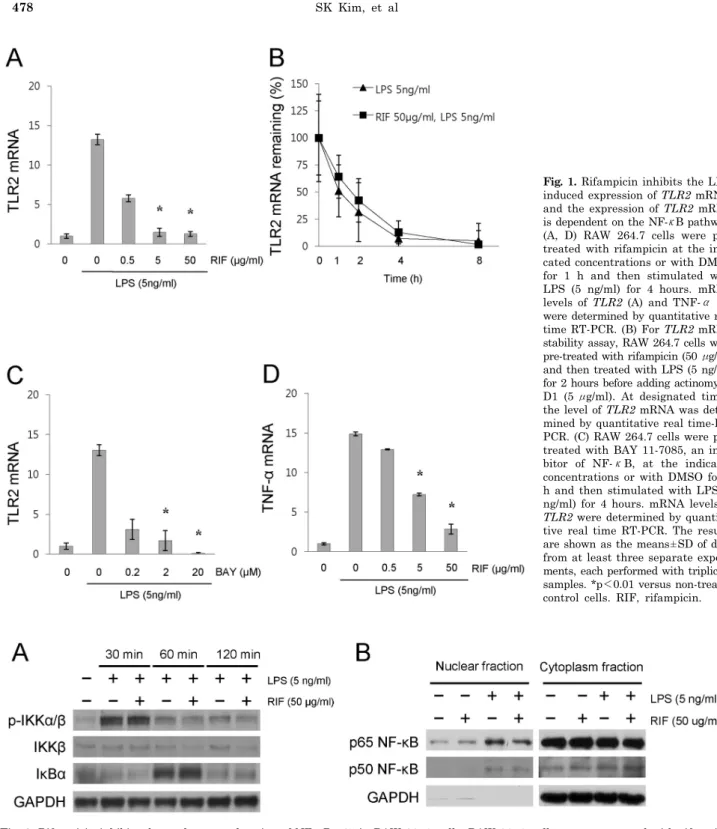
Fig. 2. Rifampicin inhibits the nuclear translocation of NF-κB p65 in RAW 264.7 cells. RAW 264.7 cells were pre-treated with rifampicin (50 μg/ml) or with DMSO for 1 h and then stimulated with LPS (5 ng/ml) for the indicated times. (A) The levels of phosphorylation of IKKα/β and the expression of IκBα were determined by immunoblot analyses. (B) The nuclear translocation of NF-κB subunits p50 and p65 were determined by immunoblot analyses. After treatment, nuclear and cytoplasmic extracts were prepared using the NE-PERⓇ Nuclear and Cytoplasmic Extraction Reagents (Pierce Biotechnology, Inc.). Similar results were observed in three independent experiments. RIF, rifampicin.
Rifampicin suppresses LPS-induced NF-κB DNA bin- ding activity
The pathway leading to the activation of NF-κB is de-
pendent on IKKβ activation. This activation leads to the degradation of IκBα and the subsequent release of NF-κB.
The NF-κB then moves to the nucleus. In the nucleus, NF- κB dimers bind to target DNA elements and activate the transcription of relevant genes (Baldwin, 1996). In order
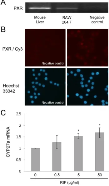
Fig. 4. Rifampicin activates PXR in RAW 264.7 cells. (A) PXR mRNA expression was determined by RT-PCR. (B) The expression pattern of PXR was determined by immunostaining. The cells were incubated overnight with an anti-PXR antibody (Santa Cruz Bio- technology) at 4oC. After washing with PBS, cells were incubated with the corresponding Cy3-conjugated secondary IgG at room temperature for 2 h. Nuclei were counterstained for 15 min with 10 μM Hoechst 33342 (Sigma-Aldrich Co. Ltd). (C) RAW 264.7 cells were treated with rifampicin at the indicated concentrations or with DMSO for 4 h. The expression of CYP27A1 mRNA was determined by quantitative real time RT-PCR. Similar results were observed in three independent experiments. *p<0.01 versus non- treated control cells. PXR, pregnane X receptor; PXR/Cy3, anti-PXR antibody/Cy3-conjugated secondary antibody; RIF, rifampicin.
Fig. 3. Rifampicin suppresses LPS-induced NF-κB DNA binding activity in RAW 264.7 cells. RAW 264.7 cells were pre-treated with rifampicin (50 μg/ml) or with DMSO for 1 h and then stimulated with LPS (5 ng/ml) for 1 h. DNA binding activity of NF-κB subunits was analyzed by EMSA. After treatment, nuclear extracts were prepared using the NE-PERⓇ Nuclear and Cytoplasmic Extraction Reagents (Pierce Biotechnology, Inc.). For the competition assay, a 200-fold excess of unlabeled probe was added together with the labeled probe. For the supershift assay, 1 μg of antibody against NF-κB p65 was added together with the nuclear extract. Similar results were observed in three independent experiments. RIF, rifampicin; Competitor oligo., unlabeled oligonucleotides probe for NF-κB p65.
to assess the effect of rifampicin on the NF-κB pathway, we examined the phosphorylation of IKKα/β, the ex- pression of IκBα, and the nuclear translocation of NF-κB subunits in LPS-activated RAW 264.7 cells in the presence and absence of rifampicin. There were no significant differ- ences in the phosphorylation of IKKα/β and the expression of IκBα between the groups with and without rifampicin (Fig. 2A). However, pre-treatment with rifampicin slightly inhibited the nuclear translocation of NF-κB p65, but not p50 in LPS-activated RAW 264.7 cells (Fig. 2B).
We then assessed the effect of rifampicin on NF-κB DNA binding activity in LPS-activated RAW 264.7 cells. As shown in Fig. 3, pre-treatment with rifampicin suppressed LPS- induced NF-κB DNA binding activity compared to that for the LPS treatment only. These results suggest that rifampi- cin inhibits the LPS-induced TLR2 expression by suppress- ing NF-κB DNA-binding activity and its suppression of NF- κB DNA-binding activity may be due to, at least in part, inhibited nuclear translocation of NF-κB p65.
Rifampicin increases the physical association between NF-κB p65 and PXR
To investigate the role of PXR in rifampicin-suppressed NF-κB DNA binding activity in LPS-activated RAW 264.7 cells, we first assessed whether PXR is expressed in RAW 264.7 cell by RT-PCR and immunostaining. PXR mRNA was found to be expressed in RAW 264.7 cells, although its level was significantly lower than that of mouse liver (Fig. 4A) and PXR protein was mainly located in the nuclei (Fig. 4B). We then assessed whether rifampicin is able to activate mouse PXR. Treatment with rifampicin increased the mRNA expression of CYP27A1, a PXR regulated gene that encodes for mitochondrial sterol 27-hydroxylase (Li et al., 2007), as compared to non-treated control cells (Fig. 4C).
We then assessed whether rifampicin is able to increase the physical association between PXR and NF-κB subunits.
As shown in Fig. 5, rifampicin increased the degree of phys-
ical association between PXR and NF-κB p65, as compared to the values for the LPS treatment only. These results sug- gest that the suppression of NF-κB DNA-binding activity by rifampicin may also be mediated through the physical association between PXR and NF-κB p65, in addition to through inhibition of nuclear translocation of NF-κB p65.
However, rifampicin-induced binding of PXR to NF-κB p65 was not dose-dependent.
Fig. 5. Rifampicin increases the physical association of PXR with NF-κB p65 in LPS-activated RAW 264.7 cells. (A, B) RAW 264.7 cells were pre-treated with rifampicin at the indicated concen- trations or with DMSO for 1 h and then stimulated with LPS (5 ng/ml) for 1 h. The physical association between PXR and NF-κB subunits was examined by a co-immunoprecipitation assay. The cell lysates were immunoprecipitated with an irrelevant rabbit IgG as a negative control and antibody against NF-κB p65, together with protein A SepharoseTM CL-4B (Amersham Biosciences, Uppsla, Sweden). The immunoprecipitates were then electrophoresed on SDS-PAGE gels, transferred onto PVDF membranes (Millipore, Bedford, MA), and immunoblotted with an antibody against PXR and NF-κB p65. Similar results were observed in three inde- pendent experiments (A). The results are shown as the means±SD of data from at least three separate experiments, each performed with triplicate samples. *p<0.05 versus the values for the LPS treatment only (B). RIF, rifampicin.
DISCUSSION
In the current study, to investigate the effects of rifampi- cin on the function of RAW 264.7 cells, it was necessary to determine whether rifampicin has the ability to activate mouse PXR, since rifampicin is a well known agonist for human PXR, but not for rodents. However, the species spe- cificity of PXR, especially, between the human and mouse seems to reflect an induction efficacy of PXR-regulated genes, not an all-or-none induction. The species specificity
of PXR is due to differences in the ligand binding domain (LBD) of PXR (Zhang et al., 1999). A previous study involv- ing an alignment analysis for PXR LBD reported that rat PXR contains nine amino acid substitutions compared with mouse PXR, whereas five of the nine amino acids in mouse PXR are conserved in human PXR, indicating that mouse PXR is more responsive to rifampicin than rat PXR, although mouse PXR is less responsive to rifampicin than human PXR (Zhang et al., 1999). In addition, a previously reported transfection assay showed that mouse PXR conferred a 2-fold induction in the mRNA expression of CYP3A4, a gene that encodes for cytochrome P450 3A4, in response to ri- fampicin, whereas the human PXR conferred a 7-fold in- crease in induction (Lehmann et al., 1998). Furthermore, an animal study showed that treatment with rifampicin caused a 3.5-fold increase in the induction of CYP3A11, a PXR regulated gene that encodes for cytochrome P450 3A11, in the mouse liver (Xu et al., 2004). Collectively, these findings suggest that rifampicin has the ability to ac- tivate, at least at a low efficacy, mouse PXR. Consistent with these findings, our results also showed that treatment with 50 μg/ml rifampicin resulted in a 1.7-fold increase in the induction of CYP27A1 mRNA in RAW 264.7 cells (Fig. 4C).
However, it was not possible to determine whether rifampi- cin is able to induce the expression of CYP3A4 and CYP3A11 mRNA in RAW 264.7 cells because CYP3A4 and CYP3A11 mRNA are not expressed in RAW 264.7 cells (data not shown).
A number of nuclear receptors, including the GC receptor (Ray and Prefontaine, 1994), the estrogen receptor (Ray et al., 1994; Stein and Yang, 1995), the progesterone receptor (Kalkhoven et al., 1996), and the androgen receptor (Palvimo et al., 1996), have been shown to inhibit NF-κB activity through physical interactions with NF-κB p65 in a ligand- dependent manner. The direct interaction between NF-κB p65 and a nuclear receptor has been proposed as a mecha- nism for controlling mutual repression between NF-κB and the nuclear receptor signaling pathway. Previous studies have shown that the activation of nuclear receptors re- presses NF-κB activity, whereas NF-κB activation recip- rocally inhibits nuclear receptors and their target gene ex- pressions (Ray et al., 1994; Stein and Yang, 1995; Kalkhoven et al., 1996; Palvimo et al., 1996). However, the issue of specifically how the nuclear receptor/NF-κB p65 complex loses its ability to bind to κB sites or to nuclear receptor response elements is not currently known.
Mutual repression also exists between PXR and the NF-κB pathway. Previous studies of intestinal inflammation and hepatic metabolism showed that PXR null mice show en- hanced expressions of NF-κB target genes and increased inflammation (Shah et al., 2007), and that the activation of NF-κB inhibited PXR function, whereas the inhibition of NF-κB increased PXR activity and the expression of its target genes (Gu et al., 2006). However, in the current study, we focused on the repression of NF-κB activity by PXR, rather than on the mutual repression between them.
Consistent with the results of previous studies (Zhou et al., 2006; Shah et al., 2007), our results showed that pre-treat- ment with rifampicin suppressed NF-κB DNA binding ac- tivity (Fig. 3) and increased the degree of physical associa- tion between PXR and NF-κB p65 (Fig. 5) in LPS-activated RAW 264.7 cells, suggesting that the suppression of NF-κB DNA-binding activity by rifampicin may be mediated through physical association between PXR and NF-κB p65.
Rifampicin-induced binding of PXR to NF-κB p65 was not
correlated with the suppressive effect of rifampicin on TLR2 expression. We thought that this discrepancy may be due to the inhibitory effect of rifampicin on the nuclear trans- location of NF-κB p65. Rifampicin not only increased bind- ing of PXR to NF-κB p65 (Fig. 5), but also inhibited the nuclear translocation of NF-κB p65 (Fig. 2B). However, NF-κB p65 has to enter the nucleus to bind PXR after stim- ulation since PXR is mainly expressed in nuclei (Fig. 4B).
Thus, although the dual function of rifampicin on NF-κB p65 activity is well focused on the suppression of NF-κB DNA binding activity and TLR2 expression, the degree of physical association between PXR and NF-κB p65 may not be correlated with the suppressive effect of rifampicin on TLR2 expression. Like rifampicin, dexamethasone, a ligand for the GC receptor, also inhibits NF-κB DNA-binding ac- tivity through physical association of the GC receptor with NF-κB p65 (Ray and Prefontaine, 1994) as well as inhibits the nuclear translocation of NF-κB p65 and p50 (Jeon et al., 2000). However, how rifampicin and dexamethasone in- hibit nuclear translocation of NF-κB p65 or whether their binding activity to NF-κB p65 affects its nuclear trans- location remained unclear.
Furthermore, the inhibitory effects of rifampicin on NF-κB pathway can also vary depending on the cell type and the ligand. Pahlevan et al. (2002) showed that rifampicin did not inhibit TNF-α or PMA-induced NF-κB DNA binding activity in Jurkat cells, a human T cell line. Although it is difficult to directly compare our results with those of the Pahlevan et al. (2002) study due to differences in cell type and ligand used, determining the reasons for the discrep- ancy would be helpful in our understanding of the in- hibitory mechanism of rifampicin on NF-κB pathway in various conditions.
The down-regulation of TLR2 and TNF-α mRNA expre- ssion were significant at rifampicin concentrations of 0.5 to 50 μg/ml (Fig. 1). These are pharmacologically relevant concentrations, as the peak concentration of rifampicin in serum 2 h after the administration of a single 600 mg oral dose is approximately 10 μg/ml and rifampicin may reach high levels in other tissues as well (Yuhas et al., 2006).
The findings reported herein suggest that the rifampi- cin-mediated inhibition of TLR2 via the suppression of NF-κB DNA-binding activity may be a novel mechanism of the immune-suppressive effects of rifampicin. However, clinical studies will be necessary in order to determine whether the rifampicin-mediated inhibition of TLR2 affects the consequences of treatment for Mycobacterium tuber- culosis, especially in cases of rifampicin-resistant strains.
ACKNOWLEDGEMENTS
This study was supported by the Korea Science &
Engineering Foundation (KOSEF) through the MRC for the Cell Death Disease Research Center at the Catholic University of Korea.
REFERENCES
Aravalli RN, Hu S, Rowen TN, Palmquist JM, Lokensgard JR.
Cutting edge: TLR2-mediated proinflammatory cytokine and chemokine production by microglial cells in response to herpes simplex virus. J Immunol 175: 4189−4193, 2005.
Baldwin AS Jr. The NF-kappa B and I kappa B proteins: new
discoveries and insights. Annu Rev Immunol 14: 649−683, 1996.
Bellahsene A, Forsgren A. Effect of rifampin on the immune response in mice. Infect Immunl 27: 15−20, 1980.
Ghosh S, May MJ, Kopp EB. NF-kappa B and Rel proteins:
evolutionarily conserved mediators of immune responses. Annu Rev Immunol 16: 225−260, 1998.
Gu X, Ke S, Liu D, Sheng T, Thomas PE, Rabson AB, Gallo MA, Xie W, Tian Y. Role of NF-kappaB in regulation of PXR- mediated gene expression: a mechanism for the suppression of cytochrome P-450 3A4 by proinflammatory agents. J Biol Chem 281: 17882−17889, 2006.
Haehnel V, Schwarzfischer L, Fenton MJ, Rehli M. Transcriptional regulation of the human toll-like receptor 2 gene in monocytes and macrophages. J Immunol 168: 5629−5637, 2002.
Jeon YJ, Han SH, Lee YW, Lee M, Yang KH, Kim HM. Dexame- thasone inhibits IL-1 beta gene expression in LPS-stimulated RAW 264.7 cells by blocking NF-kappa B/Rel and AP-1 acti- vation. Immunopharmacology 48: 173−183, 2000.
Kalkhoven E, Wissink S, van der Saag PT, van der Burg B.
Negative interaction between the RelA(p65) subunit of NF- kappaB and the progesterone receptor. J Biol Chem 271: 6217−
6224, 1996.
Lehmann JM, McKee DD, Watson MA, Willson TM, Moore JT, Kliewer SA. The human orphan nuclear receptor PXR is acti- vated by compounds that regulate CYP3A4 gene expression and cause drug interactions. J Clin Invest 102: 1016−1023, 1998.
Li T, Chen W, Chiang JY. PXR induces CYP27A1 and regulates cholesterol metabolism in the intestine. J Lipid Res 48: 373−
384, 2007.
Li T, Chiang JY. Mechanism of rifampicin and pregnane X receptor inhibition of human cholesterol 7 alpha-hydroxylase gene trans- cription. Am J Physiol Gastrointest Liver Physiol 288: G74−G84, 2005.
Mlambo G, Sigola LB. Rifampicin and dexamethasone have similar effects on macrophage phagocytosis of zymosan, but differ in their effects on nitrite and TNF-alpha production. Int Immuno- pharmacol 3: 513−522, 2003.
Moreau A, Vilarem MJ, Maurel P, Pascussi JM. Xenoreceptors CAR and PXR activation and consequences on lipid metabolism, glucose homeostasis, and inflammatory response. Mol Pharm 5:
35−41, 2008.
Mun HS, Aosai F, Norose K, Chen M, Piao LX, Takeuchi O, Akira S, Ishikura H, Yano A. TLR2 as an essential molecule for protective immunity against Toxoplasma gondii infection. Int Immunol 15: 1081−1087, 2003.
Nakamura K, Moore R, Negishi M, Sueyoshi T. Nuclear pregnane X receptor cross-talk with FoxA2 to mediate drug-induced regulation of lipid metabolism in fasting mouse liver. J Biol Chem 282: 9768−9776, 2007.
Pahlevan AA, Wright DJ, Bradley L, Smith C, Foxwell BM. Poten- tial of rifamides to inhibit TNF-induced NF-kappaB activation.
J Antimicrob Chemother 49: 531−534, 2002.
Palvimo JJ, Reinikainen P, Ikonen T, Kallio PJ, Moilanen A, Janne OA. Mutual transcriptional interference between RelA and androgen receptor. J Biol Chem 271: 24151−24156, 1996.
Ray A, Prefontaine KE. Physical association and functional anta- gonism between the p65 subunit of transcription factor NF-kappa B and the glucocorticoid receptor. Proc Natl Acad Sci U S A 91: 752−756, 1994.
Ray A, Prefontaine KE, Ray P. Down-modulation of interleukin-6 gene expression by 17 beta-estradiol in the absence of high affinity DNA binding by the estrogen receptor. J Biol Chem 269:
12940−12946, 1994.
Shah YM, Ma X, Morimura K, Kim I, Gonzalez FJ. Pregnane X receptor activation ameliorates DSS-induced inflammatory bowel disease via inhibition of NF-kappaB target gene expression. Am J Physiol Gastrointest Liver Physiol 292: G1114−G1122, 2007.
Stein B, Yang MX. Repression of the interleukin-6 promoter by estrogen receptor is mediated by NF-kappa B and C/EBP beta.
Mol Cell Biol 15: 4971−4979, 1995.
Tsankov N, Angelova I. Rifampin in dermatology. Clin Dermatol 21: 50−55, 2003.
Xu DX, Wei W, Sun MF, Wu CY, Wang JP, Wei LZ, Zhou CF.
Kupffer cells and reactive oxygen species partially mediate lipopolysaccharide-induced downregulation of nuclear receptor pregnane x receptor and its target gene CYP3a in mouse liver.
Free Radic Biol Med 37: 10−22, 2004.
Yuhas Y, Berent E, Ovadiah H, Azoulay I, Ashkenazi S. Rifampin augments cytokine-induced nitric oxide production in human alveolar epithelial cells. Antimicrob Agents Chemother 50: 396−
398, 2006.
Zhang H, LeCulyse E, Liu L, Hu M, Matoney L, Zhu W, Yan B.
Rat pregnane X receptor: molecular cloning, tissue distribution, and xenobiotic regulation. Arch Biochem Biophys 368: 14−22, 1999.
Zhou C, Tabb MM, Nelson EL, Grun F, Verma S, Sadatrafiei A, Lin M, Mallick S, Forman BM, Thummel KE, Blumberg B.
Mutual repression between steroid and xenobiotic receptor and NF-kappaB signaling pathways links xenobiotic metabolism and inflammation. J Clin Invest 116: 2280−2289, 2006.