Impaction of a tooth is a situation in which an unerupted tooth is wedged against another tooth (or teeth) or otherwise located so that it cannot erupt normally.1The mandibular third molar is the most frequently impacted. Impacted mandibular third molars are categorized according to the anterior-posterior space between the second molar and the mandibular ramus, its medial-lateral position in the body of the mandible, and the position of its long axis. Ectopic mandibular third molars, however, are rare and usually found because of the clinical symptoms. Their heterotopic positions were reported to be in the condylar area, in the ascending ramus of the mandible, or in the coronoid pro- cess.2Unilateral or bilateral ectopic impacted third molar teeth have been reported in various parts of the mandibular ramus.
This report presents an ectopic mandibular third molar in the mandibular condyle without any pathologic condi- tion.
Case Report
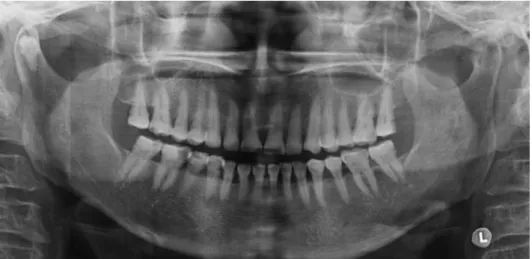
A 70-years-old woman visited the department with a complaint of discomfort on the left preauricular area during mastication and mouth opening, which had occurred 3 or 4 months before. On the clinical examination there was tenderness on the left preauricular area without any specific findings. Panoramic radiograph showed generalized alve- olar bone destruction and calculus deposition. All the teeth showed moderate attrition. Additionally, a well-formed, impacted tooth was found in the right condylar head of the mandible. The tooth crown seemed to direct into the head of the condyle (Fig. 1).
The patient had no experience of extraction of third molars, and any associated pathologic sign was not observ- ed. According to the orientation and location of the impact- ed tooth, it was considered as an ectopic impaction of a mandibular third molar.
Cone beam CT scans were performed to evaluate the position and direction of the impacted tooth in the mandi- bular condyle. Coronal image showed the upward crown position of the impacted tooth (Fig. 2). There was no path- ologic sign such as cystic change around the impacted tooth. On the axial image, an impacted tooth was found in close proximity to the front cortical bone and in the middle portion of the mandibular condyle (Fig. 3). Sagittal image
─ 135 ─
Cone beam computed tomography findings of ectopic mandibular third molar in the mandibular condyle: report of a case
Jin-Soo Kim
Department of Oral and Maxillofacial Radiology, School of Dentistry, Chosun University, Gwangju, Korea ABSTRACT
Impaction of third molar is a common developmental abnormality. However, ectopic impaction of the mandibular third molar in condylar region is an extremely rare condition. This report describes a case of impacted tooth in the mandibular condyle without any associated pathologic condition. Also, this report presents the spatial relationship of the impacted mandibular third molar to the surrounding anatomic structures using cone beam computed tomog- raphy. (Imaging Sci Dent 2011; 41 : 135-7)
KEY WORDS : Cone-Beam Computed Tomography; Tooth Eruption, Ectopic; Tooth, Impacted; Molar, Third
*This study was supported by research funds from Chosun University, 2009 Received January 24, 2011; Revised March 8, 2011; Accepted May 9, 2011 Correspondence to: Prof. Jin-Soo Kim
Department of Oral and Maxillofacial Radiology, School of Dentistry, Chosun Univer- sity, 375 Seosuk-dong, Dong-gu, Gwangju 501-759, Korea
Tel) 82-62-220-3886, Fax) 82-62-227-0270, E-mail) [email protected]
Imaging Science in Dentistry 2011; 41 : 135-7 http://dx.doi.org/10.5624/isd.2011.41.3.135
Copyright ⓒ 2011 by Korean Academy of Oral and Maxillofacial Radiology
This is an Open Access article distributed under the terms of the Creative Commons Attribution Non-Commercial License (http://creativecommons.org/licenses/by-nc/3.0) which permits unrestricted non-commercial use, distribution, and reproduction in any medium, provided the original work is properly cited.
Imaging Science in Dentistry∙pISSN 2233-7822 eISSN 2233-7830
showed a well developed impacted tooth with proximity to the outer cortical bone of the mandibular condyle (Fig.
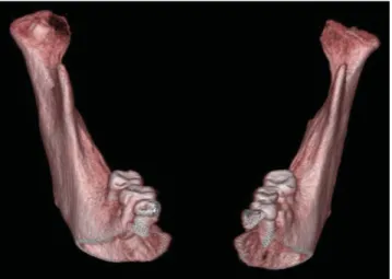
4). The follicular space and lamina dura of the tooth were not distinguished. On the frontal view of 3D volumetric image, the crown of the impacted tooth was partially cover- ed with bone (Fig. 5).
The mandibular condyle was not expanded. There was no pathologic or morphologic change on the surrounding bone. Final diagnosis was an ectopic impaction of the mandibular third molar in the mandibular condyle.
Discussion
The true incidence and etiology of ectopic impaction of mandibular third molar remain unknown. Several theories have been suggested to explain the ectopic eruption, includ- ing aberrant eruption, trauma, and ectopic formation of the germs of the teeth.2It has been suggested that an aberrant eruption pattern occurred when the tooth has been displaced by a lesion, usually an odontogenic cyst.3
Wang et al3in 2008 reported a similar case and reviewed the literatures from 1978 to 2003. They found 13 case re-
─ 136 ─
Cone beam computed tomography findings of ectopic mandibular third molar in the mandibular condyle: report of a case
Fig. 1.Panoramic radiograph shows the impacted tooth in the right mandi- bular condylar head.
Fig. 2.Coronal CBCT image shows the upward crown position of the impacted tooth. There is no cystic change around impacted tooth.
Fig. 3.Axial CBCT image shows the impacted tooth with the pro- ximity to the front cortical bone and middle portion of the mandi- bular condyle.
ports of the ectopic third molars in the subcondylar area, including 1 case of a dry skull and 1 of a cadaveric mandi- ble. The management for these conditions was described as no treatment or as surgical removal via extraoral or in- traoral access. Salmerón et al2in 2008 reported 2 cases of
third molars in the subcondylar area, which were associated with odontogenic cysts and treated through the extraoral approach, indicating that the treatment was primarily deter- mined by the experience of the surgeon or physician.
Wang et al3reviewed that eight of the 11 cases included the description of a radiolucent image around the ectopic molar on the radiograph, including 5 with the diagnosis of a dentigerous cyst. These mandibular third molars might be displaced by the lesion. The expansion of a cyst might put pressure on the crown of a tooth and displace it in the opposite direction to the path of eruption. Several teeth had an upward whereas the others had a downward inclination or even an inverted crown position. In this case, the abnor- mal position of the tooth germ might be the most likely causative factor because the impacted tooth was positioned in the mandibular condyle with an upward crown position without any pathologic change which resulted in such unusual movement.
The surgical removal of an ectopic mandibular third molar with acute inflammation or cystic lesion is recommended to prevent further complications such as diffuse osteolysis, condylar process deformity, or bone absorption.4,5In cases of symptom-free highly aberrant wisdom teeth or without urgent necessity, annual follow-up visits to monitor the growth of the lesion is appropriate.6,7In this case, the im- pacted tooth was not removed because of the absence of the associated symptom or lesion unlike other reports2,4-6 and the patient’s age.
References
1. Alling CC 3rd, Catone GA. Management of impacted teeth. J Oral Maxillofac Surg 1993; 51(1 Suppl 1) : 3-6.
2. Salmerón JI, del Amo A, Plasencia J, Pujol R, Vila CN. Ectopic third molar in condylar region. Int J Oral Maxillofac Surg 2008;
37 : 398-400.
3. Wang CC, Kok SH, Hou LT, Yang PJ, Lee JJ, Cheng SJ, et al.
Ectopic mandibular third molar in the ramus region: report of a case and literature review. Oral Surg Oral Med Oral Pathol Oral Radiol Endod 2008; 105 : 155-61.
4. Medici A, Raho MT, Anghinoni M. Ectopic third molar in the condylar process: case report. Acta Biomed Ateneo Parmense 2001; 72 : 115-8.
5. Gadre KS, Waknis P. Intra-oral removal of ectopic third molar in the mandibular condyle. Int J Oral Maxillofac Surg 2010;
39 : 294-6.
6. Wassouf A, Eyrich G, Lebeda R, Grätz KW. Surgical removal of a dislocated lower third molar from the condyle region: case report. Schweiz Monatsschr Zahnmed 2003; 113 : 416-20.
7. Park W, Lee JH, Park H, Jung HG, Kim KD. Impacted supernu- merary tooth in coronoid process: a case report. Korean J Oral Maxillofac Radiol 2010; 40 : 89-91.
─ 137 ─
Jin-Soo Kim
Fig. 4.Sagittal CBCT image shows the fully developed impacted tooth with the proximity to the outer cortical bone of the mandible.
Fig. 5. Frontal view of 3-dimensional volumetric CBCT image shows the crown of the impacted tooth partially covered with bone.