Brief Report
824 Ann Dermatol
Received August 17, 2016, Revised November 9, 2016, Accepted for publication November 30, 2016
Corresponding author: Kapsok Li, Department of Dermatology, Chung-Ang University Hospital, 102 Heukseok-ro, Dongjak-gu, Seoul 06973, Korea. Tel:
82-2-6299-1525, Fax: 82-2-823-1049, E-mail: [email protected]
This is an Open Access article distributed under the terms of the Creative Commons Attribution Non-Commercial License (http://creativecommons.org/
licenses/by-nc/4.0) which permits unrestricted non-commercial use, distribution, and reproduction in any medium, provided the original work is properly cited.
Copyright © The Korean Dermatological Association and The Korean Society for Investigative Dermatology with psoriasis in adults: a population-based study. Br J
Dermatol 2011;165:1037-1043.
3. Kim JW. Hepatitis B virus infection in South Korea: three decades after universal vaccination. Korean J Intern Med 2013;28:408-409.
4. Guadagnino V, Ayala F, Chirianni A, Picciotto L, Tiseo D, Piazza M. Risk of hepatitis B virus infection in patients with eczema or psoriasis of the hand. Br Med J (Clin Res Ed) 1982;284:84.
5. Tsai TF, Wang TS, Hung ST, Tsai PI, Schenkel B, Zhang M, et al. Epidemiology and comorbidities of psoriasis patients in a national database in Taiwan. J Dermatol Sci 2011;63:
40-46.
6. Huang HH, Shih WL, Li YH, Wu CF, Chen PJ, Lin CL, et al.
Hepatitis B viraemia: its heritability and association with common genetic variation in the interferon gamma signal-
ling pathway. Gut 2011;60:99-107.
7. Ahmad QM, Sameem F, Shah IH. Prevalence of hepa- totrophic viruses B & C in psoriasis. Indian J Dermatol 2005;50:200-202.
8. Kanada KN, Schupp CW, Armstrong AW. Association between psoriasis and viral infections in the United States:
focusing on hepatitis B, hepatitis C and human immu- nodeficiency virus. J Eur Acad Dermatol Venereol 2013;27:
1312-1316.
9. Cohen AD, Weitzman D, Birkenfeld S, Dreiher J. Psoriasis associated with hepatitis C but not with hepatitis B. Der- matology 2010;220:218-222.
10. Chae HB, Kim JH, Kim JK, Yim HJ. Current status of liver diseases in Korea: hepatitis B. Korean J Hepatol 2009;
15(Suppl 6):S13-S24.
https://doi.org/10.5021/ad.2017.29.6.824
Tattoo Granuloma Restricted to Red Dyes
Joon Seok, Sun Young Choi
1, Tae-Rin Kwon
2, Jong Hwan Kim
2, Kui Young Park, Kapsok Li, Hee Sung Kim
3, Beom Joon Kim
Department of Dermatology, Chung-Ang University College of Medicine, 1Department of Dermatology, Inje University Seoul Paik Hospital, Inje University College of Medicine, Departments of 2Medicine and 3Pathology, Chung-Ang University College of Medicine, Seoul, Korea
Dear Editor:
A tattoo is as forms of visual art, which entails insertion of an ink design into the skin. Although there have been re- ports on allergic reactions caused by tattoos of almost ev- ery color, the most common reactions are those caused by red tattoos. Here we describe a case restricted to reactions to red portion in colored tattoos.
A 34-year-old man presented with a 4-month history of el- evating plaques restricted to red tattoo portions of the tat-
too on the right thigh. These skin lesions were firm and well-demarcated. The remainder of the tattoo was un- affected (Fig. 1A). The patient had been tattooed 10 years ago without any complication since then. Recently, how- ever, he felt itching sensation and induration confined to red-tattooed area. Histopathologic finding revealed gran- ulomatous response with Swiss-cheese pattern and scat- tered exogenous tattoo pigments (Fig. 1B, C). To figure out tattoo component, biopsy specimen was analyzed via
Brief Report
Vol. 29, No. 6, 2017 825 Fig. 1. (A) Plaque lesions restricted to red tattoo areas. (B) Granulomatous reaction with Swiss-cheese appearance and granule containing black pigmented material on the superficial dermis (H&E, ×100; arrowhead: granule). (C) Multiple ovoid cavities with scattered black pigmented materials, suspicion of tattoo pigment (H&E, ×400; arrowheads: tattoo pigment). (D) Scanning electron microscopy image.
(E) In the scanning electron microscopy and energy-dispersive X-ray spectroscopy findings, zirconium was detected. The mean zirconium content (weight, %) of biopsy specimen was 30.28.
scanning electron microscopy and energy-dispersive X-ray spectroscopy (SEM-EDS). In the SEM-EDS findings, zirco- nium was detected (Fig. 1D, E). He has been receiving in- termittent steroid injection and taking topical methyl- prednisolone aceponate, antihistamine for pruritus as a part of his maintenance treatment, though his symptoms continue to wax and wane.
It has already been widely recognized in the past that the mercury content in red ink is the agent that causes re- actions related to red tattoos1. Modern alternatives such as sienna-ferric hydrate, cadmium-selenide, organic vegeta- ble dyes, sandalwood and brazilwood have largely re- placed mercury. In this case, we found zirconium, asso- ciated with allergic reaction and granuloma formation2. Zirconium could chelate with alizarin red S, forming zir- conium-alizarin red S complex, and this complex pro- duces red-violet color3. This material is used in analytical chemistry for spectrometry and anionic dye3.
Illegal tattooing that violated medical laws in Korea has occurred frequently. The majority of tattooists are not fully aware of the composition of the pigments they work with.
There is much difficulty in defining exactly which chem- icals are involved. Furthermore, there are even greater
challenges in recognizing the particular ingredients in a certain type of ink, especially with the creation of new mixtures. It appears that there is generally a lack of under- standing regarding the risk of dangerous chemicals in tattoos. These could become carcinogens. Several studies reported that benign and malignant lesions could occur in tattoos4.
The reaction occurred in this patient 10 years after tattooing. The long period might need to break the tattoo pigment to a critical size. However, in general, the black dye particles, which have a less tendency to react in- flammatory response, are the smallest, and the red dye particles have a bigger size5. Swiss-cheese pattern ap- peared in histology of this patient could be presented in the injection of oily substance. It is possible that oil sol- vent to mix tattoo substances could make granulomatous reaction synergistically in the restricted red tattoo area.
Relatively high rate of granulomatous reaction to red tat- too could be attributed to the allergen in red dye with oil solvent in comparison with inertness of carbon material in black tattoo.
Brief Report
826 Ann Dermatol
Received August 9, 2016, Revised November 28, 2016, Accepted for publication December 14, 2016
Corresponding author: Soo Hong Seo, Department of Dermatology, Korea University Anam Hospital, Korea University College of Medicine, 73 Inchon-ro, Seongbuk-gu, Seoul 02841, Korea. Tel: 82-2-920-5470, Fax:
82-2-928-7540, E-mail: [email protected]
This is an Open Access article distributed under the terms of the Creative Commons Attribution Non-Commercial License (http://creativecommons.
org/licenses/by-nc/4.0) which permits unrestricted non-commercial use, distribution, and reproduction in any medium, provided the original work is properly cited.
Copyright © The Korean Dermatological Association and The Korean Society for Investigative Dermatology
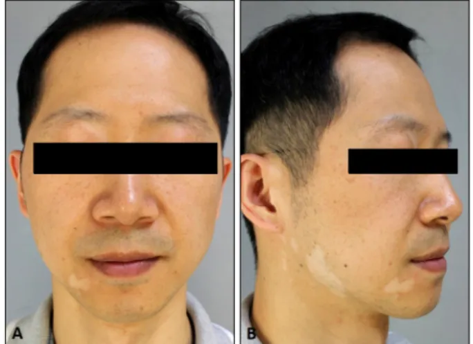
Fig. 1. Type II segmental vitiligo showing multiple irregular bordered hypopigmented patches on right facial area: upper medial eyelid and perioral area (A), mainly on mandibular and auricular are (B).
CONFLICTS OF INTEREST
The authors have nothing to disclose.
REFERENCES
1. Mortimer NJ, Chave TA, Johnston GA. Red tattoo reactions.
Clin Exp Dermatol 2003;28:508-510.
2. Skelton HG 3rd, Smith KJ, Johnson FB, Cooper CR, Tyler WF, Lupton GP. Zirconium granuloma resulting from an aluminum zirconium complex: a previously unrecognized agent in the development of hypersensitivity granulomas. J
Am Acad Dermatol 1993;28:874-876.
3. Muñoz JA, Campaña AM, Barrero FA. Effect of cationic micelles on the formation of the complex oxalate-Alizarin Red S-Zr(IV) Application to the sensitive fluorescence deter- mination of oxalate ion. Talanta 1998;47:387-399.
4. Lee JS, Park J, Kim SM, Yun SK, Kim HU. Basal cell car- cinoma arising in a tattooed eyebrow. Ann Dermatol 2009;21:281-284.
5. Høgsberg T, Loeschner K, Löf D, Serup J. Tattoo inks in general usage contain nanoparticles. Br J Dermatol 2011;165:1210-1218.
https://doi.org/10.5021/ad.2017.29.6.826
A Type II Segmental Vitiligo Developed under Infliximab Treatment for Ulcerative Colitis
Tae Hyung Ryu, Dong Won Lee, Jae Eun Choi, Hyo Hyun Ahn, Young Chul Kye, Soo Hong Seo
Department of Dermatology, Korea University College of Medicine, Seoul, Korea
Dear Editor:
Tumor necrosis factor (TNF)-α inhibitor has shown vari- ous adverse skin reactions, including alopecia areata, atopic dermatitis, leukocytoclastic vasculitis, and so forth.
Herein, we report a rare case of segmental vitiligo (SV) during infliximab therapy and like to provide academic in- formation about its pathogenesis.
A 34-year-old man presented with multiple hypopigmented patches appeared 4 months after the initiation of intra- venous infliximab therapy, as part of which he had re-
ceived 4 infusions (Fig. 1). He had been treated for ulcer- ative colitis (UC) for 9 years showing wax and wane, but did not report any symptomatic aggravation at the time of