INTRODUCTION
Dual source computed tomography (DSCT) has provided the best image on coronary CT angiography (CCTA) in order to achieve high diagnostic accuracy for coronary artery stenosis (1-4). With recent technical developments, there were no sig- nificant differences in image quality or diagnostic performance among different heart rate frequencies when an adaptive elec- trocardiography (ECG)-pulsing algorithm used. However, a trend toward increased false positives in patients with low cal- cium scores (Agatston score < 100) or heart rate variability of more than 10 was still observed (5-8). Indeed, if we obtain a
poor image for assessing the stenotic coronary artery on the di- astolic phase of CCTA because of high heart rate or high calci- um score, we often hesitate to re-check the CCTA after the heart rate has decreased, or select a proper systolic phase for evaluating the stenotic coronary artery if we cannot perform CCTA again.
Recently, several papers have focused on the diagnostic accu- racy of coronary artery stenosis or image quality of the coro- nary artery on CCTA based on an invasive coronary angiogra- phy (ICA) in the diastolic phase (2, 4, 5). If the best image for coronary artery stenosis is obtained on the systolic phase, we should compare the same stenotic portion of coronary vessel in
J Korean Soc Radiol 2012;66(3):221-228
Received September 28, 2011;
Accepted January 10, 2012
Corresponding author: Gong Yong Jin, MD
Department of Diagnostic Radiology, Chonbuk National University Hospital and Medical School, 20 Geonji-ro, Deokjin-gu, Jeonju 561-712, Korea.
Tel. 82-63-250-2307 Fax. 82-63-272-0481 E-mail: [email protected]
Copyrights © 2012 The Korean Society of Radiology
Purpose: To assess the stenotic coronary artery diameter in systole and end-diasto- le by coronary CT angiography and to evaluate the change in diameter according to heart rate and calcium score.
Materials and Methods: Twenty-seven patients with coronary artery stenosis that underwent a coronary CT angiography and invasive coronary angiography were en- rolled in the study. We assessed the percentage of diameter change in the stenotic coronary artery between the systolic and end-diastolic phase and evaluated its rela- tionship with heart rate or Agatston score using a linear regression analysis.
Results: The mean difference in the change of vessel diameter was 12.9% for a heart rate of 50–59 beats/min (bpm), 11.3% for 60–69 bpm, 10% for 70–79 bpm, 20% for 80–89 bpm, 13% for 90–99 bpm, and 6.7% for 100–109 bpm, none of which were statistically significant (p = 0.760). For the Agatston score, the mean difference in the change of vessel diameter was 8.3% for a score < 100, 12.4%
for 100-400, and 20% for > 400. The relationship between the change in diame- ter and Agatston score was statistically significant (p = 0.004).
Conclusion: The data suggest that a change in diameter of the stenotic coronary artery in systole and end-diastole might be affected by the Agatston score.
Index terms Diagnosis
Computer-Assisted
Dual Source Computed Tomography Coronary Angiography
Coronary Stenosis
Variation of the Degree of Coronary Artery Stenosis during the Cardiac Cycle: Influence of Heart Rate or Calcium Score on Coronary CT Angiography
1심장박동수 또는 석회수치에 따른 수축기와 이완기 말기에서 협착된 관상동맥의 직경 변화 측정1
Hyun Kyung Lee, MD
1, Gong Yong Jin, MD
1, Jae Keun Park, MD
1, Young Min Han, MD
1, Young Sun Lee, MD
1, Keun Sang Kwon, MD
2, Song Soo Kim, MD
3Departments of 1Radiology, 2Preventive Medicine, Chonbuk National University Hospital and Medical School, Research Institute of Clinical Medicine, Institute of Cardiovascular Research, Jeonju, Korea
3Department of Radiology, Chungnam National University Hospital, Chungnam National University School of Medicine, Daejeon, Korea
rection. Non-contrast CT scans were obtained with a collima- tion of 0.6-mm thickness; rotation time of 0.33 msec; tube volt- age of 100 or 120 kV; tube current of 100 mAs per rotation on both tubes; and pitch of 0.3-0.39 (depending on heart rate). The scan time was 5.7-8.1 seconds for a single breath hold.
CCTA was used with a tri-phasic injection protocol. Bolus tracking was performed in the ascending aorta, with an addi- tional scan delay of 7 seconds used for timing. In the first phase, a 50-60 mL bolus of 370 mg/mL iopromide contrast media (Ultravist; Schering, Erlangen, Germany) was injected into an antecubital vein at a flow rate of 4-5 mL/s. In the second phase, 50 mL of mixed contrast agent and saline (7 : 3 or 6 : 4) was in- jected at the same flow rate, followed by a 40 mL saline chasing bolus administered using an autoinjector (Stellant; Medrad, Warrendale, PA, USA). The patient held his/her breath at mild inspiration. The mean scan time was 10 seconds (range 7-12 seconds). The tube voltage was 100-120 kVp for both tubes.
Height and weight of all patients were measured and BMI was calculated. If BMI was standard or lower, 100 kVp was used, and if BMI was above standard, 120 kVp was used. The current was between 30% and 80% of the cardiac cycle if the heart rate was less than 90 beats per minute (bpm). The current was full between 10% and 100% of the cardiac cycle if the heart rate was more than 90 bpm, because good images could be obtained at the systolic phase for the majority of patients with a fast heart beat. The gantry rotation time was 0.33 seconds, and the pitch was 0.3-0.4, depending on heart rate. Using a medium soft-tis- sue convolution kernel and a mono-segment reconstruction al- gorithm that uses data from quarter rotations of both detectors, 1-mm axial images were reconstructed for the entire cardiac cycle, with reconstruction intervals obtained in 10% steps.
ECG-pulsing was applied for radiation dose reduction in all pa- tients.
A retrospective gating technique was used to synchronize the data reconstruction with the ECG signal. A mono-segment re- construction algorithm that uses the data from 1/4 rotation of both detectors was used for image reconstruction. In each pa- tient, images were reconstructed at 70-75% of the RR interval for diastolic images and 30-35% of the RR interval for systolic images. If considered necessary, additional images were recon- structed in 5% steps of the RR interval.
For CCTA, images were reconstructed from the contrast-en- systolic phase on CCTA with the diastolic phase on ICA. For
this reason, it is important to understand changes in the stenotic coronary vessel using DSCT during diastole and systole at the same stenotic site with respect to heart rate and calcium score.
This study was designed to assess the difference in diameter of the stenotic coronary artery between the in-systolic and end-dia- stolic phase on CCTA according to heart rate and calcium score.
We also evaluated the diagnostic accuracy of CCTA for coronary artery stenosis during systolic and end-diastolic phase base by comparing it with invasive ICA as a reference value.
MATERIALS AND METHODS
Study Population
This study was performed prospectively with the approval of the institutional ethics committee at our institution. Written in- formed consent was obtained from all patients. From Novem- ber 2007 to January 2008, 347 patients with chest pain under- went DSCT examinations. Among these patients, 27 (13 men and 14 women; age range 46-78 years, mean age 63.1 ± 8.4 years) with coronary artery stenosis on DSCT images and ICA were enrolled. The mean time interval between CCTA and ICA was 5.2 ± 2.1 days (range, 0-8 days). Patients who were pregnant or who had unstable clinical conditions, severe renal failure, previ- ous allergic reactions to the use of an iodinated contrast agent, hyperthyroidism, any circumstances that would not allow the pa- tient to lie in the supine position or patients who had inserted coronary stents or who had undergone previous bypass sur- gery, were excluded from the study. In addition, image quality of a stenotic coronary segment that was assessed for moderate artifacts with diagnostic or severe artifacts impairing the accu- rate evaluation on CCTA, was excluded.
DSCT-Calcium Scoring and Coronary CT Angiography All CT examinations were performed on a DSCT scanner (Somatom Definition, Siemens Medical Solutions, Forchheim, Germany). The patients were centrally placed in the scanner to ensure that the entire heart was covered by the smaller field-of- view of the second tube detector array. No beta-blockers were administered prior to scanning. Non-enhanced DSCT for cal- cium scoring was performed from 1 cm below the level of the tracheal bifurcation to the diaphragm in the cranio-caudal di-
patients were divided into cardiac contraction of the end-diastol- ic and systolic phase as well as for mean heart rate of ≤ 80 bpm and > 80 bpm.
Invasive Coronary Angiography (ICA)
ICA was performed according to standard techniques, and multiple views were stored on a CD-ROM. One experienced cardiologist who was blinded to the results of the CCTA evalu- ated the angiograms. Coronary artery segments were defined according to the guidelines described above (9). Each vessel segment was scored as being significantly stenosed, which was defined as a diameter reduction of > 50%. A coronary artery analysis was performed in all vessels with a luminal diameter of at least 1.5 mm, excluding vessels distal to complete occlusions.
The narrowest stenotic area at the end-diastolic phase was se- lected. The diameter was measured three times continuously to get an average value at the most stenotic area and the mean val- ue of the stenotic vessel’s diameter was calculated (10).
Statistical Analyses
Statistical analyses were performed using commercially avail- able software (SPSS 12.0, SPSS, Chicago, IL, USA). We evaluat- ed the percent difference in the stenotic coronary artery between systolic and end-diastolic phases with respect to heart rate or calcium score and correlated the diagnostic accuracy with heart rate in the end-diastolic phase using a linear regression analy- sis. The sensitivity, specificity, positive predictive value (PPV), and negative predictive value (NPV), and accuracy on a per- vessel basis of CCTA in diastole in terms of the heart rate or Agatston score were calculated using the Chi-square test for contingency. The ICA findings were considered the standard of reference. A correlation between the degree of coronary artery stenosis on DSCT and ICA was evaluated by Pearson’s correla- tion analysis depending on overall end-diastolic phase, heart rate ≤ 80 bpm, and heart rate > 80 bpm.
RESULTS
The clinical characteristics of the enrolled patients are sum- marized in Table 1. ICA demonstrated coronary artery stenosis in all patients. Among all of the coronary arteries, 51 vessels (62.9%) had stenoses. Among the vessels with a stenosis, 23 hanced DSCT scans with a slice thickness of 0.75 mm, a recon-
struction increment of 0.5 mm, and using a medium soft-tissue convolution kernel (B26f). Depending on each patient’s anatomy, the reconstructed field-of-view (FOV) was adjusted to exactly encompass the heart (mean FOV, 167 ± 19 mm; range, 131-187 mm; image matrix, 512 × 512 pixels). After the patient was re- moved and the ECG information was obtained, all reconstructed images were transferred to a dedicated workstation (Wizard, Sie- mens Medical Solutions, Forchheim, Germany) equipped with dedicated cardiac post-processing software (Syngo Circulation, Siemens Medical Solutions, Forchheim, Germany).
Data Analyses
For analysis of CCTA data, the coronary arteries were seg- mented according to the guidelines of the American Heart As- sociation (10) as follows. All segments with a diameter of at least 1.5 mm at the origin were included. Image quality of a stenotic coronary segment was assessed semiquantitatively on a four- point ranking scale by one radiologist as follows: 1, excellent (no artifacts); 2, good (minor artifacts); 3, adequate (moderate arti- facts, diagnostic), and 4, not assessable (severe artifacts impair- ing accurate evaluation). The mean Agatston score was calculat- ed for each patient using a semi-automated software (Syngo Calcium Scoring, Siemens Medical Solutions, Forchheim, Ger- many). Diameter measurements were performed using an elec- tronic caliper by one radiologist. All reconstructed images were simultaneously evaluated by two radiologists using axial source images, multi-planar reformations, maximum intensity projec- tions, and volume rendering on a per-segment basis. All of the coronary artery segments were assessed for the presence of he- modynamically significant stenoses. Significant stenosis was de- fined as narrowing of the coronary luminal diameter that ex- ceeded 50%. The diameter of the stenotic vessel diameters was measured perpendicular to the vessel course. For the initial mea- surement, the narrowest stenotic area at the same site in systolic and end-diastolic phase was selected. The diameter was mea- sured three times continuously to get an average value at the most stenotic area and the mean value of the stenotic vessel’s di- ameter was calculated. The percentage of diameter change in ste- notic coronary artery was calculated as: [(diameter of stenotic portion in diastole – diameter of stenotic portion in systole) ÷ di- ameter of stenotic portion in diastole] × 100%. In a sub-analysis,
(p < 0.001) determined during the diastolic phase. The correla- tion between the use of ICA and DSCT was 0.867 (p < 0.001) for a heart rate ≤ 80 bpm, and 0.817 (p < 0.001) and for a heart rate
> 80 bpm, both determined during the diastolic phase (Fig. 2).
The mean Agatston score was 238.5 ± 196.2 and the mean difference in the stenotic coronary artery diameter between sys- tole and end-diastole according to calcium score was 8.3% for less than 100, 12.4% for 100-400, and 20% for ≥ 400 (Figs. 3-5).
There was a significant correlation between the diameter change of the stenotic portion of the coronary artery and Agatston score (r = 0.942, p = 0.004).
In patients with calcium scores < 100, sensitivity, specificity, PPV, NPV, and accuracy on a per-vessel basis were 97.3%, 97.9%, 88.8%, 99.0%, and 97.6%, respectively. These measurements were 96.6%, 95.3%, 82.6%, 95.2%, and 94.1%, respectively in patients with calcium scores between 100 and 400; and 90.3%, 91.2%, 81.7%, 90.7%, and 88.2%, respectively in patients with calcium scores of > 400.
DISCUSSION
High calcium burdens make it difficult to diagnose the coro- (45.0%) were significantly stenosed, which is defined as having
a diameter reduction ≥ 50%.
Image quality of stenotic coronary segments, assessed semi- quantitatively, was excellent (mean score, 1.2 ± 0.6). The mean difference in the stenotic coronary artery diameter between systole and end-diastole according to heart rate was 12.9% for <
60 bpm, 11.3% for 60-69 bpm, 10.0% for 70-79 bpm, 20.0% for 80-89 bpm, 13.0% for 90-99 bpm, and 6.7% for ≥ 100 bpm. The stenotic site of the coronary arteries in the systolic phase was narrower than in the diastolic phase. However, there was no statistically significant correlation between the diameter change for the stenotic portion of coronary artery and heart rate (r = -0.198, p = 0.760) (Fig. 1).
The overall sensitivity, specificity, PPV, NPV, and accuracy on a per-vessel basis of CCTA determined during diastole were 100%, 84%, 87%, 100%, and 92%, respectively. For a heart rate
≤ 80 bpm (n = 26, 50.9%), the sensitivity, specificity, PPV, NPV, and accuracy of CCTA determined during diastole were 100%, 88%, 83%, 100%, and 92%, respectively. For a heart rate > 80 bpm (n = 25, 49.1%), these values were 100%, 78%, 89%, 100%, and 92%, respectively.
The correlation between the use of ICA and DSCT was 0.805
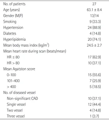
Table 1. Patients Characteristics
No. of patients 27
Age (years) 63.1 ± 8.4
Gender (M/F) 13/14
Smoking 9 (33.3)
Hypertension 24 (88.9)
Diabetes 4 (14.8)
Hyperlipidemia 20 (74.1)
Mean body mass index (kg/m3) 24.5 ± 2.7 Mean heart rate during scan (beats/mean)
HR ≤ 80 17 (62.9)
HR > 80 10 (37.1)
Mean Agatston score
0-100 15 (55.6)
101-400 7 (25.9)
> 400 5 (18.5)
No. of diseased vessel
Non-significant CAD 10 (37.1)
Single vessel 12 (44.4)
Two vessel 4 (14.8)
Three vessel 1 (3.7)
Data in parentheses are percentages.
Note.-CAD = coronary artery disease, HR = heart rate
Fig. 1. This graph shows the mean difference in the stenotic coronary artery diameter between the systole and end-diastole according to heart rate. There was no statistically significant correlation between the change in diameter of coronary artery stenosis and heart rate (r = -0.198, p = 0.760). However, the change in diameter of coronary ar- tery stenosis and Agatston score were correlated (r = 0.942, p = 0.004).
Note.-HR = heart rate 0
50 100 150 200 250 300 350 400 450
5% 10% 11.3% 12.9% 13% 20%
Change in diameter of coronary artery stenosis HRAgatston score
A
A
A
B
B
B
C
C
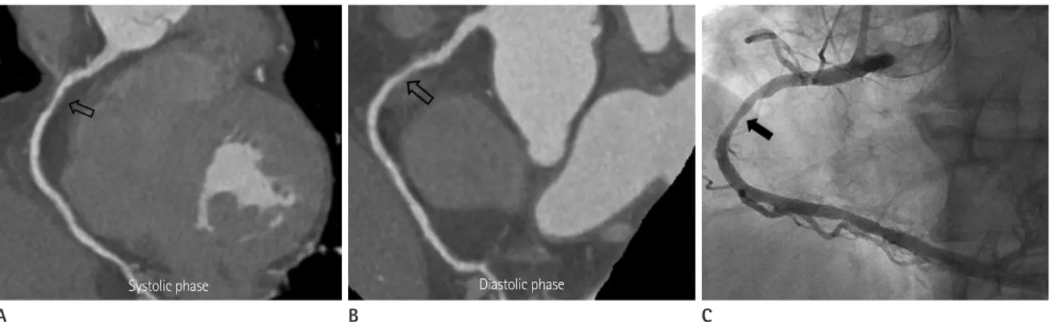
C Fig. 2. A 65-year-old man diagnosed with unstable angina (heart rate of 62 bpm and Agatston score of 40).
A, B. On coronary CT angiography, there is diffuse, moderate stenosis with a plaque in the diastolic phase and diffuse, severe stenosis with a plaque at the middle RCA in the systolic phase. The difference in stenotic coronary vessel diameter between the systolic and diastolic phase was 5%.
C. Invasive coronary angiography shows diffuse, moderate stenosis (50%) at the middle RCA.
Note.-RCA = right coronary artery
Fig. 3. A 69-year-old man who had chest pain (heart rate of 65 bpm and Agatston score of 345).
A, B. On coronary CT angiography, there is diffuse, severe stenosis with mixed plaque at the proximal LAD in the diastolic phase and had tight stenosis in the systolic phase. The difference in the stenotic coronary vessel diameter between systolic and diastolic phase was 15%.
C. Invasive coronary angiography shows diffuse, moderate stenosis (70-80%) at the proximal LAD.
Note.-LAD = left anterior descending artery
Fig. 4. A 76-year-old woman diagnosed with stable angina (heart rate of 69 bpm and Agatston score of 2429.7).
A, B. On coronary CT angiography, there is diffuse, moderate to severe stenosis with calcified plaque at the proximal LCX in the diastolic phase and had severe to tight stenosis in the systolic phase. The difference in the stenotic coronary vessel diameter between systolic and diastolic phase was 52%.
C. Invasive coronary angiography shows diffuse, severe stenosis (70%) at the proximal LCX.
Note.-LCX = left circumflex artery
Systolic phase Diastolic phase
Systolic phase Diastolic phase
CCTA is typically performed in mid to late diastole (6, 11). Be- cause the diastolic phase is relatively long in patients with low heart rates, we usually select the diastolic phase for image re- construction in such cases. However, the diastolic phase dra- matically shortens with increasing heart rates, and even ceases to exist at heart rates > 80 bpm. In contrast to the duration of diastole, the duration of systole is less affected by changes in heart rate (11), thus explaining why image quality in systolic re- constructions did not deteriorate as much with increasing heart rate as observed in diastolic reconstructions. The transition of the optimal reconstruction interval from the diastole to systole in this study was around 80 bpm, a finding that is supported by an early report by Johnson et al. (1) on DSCT of the coronary arteries. In our study, the diagnostic accuracy of the stenotic coronary artery was always lower in the systolic phase than the diastolic phase. Hence, although we could obtain the best im- age of the stenotic coronary artery in the systolic phase, this might not provide the best diagnostic accuracy.
Generally, stenosis less than 50% are revascularized only in the presence of clinical symptoms, whereas stenosis greater than 70% are typically treated, even in the absence of symp- toms (12). To understand the change in stenotic vessel diame- ter between the systolic and diastolic phase on CCTA, it is sometimes helpful to determine the severity of coronary artery stenosis based on ICA. ICA is the standard for the analysis of the severity of coronary artery stenosis in the diastolic phase. In clinical practice, when we obtain a best image in the systolic phase, we should compare the severity of coronary artery steno- sis in the systolic phase on CCTA with the diastolic phase on ICA. In our study, the mean difference in stenotic coronary ves- sel diameter between systolic and diastolic phase was on average 12%, although this varied according to Agatston score. Herzog et al. (12) reported that marked calcifications did not necessarily compromise image quality but were causes of overestimation of moderate stenosis in the diastolic phase, hence the specificity decreased. Consequently, because stenotic coronary vessels with marked calcification in the systolic phase could be overestimat- ed based on ICA, radiologists and cardiologists should be cau- tious when diagnosing significant stenosis in such vessels.
A limitation of our study was the small number of patients (n
= 27) included who had stenotic coronary vessels. Accordingly, a further study with a large number of patients is needed. In nary artery stenosis accurately and which significantly reduce
the sensitivity and specificity of CCTA using DSCT (4, 10).
Meng et al. (10) reported that on a per-artery basis of CCTA for coronary artery stenosis in diastolic phase, a specificity of 99%, PPV of 96%, and accuracy of 98% was achieved in the low cal- cium group, and reduced to 79%, 69%, and 85%, respectively in the high calcium group. In our study, we found that the mean difference in the stenotic coronary artery diameter between systole and end-diastole was 8.3% for an Agatston score < 100, but increased approximately three-fold when the mean Ag- atston score was > 400. According to these results, we expect that the diagnostic accuracy of coronary artery stenosis could be possible with the best image in the systolic phase if the Ag- atston score is less than 100, because the mean difference in the stenotic coronary artery diameter between end-diastole and systole is low. However, if the Agatston score is > 400, on the basis of our study, we suggest that coronary artery stenosis in the systolic phase could be overestimated compared with the diastolic phase.
Many studies have reported the technical capacity of DSCT to provide diagnostic quality images of coronary arteries in pa- tients regardless of heart rate (1-4). Image reconstruction in Fig. 5. This graph shows the correlation between the observers of ICA and DSCT for the evaluation of coronary artery stenosis during the di- astolic phase.
Note.-DSCT = dual source computed tomography, ICA = invasive coronary angiography
0 20 40 60 80 100
0 20 40 60 80 100
DSCT r = 0.805 (p < 0.001)
ICA
and calcium score in a clinical perspective. Acta Radiol 2010;51:727-740
5. Weustink AC, Neefjes LA, Kyrzopoulos S, van Straten M, Neoh Eu R, Meijboom WB, et al. Impact of heart rate fre- quency and variability on radiation exposure, image qual- ity, and diagnostic performance in dual-source spiral CT coronary angiography. Radiology 2009;253:672-680 6. Leschka S, Husmann L, Desbiolles LM, Gaemperli O, Schepis
T, Koepfli P, et al. Optimal image reconstruction intervals for non-invasive coronary angiography with 64-slice CT.
Eur Radiol 2006;16:1964-1972
7. Seifarth H, Wienbeck S, Püsken M, Juergens KU, Maintz D, Vahlhaus C, et al. Optimal systolic and diastolic recon- struction windows for coronary CT angiography using du- al-source CT. AJR Am J Roentgenol 2007;189:1317-1323 8. Matt D, Scheffel H, Leschka S, Flohr TG, Marincek B,
Kaufmann PA, et al. Dual-source CT coronary angiogra- phy: image quality, mean heart rate, and heart rate vari- ability. AJR Am J Roentgenol 2007;189:567-573
9. Austen WG, Edwards JE, Frye RL, Gensini GG, Gott VL, Griffith LS, et al. A reporting system on patients evaluated for coronary artery disease. Report of the Ad Hoc Com- mittee for Grading of Coronary Artery Disease, Council on Cardiovascular Surgery, American Heart Association. Cir- culation 1975;51:5-40
10. Meng L, Cui L, Cheng Y, Wu X, Tang Y, Wang Y, et al. Effect of heart rate and coronary calcification on the diagnostic accuracy of the dual-source CT coronary angiography in patients with suspected coronary artery disease. Korean J Radiol 2009;10:347-354
11. Chung CS, Karamanoglu M, Kovács SJ. Duration of diastole and its phases as a function of heart rate during supine bicycle exercise. Am J Physiol Heart Circ Physiol 2004;287:
H2003-H2008
12. Herzog C, Zwerner PL, Doll JR, Nielsen CD, Nguyen SA, Savi- no G, et al. Significant coronary artery stenosis: comparison on per-patient and per-vessel or per-segment basis at 64-section CT angiography. Radiology 2007;244:112-120 addition, although we measured the stenotic coronary vessel
diameter in the systolic and diastolic phases on CCTA, we mea- sured the stenotic coronary vessel diameter only in the end-dia- stolic phase on ICA. We recommend an additional study in which the change in stenotic coronary vessel diameter in the systolic and diastolic phase on CCTA and ICA will be mea- sured simultaneously to obtain the standard reference for sys- tolic phase on CCTA. Patients with irregular heart rates were excluded, especially on atrial fibrillation or premature ventricu- lar contraction because image quality is related to heart rate variability.
In conclusion, the calcium burden in stenotic coronary ves- sels might markedly affect the change in vessel diameter ac- cording to the cardiac cycle. In particular, stenotic coronary vessels with a high calcium burden in the systolic phase could lead to the overestimation of the severity of coronary artery ste- nosis based on ICA. Therefore, a radiologist should be cautious when interpreting the severity of coronary artery stenosis on CCTA when they obtain the best image of the coronary artery in the systolic phase only along with a high Agatston score.
REFERENCES
1. Johnson TR, Nikolaou K, Wintersperger BJ, Leber AW, von Ziegler F, Rist C, et al. Dual-source CT cardiac imaging: ini- tial experience. Eur Radiol 2006;16:1409-1415
2. Scheffel H, Alkadhi H, Plass A, Vachenauer R, Desbiolles L, Gaemperli O, et al. Accuracy of dual-source CT coronary angiography: first experience in a high pre-test probability population without heart rate control. Eur Radiol 2006;16:
2739-2747
3. Achenbach S, Ropers D, Kuettner A, Flohr T, Ohnesorge B, Bruder H, et al. Contrast-enhanced coronary artery visual- ization by dual-source computed tomography--initial ex- perience. Eur J Radiol 2006;57:331-335
4. Zhang LJ, Wu SY, Wang J, Lu Y, Zhang ZL, Jiang SS, et al.
Diagnostic accuracy of dual-source CT coronary angiogra- phy: the effect of average heart rate, heart rate variability,
심장박동수 또는 석회수치에 따른 수축기와 이완기 말기에서 협착된 관상동맥의 직경 변화 측정1
이현경
1· 진공용
1· 박재근
1· 한영민
1· 이영선
1· 권근상
2· 김성수
3목적: CT 관상동맥조영술을 이용하여 좁아진 관상동맥의 수축기와 이완기 말기에서의 직경 변화를 측정하고 심장박동 수 또는 석회수치와의 관련성에 대해서 알아 보았다.
대상과 방법: 관상동맥협착이 있는 환자 중 CT 관상동맥조영술과 침습적 관상동맥조영술을 모두 시행한 27명의 환자 를 대상으로 하였다. CT 관상동맥조영술에서 협착된 동맥의 수축기와 이완 말기에서의 관상동맥 직경의 백분율 변화를 측정한 후 심장박동수 또는 Agatston 점수와의 관련성을 단순선형회귀분석을 이용하여 조사하였다.
결과: 수축기와 이완 말기에서의 좁아진 관상동맥 직경의 변화는 분당 심장박동수 50~59회에서 12.9%, 60~69회에서 11.3%, 70~79회에서 10%, 80~89회에서 20%, 90~99회에서 13%, 100~109회에서 6.7%의 차이를 보였다. 그러나 심장박동수와 관상동맥 직경 변화의 차이는 통계적으로 유의하지 않았다(p = 0.760). Agatston 점수에 따른 관상동맥 직경 변화의 차이는 100 미만에서 8.3%, 100~400에서 12.4%, 400 이상에서 20%를 보였고, Agatston 점수와 관상동 맥 직경 변화의 차이는 통계적으로 유의하였다(p = 0.004).
결론: 수축기와 이완 말기에서 좁아진 관상동맥 직경 변화는 Agatston 점수에 영향을 받는다.
전북대학교 의학전문대학원 전북대학교병원 임상의학연구소 심혈관연구소 1영상의학과학교실, 2예방의학과학교실,
3충남대학교 의학전문대학원 충남대학교병원 영상의학과학교실