■Received: 2015. 11. 5. ■ Revised: 2015. 12. 3.
■Accepted: 2016. 1. 25.
■Address reprint requests to Jeong Hun Kim, MD, PhD
Department of Ophthalmology, Seoul National University Hospital, #101 Daehak-ro, Jongno-gu, Seoul 03080, Korea Tel: 82-2-2072-2438, Fax; 82-2-741-3187, E-mail: [email protected]
* This study was presented as a narration at the 113th Annual Meeting of the Korean Ophthalmological Society 2015.
* This research was supported by the Bio & Medical Technology Development Program of the NRF funded by the Korean government, MSIP(2015M3A9E6028949) and the Pioneer Research Center Program through the National Research Foundation of Korea funded by the Ministry of Science, ICT & Future Planning (2012-0009544).
ISSN 0378-6471 (Print)⋅ISSN 2092-9374 (Online)
http://dx.doi.org/10.3341/jkos.2016.57.3.499
Original Article
생쥐의 망막혈관 발생 과정에서 혈관내피세포 및 혈관주위세포의 네스틴 발현 분석
Expression of Nestin on Endothelial Cells and Pericytes During Retinal Vascular Development in Mouse
김진수1⋅박성욱2,3⋅황인영4⋅김용우1⋅김진형2⋅김정훈1,2,3
Jin Soo Kim, MD1, Sung Wook Park, MD2,3, In Young Hwang, MD4, Yong Woo Kim, MD1, Jin Hyoung Kim, PhD2, Jeong Hun Kim, MD, PhD1,2,3
서울대학교 의과대학 안과학교실1, 서울대학교병원 의생명연구원 망막혈관실험실2, 서울대학교 의과대학 의과학과3, 서울대학교 의과대학 의학과4
Department of Ophthalmology, Seoul National University College of Medicine1, Seoul, Korea
Fight against Angiogenesis-Related Blindness Laboratory, Biomedical Research Institute, Seoul National University Hospital2, Seoul, Korea Department of Biomedical Sciences, Seoul National University College of Medicine3, Seoul, Korea
Department of Medicine, Seoul National University College of Medicine4, Seoul, Korea
Purpose: Nestin, a marker of neural stem cells, is expressed in Müller cells during retinal development. However, the role of nes- tin in retinal vascular development is not well established. Thus, we investigated the expression of nestin in developmental mouse retina and identified which retinal cells are related to the expression of nestin during the retinal vascular development.
Methods: Eyes were enucleated from C57BL/6 mice on postnatal day (P) 4, P8, P12, P16 and P26. Immunofluorescence was used to evaluate nestin expression in relation to endothelial cells (isolectin B4), pericytes (neural/glial antigen 2) and astrocytes (glial fibrillary acidic protein).
Results: Nestin was strongly expressed from the ganglion cell layer to retinoblast layer at P4. At P8, P12 and P16, the expression of nestin was observed from the upper border of the ganglion cell layer, and vertically penetrating to outer nuclear layer. At P26, the expression of nestin was decreased and confined to the ganglion cell layer and inner nuclear layer. Interestingly, there was strong vascular shape expression of nestin at all stages. The superficial, deep and intermediate vascular plexus was completely merged with nestin expression at P4, P8, P12 and P16. In addition, the nestin expression merged with pericytes but not with astrocytes.
Conclusions: Nestin was expressed in endothelial cells and pericytes during retinal vascular development in the retina. These results suggest that nestin could play an important role in developmental angiogenesis via interplay with endothelial cells and pericytes.
J Korean Ophthalmol Soc 2016;57(3):499-506
Keywords: Endothelial cell, Mouse retina, Nestin, Pericyte, Retinal vascular development
ⓒ2016 The Korean Ophthalmological Society
This is an Open Access article distributed under the terms of the Creative Commons Attribution Non-Commercial License (http://creativecommons.org/licenses/by-nc/3.0/) which permits unrestricted non-commercial use, distribution, and reproduction in any medium, provided the original work is properly cited.
혈관신생(angiogenesis)은 기존의 혈관으로부터 새로운 혈관이 만들어지는 과정으로, 정상적인 기관의 발달뿐만 아니라 상처 치유과정이나 다양한 신생혈관성 질환, 악성 종양의 신생혈관 형성에도 관여한다.1 안구 내 신생혈관으 로 인해 발생하는 나이관련황반변성, 당뇨망막병증, 그리고 미숙아망막병증은 가장 중요한 실명의 원인 질환으로 꼽히 고 있으며, 특히 선진국에서 나이관련황반변성과 당뇨망막 병증은 각각 실명한 노인 환자의 20.9%, 2.7%에서 원인이 되는 질환으로 알려져 있다.2 우리나라에서 시행된 역학 조 사에 따르면 40세 이상 인구에서 초기 및 후기 나이관련황 반변성은 각각 5.1% 및 0.3%, 당뇨를 가진 40세 이상 인구 에서 당뇨망막병증은 13.4%의 유병률을 가지는 것으로 조 사된 바 있으며, 최근 들어 유병률이 더 증가하고 있는 만 큼 임상적인 중요성도 함께 커지고 있다고 할 수 있다.3-5 따라서 정상적인 발달과정에서의 혈관신생을 이해하는 것 은 신생혈관성 질환의 발생과정을 이해하고 궁극적으로는 적절한 치료법을 찾아 이로 인한 실명의 위험을 낮추는 데 매우 중요하다고 할 수 있겠다.
망막혈관의 발생과정은 크게 1차 혈관형성(primary vas- culogenesis)과 2차 혈관신생으로 나뉜다. 혈관형성은 혈구 모세포(angioblast)로부터 혈관이 새로 생성되는 과정을 뜻 하며, 얕은 혈관 얼기(superficial vascular plexus)는 1차 혈 관형성을 통해 시신경유두에서부터 망막의 주변부를 향해 방사형으로 자라나가는 혈관들로 만들어진다고 알려져 있 다. 혈관신생은 이미 존재하는 혈관으로부터 새로운 혈관 이 생겨나는 과정을 뜻하며, 중간 및 깊은 혈관 얼기 (intermediate and deep vascular plexus)는 얕은 혈관 얼기 에서 혈관신생을 통해 수직으로 자라 들어가는 혈관들로부 터 생겨나는 것으로 이해된다.6 이러한 망막혈관의 발생 과 정은 포유류의 망막혈관 발생과정에서 비교적 유사한 형태 로 일어나고 있으며, 적어도 생쥐에서는 시간적, 그리고 공 간적으로 일정한 과정을 취하고 있다.7,8 이러한 사실은 망막 혈관의 발생과정에서 특정한 신생혈관 유도 기전이 작용하 고 있다는 것을 강하게 암시하며, 혈관내피세포(vascular en- dothelial cell), 혈관주위세포(pericyte), 성상교세포(astrocyte) 등의 세포가 긴밀하게 관련되어 있는 것으로 생각되고 있다.9
제6형 중간섬유로 분류되는 네스틴(nestin)은 중추신경계 에서 신경계 줄기세포의 표지자로 알려져 있으며,10,11 신경 계 외에 다른 조직의 줄기세포에서도 발현된다.12-14 최근에 는 네스틴이 혈관신생과정의 표지자로서 주목 받고 있는데, 췌장, 황체, 태반 등 다양한 기관뿐만 아니라 다양한 종류 의 암의 혈관신생과정에서 혈관내피세포로부터 네스틴이 발현되는 것으로 알려져 있다.15,16 이러한 사실은 네스틴이 혈관신생과정에서 중요한 역할을 담당하고 있음을 암시하
고 있지만, 망막의 혈관신생과정에서의 네스틴의 역할에 대해서는 명확히 알려진 바가 없다. 본 연구에서는 망막혈 관 발생과정에서의 네스틴의 발현에 대해 알아보았으며, 특히 어떤 종류의 망막세포에서 네스틴을 발현하고 있는지 를 isolectin B4 (IB4), neural/glial antigen 2 (NG2), glial fi- brillary acidic protein (GFAP)에 대한 면역형광염색을 통해 분석하였다.
대상과 방법
대상
외견상 외안부 질환이 없는 C57BL/6 생쥐(Central Lab.
Animal, Korea)를 대상으로 하였으며, 안과 및 시과학 연구 회(Association for Research in Vision and Ophthalmology) 에서 규정하고 있는 동물사용에 대한 지침을 준수하였다. C57BL/6 생쥐는 12시간의 낮/밤 주기와 약 23℃의 상온에 서 돌보았으며, 출생 후 4일, 8일, 12일, 16일 그리고 26일 에 각각 5마리의 C57BL/6 생쥐의 안구를 적출하였다.
조직의 고정 및 표본 제작
생쥐를 희생시킨 후, 안구를 적출하여 4% 파라포르말린 용액에서 4시간 동안 고정시켰다. 고정된 안구를 파라핀 포 매한 후 각막의 중심부에서부터 시신경을 가로지르는 방향 으로 잘라 블록을 제작하였다. 안구 블록을 4 μm 두께의 절편으로 만들어 유리 슬라이드에 표본 제작하였다.
면역형광염색
위에서 만든 조직 절편 슬라이드 중 가능한 시신경 단면 을 포함하는 슬라이드를 골라 실험에 사용하였다. 조직 절편 슬라이드를 60℃ 오븐에서 2시간 동안 반응시켜 파라핀을 녹인 후 자일렌(xylene)에 5분씩 4회, 100%-100%-95%- 80%-70% 에탄올에 순서대로 5분씩 반응시켜서 파라핀을 제 거하고 5분간 흐르는 증류수에 세척하였다. 이후 pH 6.0의 0.01 M 구연산나트륨 완충용액(sodium citrate buffer sol- ution)에 담가 고압멸균기(autoclave)를 이용하여 10분간 120℃로 반응시키고 30분간 4℃에서 냉각시켰다. 슬라이 드를 인산완충식염수(phosphate buffer solution)로 5분간 2 회 세척한 뒤 0.2% Triton X-100 용액으로 상온에서 15분 간 처리하였다. 다시 인산완충식염수로 5분간 5회 세척 후 차단용액(blocking solution)으로 상온에서 1시간 처리하고, 각 슬라이드마다 정해 놓은 1차 항체를 인산완충식염수로 희석시킨 용액에 4℃에서 하룻밤 동안 반응시켰다. 이후 다시 인산완충식염수로 10분간 5회 세척한 후 정해 놓은 2 차 항체를 인산완충식염수로 희석시킨 용액에 상온에서 2
Peripapillary Mid periphery Far periphery
Peripapillary Mid periphery Far periphery
Peripapillary Peripapillary Peripapillary
A
B
C
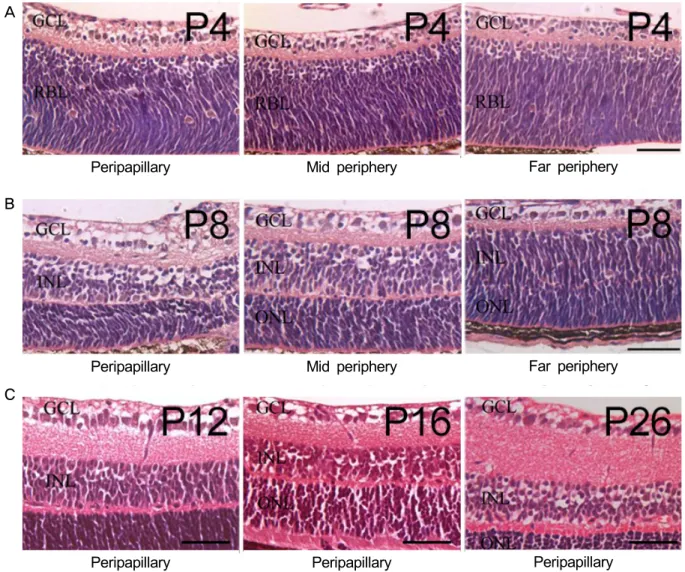
Figure 1. Retinal vascular development in normal mouse retina. On P4, superficial vascular plexus originating from the optic nerve
head extended radically, which reached to the mid-peripheral retina (A) and extended to far periphery on P8 (B). P12 shows the ver- tical sprouting vessels toward the deep vascular plexus, which is secondary angiogenesis (C). Further maturation of vessels, espe- cially intermediate vascular plexuses, occurred during P16 to P26 (C). Scale bar means 50 μm. P = postnatal day; GCL = ganglion cell layer; RBL = retinoblast layer; INL = inner nuclear layer; ONL = outer nuclear layer.시간 동안 반응시켰다. 다시 인산완충식염수로 10분간 2회 세척 후 4’,6-diamidino-2-phenylindole (DAPI, Sigma, St.
Louis, MO, USA)을 이용하여 세포 내 핵의 대조염색을 시 행하였다.
항체
1차 항체로서 생쥐 항-네스틴 IgG1 단일클론항체 (1:1,000, Merk Millipore, Temecula, CA, USA), 항-IB4 항 체(1:200, Alexa Fluor® 488 conjugated, Life Technologies, Camarillo, CA, USA), 토끼 항-NG2 다클론항체(1:1,000, Abcam, Cambridge, MA, USA), 쥐 IgG2 항-GFAP 단일클 론항체(1:1,000, Novex, Life technologies, USA)가 사용되 었다. 2차 항체로서는 알렉사 플루오르® 594 염소 항-생쥐
항체(Alexa Fluor® 594 goat anti-mouse, 1:500, Life tech- nologies), 알렉사 플루오르® 488 당나귀 항-토끼 항체 (Alexa Fluor® 488 donkey anti-rabbit, 1:500, Life tech- nologies), 알렉사 플루오르® 647 염소 항-쥐 항체(Alexa Fluor® 647 goat anti-rat, 1:500, Life technologies)가 사용 되었다.
이미지 분석
면역형광염색을 시행한 조직 절편 슬라이드를 형광 현 미경(Nikon Eclipse 80i, Nikon, Tokyo, Japan)을 이용하여 관찰하였으며, 이미지는 NIS-Elements Microscope Imaging Software ver. 4.00 (Nikon, Tokyo, Japan)을 이용하여 얻었 다.
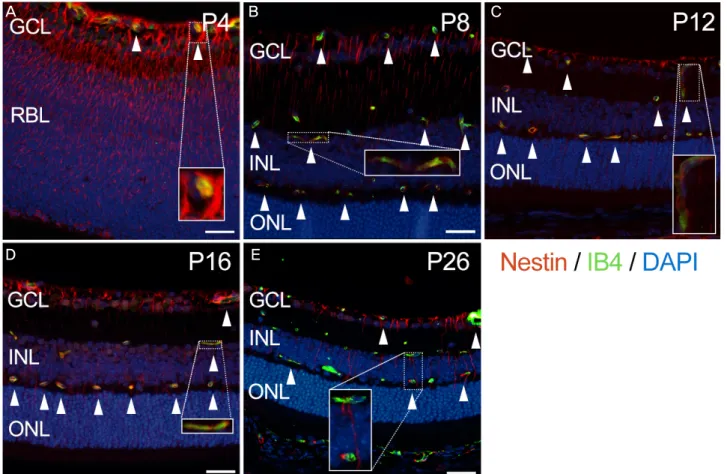
Figure 2. The nestin expression merged with the expression of IB4 in developing mouse retina. At P4, nestin (red) expression is ob-
served as a fibrous bundle penetrating the retina vertically, from the upper border of the ganglion cell layer to outer nuclear layer (A). This pattern of expression decreased as the mouse grew and finally confined to GCL and INL at P26 (B-E). The other observed pattern of nestin expression is an intracytoplasmic aggregation showing vascular shape at all stages. From P4 to P16, this pattern of nestin expression is completely merged with expression of IB4 (green) at the location of superficial, intermediate and deep vascular plexuses (A-D: arrowheads). At P26, intracytoplasmic expression of nestin is partially merged with expression of IB4 (E: arrow- heads). Scale bar means 20 μm. IB4 = isolectin B4; DAPI = 4’,6-diamidino-2-phenylindole; GCL = ganglion cell layer; RBL = retinoblast layer; INL = inner nuclear layer; ONL = outer nuclear layer; P = postnatal day.결 과
정상 생쥐 망막혈관 발생
생후 4일, 시신경유두에서부터 뻗어나가기 시작한 얕은 혈관 얼기는 중간 주변부 망막까지 뻗어 있는 양상이었으 며(Fig. 1A), 생후 8일에는 먼 주변부 망막에 도달하는 모 습이었다(Fig. 1B). 생후 12일에는 얕은 혈관 얼기에서 분 지하여 망막 깊은 쪽으로 자라 들어가는 혈관이 관찰되었 으며, 생후 16일 및 26일에는 중간 혈관 얼기 및 깊은 혈관 얼기가 형성되어 있는 것이 관찰되었다(Fig. 1C). 이상의 생쥐 망막혈관 발생과정을 종합하여 볼 때, 시신경유두에 서 주변부 망막으로 뻗어나가는 방사형의 얕은 혈관 얼기 를 만드는 일차 혈관형성 과정은 생후 8일까지 일어났으며, 이후에는 수직으로 망막의 내과립층까지 뻗어 들어가는 이 차 혈관신생과정이 일어났다.
생쥐의 망막혈관 발생 과정에서 네스틴의 발현 생후 4일, 네스틴은 신경절세포층(ganglion cell layer)부 터 망막모세포층(retinoblast layer)에 걸쳐 강하게 발현되는 모습이었다(Fig. 2A). 생후 8일에는 네스틴의 발현이 전체 적으로 약해져 있는 양상이며, 신경절세포층의 위경계선에 서 시작하여 외과립층(outer nuclear layer)까지 수직으로 뻗 어 있었다(Fig. 2B). 이후 생후 12일, 16일을 거치며 점점 수직으로 뻗어 있는 네스틴의 발현은 줄어드는 양상이었으 며(Fig. 2C, D), 생후 26일에는 네스틴의 발현이 감소되어 신경절세포층과 내과립층(inner nuclear layer)에 국한되어 있었다(Fig. 2E). 또한 모든 망막 발생 단계에서 망막혈관 과 유사한 모양으로 발현되는 네스틴이 관찰되었는데, 세 포질 내에 응집된 형태로 발현되고 있었다.
A B C
D E
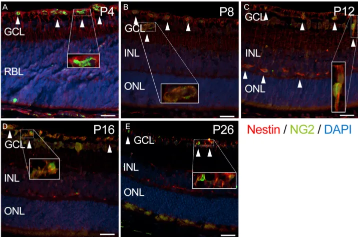
Figure 3. The nestin expression merged with the expression of NG2 in developing mouse retina. NG2 (green) expression is observed
at the level of retinal vascular plexuses and partially merged with expression of nestin (red) from P4 to P26 (A-E: arrowheads). Scale bar means 20 μm. NG2 = neural/glial antigen 2; DAPI = 4’,6-diamidino-2-phenylindole; GCL = ganglion cell layer; RBL = reti- noblast layer; INL = inner nuclear layer; ONL = outer nuclear layer; P = postnatal day.생쥐 망막혈관 발생 과정에서 IB4, NG2, GFAP의 발현과 네스틴 발현과의 관계
IB4의 발현은 생후 4일에는 얕은 혈관 얼기가 위치한 신 경절세포층의 위경계선에 국한되어 있었으며(Fig. 2A), 생 후 8일 이후에는 중간 및 깊은 혈관 얼기에 해당하는 내과 립층의 위경계선 및 아래경계선 바로 바깥쪽에서 관찰되었 다(Fig. 2B-E). 생후 4일부터 생후 16일까지 IB4의 발현은 네스틴의 발현과 완전히 어우러지는 양상이었으며(Fig.
2A-D), 생후 26일에는 네스틴의 발현과 부분적으로 어우러 지는 양상이었다(Fig. 2E).
NG2의 발현은 생후 8일까지 얕은 혈관 얼기가 위치한 신경절세포층의 위경계선에 국한되어 있었다(Fig. 3A, B).
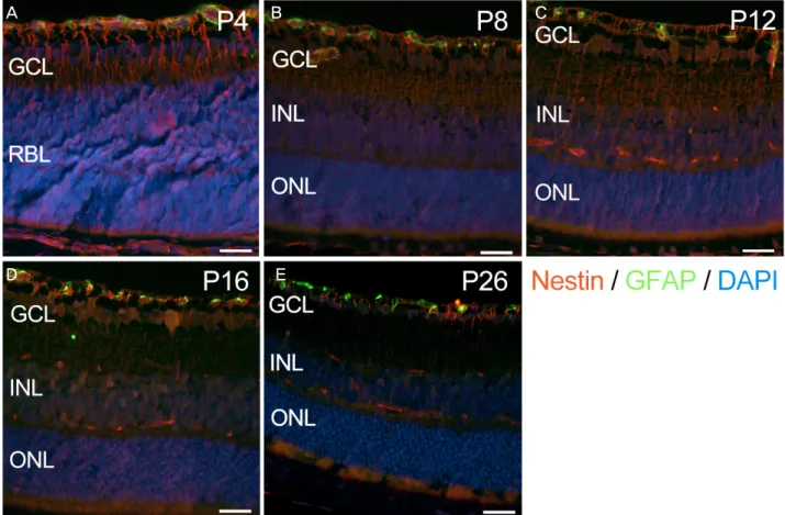
생후 12일째 이후에는 중간 및 깊은 혈관 얼기의 위치에 해 당하는 부위에서 NG2의 발현이 관찰되었으며(Fig. 3C-E), 모든 단계에서 NG2의 발현은 네스틴의 발현과 부분적으로 어우러지는 양상이었다. GFAP의 발현은 생후 4일부터 26 일까지 본 연구에서 관찰한 모든 생쥐의 망막혈관 발달단 계에서 신경절세포층에 국한되어 있는 모습이었으며 네스 틴의 발현과는 어우러지지 않았다(Fig. 4).
고 찰
망막혈관의 정상적인 발달 및 유지는 정상적인 망막 기 능의 유지에 필수적이다. 선진국의 경우, 노인층에서 실명 의 원인이 되는 많은 질환이 비정상적인 혈관의 발생에 기 인한다.2 인구구조가 점점 고령화되어 가는 우리나라 역시 선진국의 이러한 흐름에 따를 것이 예상되므로, 망막혈관 의 정상적인 발생에 대한 이해는 나이관련황반변성, 당뇨 망막병증, 미숙아망막병증 등 안구의 신생혈관성 질환을 이해하고 나아가 새로운 치료법을 찾는데 필수적이라고 할 수 있다.
생쥐의 망막혈관의 발생 과정은 시간적, 공간적으로 일 정한 과정을 취하고 있다는 것이 많은 연구를 통해 알려져 있다.7,8 본 연구에서 생쥐 망막의 얕은 혈관 얼기는 생후 8 일이 되어서야 망막의 주변부까지 뻗어나가는 것이 확인되 었다. 이것은 생쥐의 얕은 혈관 얼기의 형성이 출생 직후 시신경유두에서 시작되어 생후 첫 1주에 걸쳐 완성된다는 Dorrell et al8,17의 보고와 일치하는 결과이다. 또한 생후 8 일부터 얕은 혈관 얼기에서 망막을 수직으로 침투하는 양
A B C
D E
Figure 4. The nestin expression (red) merged with the expression of GFAP (green) in developing mouse retina. Expression of GFAP
is confined to GCL and not merged with expression of nestin at any stage (A-E). Scale bar means 20 μm. GFAP = glial fibrillary acidic protein; DAPI = 4’,6-diamidino-2-phenylindole; GCL = ganglion cell layer; RBL = retinoblast layer; INL = inner nuclear layer; ONL = outer nuclear layer; P = postnatal day.상의 혈관내피세포가 관찰되어 깊은 혈관 얼기의 생성이 시작되었다는 점 역시 Dorrell et al8,17의 보고와 일치하는 결과였다. 따라서 본 연구에서 관찰된 네스틴 발현의 시간 적, 공간적 특성을 기존에 알려진 망막혈관 발생과정과 비 교하여 분석함으로써 네스틴의 기능에 대한 실마리를 얻을 수 있을 것이다.
본 연구에서 망막혈관의 발생과정 중 관찰된 네스틴의 발현은 크게 두 종류로 나눠볼 수 있는데, 첫 번째는 망막 을 수직으로 관통하는 방향으로 뻗어 있는 양상의 다발 형 태의 발현, 두 번째는 세포질 내에서 응집되어 있는 형태의 발현이다. 전자의 경우 비교적 초기인 생후 4일부터 망막의 전층에 걸쳐서 강하게 발현되었으며, 생후 8일에는 신경절 세포층의 위경계선에서 시작하여 외과립층까지 수직으로 뻗어 있는 양상이었다. 이러한 형태의 네스틴의 발현은 시 간이 지나면서 그 세기 및 범위가 점점 줄어들어서 생후 26 일에는 신경절층과 내과립층에 국한되어 약하게 발현되었 다. 이는 네스틴이 생쥐의 망막혈관 발생 과정에서 방사아 교세포(radial glial cell)로부터 새로 생성된 신경세포의 이 동에 관여하며, 시간이 지날수록 발현이 줄어들어 뮬러세
포의 발 끝 부분에 국한된다고 보고한 Lee et al18의 발표와 부분적으로 일치한다.
본 연구에서 생쥐의 망막혈관 발생과정 중 세포질 내에 응집된 형태로 나타나는 네스틴 발현은 생후 26일째를 제 외하면 IB4의 발현과 완전히 겹치는 양상이었으며, NG2의 발현과도 부분적으로 겹쳐지는 양상이었다. IB4와 NG2는 각각 혈관내피세포 및 혈관주위세포의 표지자로 알려져 있 으므로, 네스틴은 이들 세포에서 발현되고 있을 가능성이 있다. 네스틴이 혈관내피세포에서 발현된다는 점은 앞에서 도 언급했듯이 많은 선행연구에서 보고된 사실이나,15,18 혈 관주위세포에서의 네스틴의 발현에 대해서는 잘 알려져 있 지 않다. Wohl et al19은 생쥐의 망막에서 네스틴과 NG2를 함께 발현하는 미세아교세포(microglia)를 보고하였으며, Lee et al18은 전자현미경을 통하여 네스틴을 부분적으로 발 현하고 있는 혈관주위세포를 보고하였다. 특히 Lee et al18 은 전형적으로 네스틴을 전구세포의 표지자의 관점에서 발 달 중인 망막 조직에 접근하여 네스틴 발현 양상을 Bromodeoxyuridine (BrdU)와 함께 분석하여 망막에서 증 식 및 이동하는 신경전구세포의 표지자로 네스틴을 사용할
A B C
D E
수 있음을 제시하였다. 하지만 면역조직화학염색 및 면역 전자현미경을 이용하여 혈관내피세포와 혈관주위세포에서 네스틴의 발현을 관찰하였으나, 단지 네스틴은 전구세포의 표지자의 관점에서 혈관내피세포 및 혈관주위세포의 전구 세포에서 네스틴이 발현된다고 보고하였다. 본 연구에서는 네스틴의 발현을 전구세포의 표지자로서의 관점보다는 망 막혈관 발생에서의 역할에 초점을 맞추어 혈관내피세포 및 혈관주위세포가 생쥐의 망막혈관 발생 과정에서 밀접하게 연관되어 있음을 제시하였다.
제6형 중간섬유인 네스틴은 중추신경계에서 신경계 줄기 세포의 표지자로 처음 알려졌지만,10,11 이후 신경계 외의 다른 조직의 줄기세포에서도 발현되는 것이 보고되고 있으며,12-14 최근에는 다양한 종류의 암에서 신생혈관의 표지자로 주목 받고 있다.15,16 네스틴은 짧은 N-말단과 상대적으로 긴 C- 말단을 가지고 있는데, C-말단은 비멘틴(vimentin), 데스민 (desmin), 인터넥신(internexin)과 같은 다른 중간섬유들과 상호작용하여 이종이합체(heterodimer)를 이룬다.20,21 최근 Liang et al22은 네스틴을 통해 혈관내피세포의 세포골격 재 배치가 일어나는 기전을 제시하였는데, 혈관내피성장인자 (vascular endothelial growth factor, VEGF)의 유도로 혈관 내피세포의 극화(polarization) 및 사상위족(filopodia)의 형 성이 일어난다고 설명하였다. 혈관주위세포 역시 네스틴의 발현과 관계가 있다는 본 연구 결과로 미루어 볼 때 혈관주 위세포의 이동 및 세포골격의 재배치 과정에서도 네스틴이 관여하고 있을 가능성이 있으며, 더 나아가 네스틴이 두 세 포 사이에서 중간자의 역할을 담당하고 있을 가능성도 생 각해 볼 수 있다. 이에 대한 검증 및 조절인자를 밝히는 후 속 연구가 필요할 것으로 생각되며, 네스틴을 통한 혈관내 피세포 및 혈관주위세포의 조절 과정은 잠재적으로 신생혈 관성 질환을 조절하는 약물 개발에 좋은 표적이 될 것으로 기대된다.
결론적으로 생쥐의 망막혈관 발생과정에서의 네스틴의 발현은 크게 두 가지의 형태를 보이고 있었는데, 망막을 수 직으로 관통하는 다발 형태의 발현과 망막혈관과 유사한 형 태를 보이는 세포질 내의 발현을 보였다. 후자에 해당하는 네스틴의 발현은 전체 망막혈관 발달단계에서 혈관내피세 포 및 혈관주위세포와 밀접한 연관이 있었으며, 성상교세포 와는 관련이 없었다. 이러한 결과는 네스틴이 혈관내피세 포와 혈관주위세포 사이에서 중간자 역할을 함으로써 혈관 발생과정에서 중요한 역할을 담당할 수 있음을 시사한다.
REFERENCES
1) Guillemin K, Krasnow MA. The hypoxic response: huffing and HIFing. Cell 1997;89:9-12.
2) Muñoz B, West SK, Rubin GS, et al. Causes of blindness and visual impairment in a population of older Americans: The Salisbury Eye Evaluation Study. Arch Ophthalmol 2000;118:819-25.
3) Yoon KC, Mun GH, Kim SD, et al. Prevalence of eye diseases in South Korea: data from the Korea National Health and Nutrition Examination Survey 2008-2009. Korean J Ophthalmol 2011;25:
421-33.
4) Jee D, Lee WK, Kang S. Prevalence and risk factors for diabetic retinopathy: the Korea National Health and Nutrition Examination Survey 2008-2011. Invest Ophthalmol Vis Sci 2013;54:6827-33.
5) Park SJ, Lee JH, Woo SJ, et al. Age-related macular degeneration:
prevalence and risk factors from Korean National Health and Nutrition Examination Survey, 2008 through 2011. Ophthalmology 2014;121:1756-65.
6) Chan-Ling T, Gock B, Stone J. The effect of oxygen on vaso- formative cell division. Evidence that 'physiological hypoxia' is the stimulus for normal retinal vasculogenesis. Invest Ophthalmol Vis Sci 1995;36:1201-14.
7) Stone J, Itin A, Alon T, et al. Development of retinal vasculature is mediated by hypoxia-induced vascular endothelial growth factor (VEGF) expression by neuroglia. J Neurosci 1995;15(7 Pt 1):
4738-47.
8) Dorrell MI, Aguilar E, Friedlander M. Retinal vascular develop- ment is mediated by endothelial filopodia, a preexisting astrocytic template and specific R-cadherin adhesion. Invest Ophthalmol Vis Sci 2002;43:3500-10.
9) Kim JH, Kim JH, Yu YS, et al. Recruitment of pericytes and as- trocytes is closely related to the formation of tight junction in de- veloping retinal vessels. J Neurosci Res 2009;87:653-9.
10) Lendahl U, Zimmerman LB, McKay RD. CNS stem cells express a new class of intermediate filament protein. Cell 1990;60:585-95.
11) Hockfield S, McKay RD. Identification of major cell classes in the developing mammalian nervous system. J Neurosci 1985;5:3310-28.
12) Terling C, Rass A, Mitsiadis TA, et al. Expression of the inter- mediate filament nestin during rodent tooth development. Int J Dev Biol 1995;39:947-56.
13) Kachinsky AM, Dominov JA, Miller JB. Myogenesis and the inter- mediate filament protein, nestin. Dev Biol 1994;165:216-28.
14) Sejersen T, Lendahl U. Transient expression of the intermediate fil- ament nestin during skeletal muscle development. J Cell Sci 1993;106(Pt 4):1291-300.
15) Suzuki S, Namiki J, Shibata S, et al. The neural stem/progenitor cell marker nestin is expressed in proliferative endothelial cells, but not in mature vasculature. J Histochem Cytochem 2010;58:721-30.
16) Yamahatsu K, Matsuda Y, Ishiwata T, et al. Nestin as a novel ther- apeutic target for pancreatic cancer via tumor angiogenesis. Int J Oncol 2012;40:1345-57.
17) Dorrell MI, Friedlander M. Mechanisms of endothelial cell guid- ance and vascular patterning in the developing mouse retina. Prog Retin Eye Res 2006;25:277-95.
18) Lee JH, Park HS, Shin JM, et al. Nestin expressing progenitor cells during establishment of the neural retina and its vasculature. Anat Cell Biol 2012;45:38-46.
19) Wohl SG, Schmeer CW, Kretz A, et al. Optic nerve lesion increases cell proliferation and nestin expression in the adult mouse eye in vivo. Exp Neurol 2009;219:175-86.
20) Steinert PM, Chou YH, Prahlad V, et al. A high molecular weight intermediate filament-associated protein in BHK-21 cells is nestin,
= 국문초록 =
생쥐의 망막혈관 발생 과정에서 혈관내피세포 및 혈관주위세포의 네스틴 발현 분석
목적: 신경계 줄기세포 표지자인 네스틴은 망막 발생 과정에서 뮬러세포에서 발현되는 것으로 알려져 있다. 하지만 망막혈관 발달에 서 네스틴의 역할은 잘 알려져 있지 않다. 본 연구에서는 생쥐 망막혈관 발생 과정에서 네스틴의 발현에 대해 분석하고, 어떠한 세포 가 네스틴의 발현과 연관되어 있는지를 확인하고자 하였다.
대상과 방법: C57BL/6 생쥐의 안구를 생후 4일, 8일, 12일, 16일, 26일째 적출하였다. 면역화학염색법을 이용하여 망막 발생 과정에서 네스틴의 발현과 혈관내피세포(이소렉틴 B4 염색), 혈관주위세포(신경-아교세포 항원 2 염색), 그리고 성상교세포(아교섬유산성단백 질 염색)의 연관성을 확인하였다.
결과: 네스틴은 생후 4일째 신경절세포층부터 망막모세포층에 걸쳐 강하게 발현되었으며, 생후 8일부터 16일까지는 신경절세포층의 위경계선에서 외과립층까지 수직으로 뻗어 있는 양상이었다. 생후 26일에는 네스틴의 발현이 감소되어 신경절세포층과 내과립층에 국한되어 있었다. 또한 모든 망막 발생 단계에서 망막혈관과 유사한 모양으로 발현되는 네스틴이 관찰되었는데, 특히 생후 4일부터 16일까지는 네스틴의 발현과 망막혈관이 완전히 어우러지는 양상이었다. 또한 네스틴의 발현은 혈관주위세포와 부분적으로 어우러졌 으나 성상교세포와는 어우러지지 않았다.
결론: 생쥐의 망막혈관 발달 과정에서 혈관내피세포와 혈관주위세포에서 발현되는 네스틴은 혈관내피세포와 혈관주위세포 사이에서 중간자 역할을 함으로써 혈관 발생 과정에서 중요한 역할을 할 수 있음을 시사한다.
<대한안과학회지 2016;57(3):499-506>
a type VI intermediate filament protein. Limited co-assembly in vi- tro to form heteropolymers with type III vimentin and type IV al- pha-internexin. J Biol Chem 1999;274:9881-90.
21) Marvin MJ, Dahlstrand J, Lendahl U, McKay RD. A rod end dele- tion in the intermediate filament protein nestin alters its subcellular localization in neuroepithelial cells of transgenic mice. J Cell Sci
1998;111(Pt 14):1951-61.
22) Liang ZW, Wang Z, Chen H, et al. Nestin-mediated cytoskeletal re- modeling in endothelial cells: novel mechanistic insight into VEGF-induced cell migration in angiogenesis. Am J Physiol Cell Physiol 2015;308:C349-58.