서 론
최종당화산물 (advanced glycation end-products, AGEs) 은 당뇨를 가속화하고, 당뇨의 합병증 예를 들어 망막병 증 (retinopathy)과 같은 합병증과도 연관이 큰 거대 단 백질이다 (Stitt and Curtis 2005). 많은 혼합물 중 pensosi-
dine과 methylglyoxal 유도체인 N-carboxymethy-lysine (CML), N-carboxyethyl-lysine (CEL)은 잘 알려지고 연구
되어진 AGEs 중 하나이다 (Moore et al. 2003). 최근 연구 에서 AGEs와 AGEs의 수용체인 RAGE는 당뇨병성 혈관 합병증의 병인으로서 큰 역할을 하는 것으로 알려졌 다 (Wautier and Guillausseau 2001; Goldin et al. 2006).
AGEs는 bovine serum albumin (BSA)과 콜라겐과 같
은 단백질을 변형하고 또한 혈관형성에 관여하는 혈관 내피 성장인자 (vascular endothelial growth factor, VEGF) 의 유도를 통하여 미세혈관 내피세포의 성장을 자극한 다 (Tanaka et al. 2000). AGEs는 당뇨병 환자의 망막 혈 관에서 높게 나타나고 이것은 혈액망막장벽 (blood-retinal barrier) 기능 장애인 망막병증의 증세와 연관이
깊다 (Stitt et al. 2000). 그러나 현재 AGEs로 유도된 망막 혈관 누출의 기작은 아직까지 알려진 바가 없다.현재까지 개발되어 검토되고 있는 항혈관 형성 (anti-
angiogenic) 약물들은 모두가 유기 화합물들이며 이들은
─
─ 8 ─ ─
소망막내피세포에서 금 나노입자의 최종당화산물에 의한 세포 이동 및 침윤성 억제 효과
채 수 철*
전남대학교 생물학과
Gold Nanoparticles Inhibit AGEs Induced Migration and Invasion in Bovine Retinal Endothelial Cells
Soo-Chul Chae*
Department of Biology, Chonnam National University
Abstract -
- This study aimed the role of gold nanoparticles (AuNP) in advanced glycation end-pro-ducts (AGEs) induced migration and invasion in bovine retinal endothelial cells (BRECs). BRECs were isolated from the retina. Cell viability was confirmed by the MTT assay. In vitro wound migra- tion assay was performed to investigate the migration of BRECs. In vitro tube formation was mea- sured by on-gel system. Apoptosis induced by AuNP was confirmed by caspase-3 assay. AGE- bovine serum albumin (BSA) demonstrated increase of cell migration and proliferation in BRECs.
In addition, AuNP regardless of the existence of AGE-BSA suppressed proliferation, migration, and angiogenesis. AuNP suppressed AGE-BSA induced migration and invasion, and induced apop- tosis through caspase-3. As a results, AuNP have a potential anti-angiogenic effect for AGE-induced angiogenesis in vitro and offer possibility for the treatment of diabetic retinopathy.
Key words : AGEs, AuNP, BRECs
* Corresponding author: Soo-Chul Chae, Tel. 062-530-3390,
Fax. 062-530-0304, E-mail. [email protected]
대부분 위 장관 천공 같은 심각한 독성을 가진 물질들 이다. 최근 천연 무기물에서 항혈관 형성 약물을 개발하 려는 시도에서 금 나노입자 (gold nanoparticles, AuNP)가 주목을 받고 있다 (Grabar et al. 1995; Cho et al. 2005; Li
et al. 2006). AuNP는 생체적합성을 가지며, 표면 플라스
몬 공명 흡수대에 의한 특성분석과 합성이 쉬운 점 등 에서 가능성이 큰 물질이다. 특히 금은 약한 산성으로서 티올, 아민과 같은 약한 염기와 강하게 결합하며, 시스테 인과 라이신 잔기를 가진 단백질들이 AuNP와 결합하여 그들의 생물학적 기능이 변하게 된다 (Resham and Pri-byabrata 2008).
본 연구에서는 소망막내피세포 (bovine retinal endothe-
lial cells, BRECs)에서 AGE-BSA로 유도된 세포의 이동
및 침윤과 같은 신생혈관 형성 기작에 AuNP의 영향을 확인하고자 하였다.재료 및 방법
1.
세포의 분리BRECs의 분리는 Frye and Patrick (2002)의 방법에 따
라 수행하였다. 소 안구를 광주의 삼호축산 도살장에서 구입하여 냉동과 무균상태로 실험실로 가져왔다. 그리고 안구를 멸균수로 2회 세척한 후 2.5μg mL-1의 ampho-tericin-B (Sigma, USA)와 100 U mL
-1의 penicillin-G(Sigma, USA), 100
μg mL-1의 streptomycin (Calbiochem,Germany)이 들어있는 1%의 항생제로 세척한 후 모양체
근 뒤로 절개를 하였다. 안구의 앞쪽 절반과 수정체와 유리체를 제거하고 망막은 망막색소상피층 (retinal pig-ment epithelial, RPE)으로부터 조심스럽게 적출하였다.
멸균된 100 mm dish로 옮긴 후 남아있는 RPE층을 부드 러운 약수저로 제거하였다. 그런 다음 PBS으로 두 번 세척하고, 망막 조직은 해부용 가위를 이용하여 1 mm 두 께로 잘게 잘랐다. 이 조직을 15 mL 튜브로 옮기고 300
rpm에서 10분간 원심분리를 하였다. 조직은 penicillin-G
와 streptomycin이 들어있는 MEM (Sigma, USA) 배지로 두 번 세척을 하였다.2.
세포 배양미세혈관이 있는 조직을 최종적으로 Isvove’s modified
dulbeccos’s medium (IMDM: Sigma, USA) 배지에서 배양
하는데 이 배지에는 추가적으로 fetal bovine serum (FBS:Gibco, USA)과 150
μg mL-1의 heparin (Sigma, USA), 10 μg mL-1의 ascorbic acid, 100 U mL-1의 penicillin-G, 100μg mL-1의 streptomycin, 2.5μg mL-1의 amphotericin-B 를 추가하였다. 그런 다음 37�
C, 5% CO
2배양기에서 배 양하였다. 24시간 후에 배지를 제거하고 혈관을 떼어낸 다음 세포 부유물과 죽은 세포를 MEM 배지로 제거한 다음 매 3일마다 IMDM 배지로 교환해 주었다.3.
세포의 생존율 측정MTT assay는 Mosmann (1983) 방법에 따라 시행하였
다. BRECs를 2×10³cells mL
-1세포 수로 계수한 다음96 well plate (Nunc, Denmark)에 200
μL씩 분주하여 24 시간 동안 배양하고 0.5% serum이 들어있는 IMDM 배 양액으로 교환하고 24시간 추가 배양하였다. 배양액을200
μg mL-1의 MTT (sigma, USA)가 포함된 배양액으로 교환하고 3시간 반응시켰다. 3시간 후 배양액을 버리고dimethylsulfoxide (Sigma, USA)를 200
μL well-1씩을 넣 어 5분간 실온 방치하여 푸른색 결정인 formazan을 용 해시킨 후 분광광도계 (microplate spectorphotometer: Bio-Rad, USA)로 570 nm에서 흡광도를 측정하여 대조군과
비교하였다.4.
세포의 이동성 측정Plate 상에서 BRECs의 이동성을 알아보기 위해 plate
에 scraper를 이용하여 배양한 세포의 일부분을 제거하 였다 (Dona et al. 2004). PBS로 2회 세척 후 시간별로BRECs의 이동을 보는 cell migration assay를 시행하였
다. BRECs의 이동성은 위상차현미경으로 관찰하였고, 상처 가장자리에서 세포 단일층의 증식을 막는 5-fluro-uracil (Sigma, USA)의 존재 하에 시행되었다.
5.
세포의 맥관 형성24 well plate에 10 mg mL
-1의 matrigel을 코팅시켜 30 분간 실온에서 배양하였다. 2 mL의 BRECs는 matrigel이 코팅된 well에 첨가한 후 37�C에서 24시간 배양하였다 (Moon and Kim 2000). 이후 세포를 PBS로 3회 세척 후 70% 에탄올로 4� C에서 30분 이상 고정하였다. Tubular length는 Axiovision FRET, Rel 4.6 software (Zeiss, Ger- many)를 이용하여 측정하였다.
6. Caspase-3
활성 측정BRECs를 lysis buffer로 얼음에서 10분간 두었다가
12,000 g로 10분간 4� C에서 원심분리 시켰다. 상층액을
취해 BCA protein assay kit (Pierce, USA)로 단백질 농도 를 측정하여 80μg의 단백질을 96 well plate에 넣고 100μM의 AC-DEVD-pNA (caspase-3 substrate)를 첨가하여
37� C에서 2시간 동안 반응시켰다 (Nicholson 1995). 그
후 방출되는 형광을 형광광도계 (Perkin Elmer, USA)를 이용하여 405 nm에서 측정하였다.7.
통계 분석실험 결과의 유의성 검증은 Student’s t test 방법에 따 라 실시하였으며, p⁄0.05 수준에서 유의성을 검증하였 다. 모든 실험은 3회 이상 실시한 실험 결과를 토대로 분석하였다.
결 과
1.
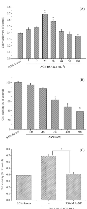
세포의 생존율BRECs에서 AGE-BSA의 신생혈관 형성 효과를 확인
하기 위해 AGE-BSA 처리 전 무혈청 배지에서 세포 생 존율을 측정하였다. BRECs가 무혈청 배지에서 자라게 했을 경우 24시간 내에 그 수가 감소하였다. BRECs를 여러 농도 (5, 10, 20, 30, 40, 50, 100μg mL-1)의 AGE-BSA
에 노출시킨 결과, AGE-BSA는 BRECs를 농도 의존적 으로 세포의 증식을 일으켰다. 세포의 증식은 20μg mL-1 의 농도에서 최고값을 나타냈다. 또한 고농도의 AGE-BSA의 경우 세포의 증식이 억제됨을 알 수 있었다 (Fig.
1A). Fig. 1B는 BRECs에서 AuNP의 효과를 알아보기
위해 MTT assay를 실시하였다. BRECs는 24시간 동안 여러 농도 (100, 200, 300, 400, 500 nM)로 AuNP를 처리 한 결과, 300, 400, 500 nM의 AuNP 농도에서 BRECs의 성장을 유의적으로(p⁄0.05) 억제하였다. Fig. 1C는 AuNP 의 항혈관 형성능을 측정하기 위해, AGE-BSA로 유도된BRECs
증식을 MTT assay를 실시하였다. AGE-BSA와300 nM의 AuNP를 동시에 처리한 결과, AGE-BSA 단독
처리한 경우와 비교하여 세포의 증식이 유의적인 (p⁄0.05) 감소를 보였다.
2.
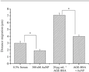
세포의 이동성세포 이동에 있어서 AuNP와 AGE-BSA의 효과를 알 기 위해 wound migration assay를 실시하였다. 본 실험은 다양한 조건에서 세포 이동을 양적으로 표현한 것으로
AGE-BSA에 의한 세포의 이동능력은 AuNP 단독 처리
한 경우와 비교하여 높게 나타났다(Fig. 2). 그러나 AuNP 와 AGE-BSA를 동시에 처리한 경우, AGE-BSA 단독 처 리한 경우와 비교하여 세포의 이동능력이 유의적인 (p⁄0.05) 감소를 보였다. 따라서 위 결과는 300 nM의 AuNP
0.5% Serum
0.5% Serum 0.0 0.1 0.2 0.3 0.4 0.5 0.6 0.7
0.8 (A)
(B)
*
*
Cell viability (% of control) Cell viability (% of control)
100 200 300 400 500
0 20 40 60 80 100
*
*
*
AuNP(nM)
5 10 20 30 40 50 100
AGE-BSA ( μg mL
-1)
Fig. 1. Effect of AuNP or AGE-BSA on cell viability of BRECs.
(A) AGE-BSA induced cell viability in BRECs. AGE-BSA showed the maximal cell proliferation at 20 μg mL
-1com- pared to control. (B) AuNP inhibited BRECs survival in a dose-dependent manner. (C) The addition of AuNP with AGE-BSA in BRECs decreased cell proliferation. Each bar represents the mean±S.D. calculated from three indepen- dent experiments. *Significantly different from control at p⁄0.05.
0.8 0.7 0.6 0.5 0.4 0.3 0.2 0.1 0.0
*
Cell viability (% of control)
0.5% Serum - 300 nM AuNP
20 μg mL
-1AGE-BSA
(C)
처리가 BRECs에서 AGE-BSA에 의한 세포의 이동능력 을 억제함을 알 수 있었다.
3.
세포의 맥관 형성신생혈관 형성에 있어서 AuNP의 역할을 알아보기 위 해 tube formation assay를 실시하였다 (Fig. 3). 그 결과,
AGE-BSA를 처리한 BRECs는 모세혈관망을 형성하였음
을 알 수 있었다. 이와 반대로 AuNP를 단독 처리한 결과 모세혈관망의 배열을 현저히 억제하였다. 또한 AuNP와AGE-BSA를 동시에 처리한 경우, AGE-BSA 단독 처리
한 경우와 비교하여 모세혈관망의 배열이 유의적인 (p⁄0.05)
감소를 보였다. 이러한 결과들을 종합해 볼 때,AuNP가 AGE-BSA에 의해 유발된 BRECs의 이동성 억
제 및 침윤 억제작용을 하고 있는 것으로 사료된다.4. Caspase-3
활성AuNP의 이러한 BRECs 세포 이동성 및 맥관형성 억
제가 어떠한 기전에 의해 일어나는지 확인하기 위해apoptosis의 대표적인 caspase-3 효소 활성을 측정하였
다 (Fig. 4). 그 결과, AGE-BSA의 존재여부와 상관없이AuNP는 caspase-3 활성의 증가를 유도하였다. AuNP에
의해 유도된 apoptosis는 caspase-3 억제제인 Ac-DEVD-CHO를 처리한 결과 효과적으로 억제되었다.
고 찰
본 연구는 BRECs에서 고농도의 AuNP 처리시 AGE-
BSA로 유도된 세포 침윤 및 이동성에 대한 연구와 cas- pase-3을 통한 apoptosis를 밝히고자 하였다.
망막내피세포 배양은 망막내피세포의 기능을 이해함 에 있어 많은 도움을 주었다. 실험실의 조건에서 내피세 포의 배양은 많은 문제점이 있었고 분리 효율이 낮았다
(Su et al. 2003). 혈관 주변 세포의 제거는 다양한 세포 Fig. 2. AuNP inhibits AGE-BSA induced cell migration in BRECs.
The quantitative assessments of cell migration, in which AuNP blocked the AGE-BSA induced the cell migration calculated from the control. Each bar represents the mean
±S.D. calculated from three independent experiments.
*Significantly different from control at p ⁄0.05.
8 7 6 5 4 3 2 1 0
*
*
0.5% Serum 300 nM AuNP 20 μg mL
-1AGE-BSA AGE-BSA
++AuNP
Distance migration (μ m)
Fig. 3. Effect of AuNP inhibits AGE-BSA induced tube formation in BRECs. The quantitative measurement of tube length expressed in mm mm
-2. Each bar represents the mean±S.D.
calculated from three independent experiments. *Signifi- cantly different from control at p⁄0.05.
8 7 6 5 4 3 2 1 0
*
*
0.5% Serum 300 nM AuNP 20 μg mL
-1AGE-BSA AGE-BSA
++AuNP Tube length (mm mm
-2)
Fig. 4. AuNP induces apoptosis in BRECs by caspase-3 activation.
Quantitative measurement of caspase-3 activity for various treatments. Each bar represents the mean±S.D. calculated from three independent experiments.
1.0
0.8
0.6
0.4
0.2
0.0
0.5% Serum
Caspase-3 (
++) control
Caspase-3 inhibitor 300
nM AuNP
20 μg mL
-1
AGE-BSA AGE-BSA
+ +
AuNP
AuNP
+
+
Caspase-3 inhibitor
Caspase-3 activity (μ M pNA min
-1)
부착 방법과 트립신의 처리를 하여 얻었고, 그 결과 주 변세포의 제거를 할 수 있었다. 그리고 여러 과정을 통 해 BRECs를 얻었다 (Frye and Patrick 2002).
안구에서 나노입자는 정상의 조건에서 신생혈관 형성 을 억제한다(Ren et al. 2005). 인체의 방수(aqueous humor) 와 유리체 (vitreous body)에서 나노입자의 농도는 0.5~3 μg mL-1로 유지되지만, 신생혈관 형성과 연관된 안구 질환이 생길 경우 감소된다 (Spranger et al. 2002). 나노입 자의 항혈관형성 활성에 대한 기작은 분명치 않다. 본 연구에서 고농도의 AuNP는 BRECs의 증식을 농도 의 존적으로 억제하였다. 이는 AuNP가 BRECs에 직접 작 용하여 억제한다는 것을 알려준다. 또한 세포독성을 일 으키지 않는 AuNP 농도에서는 BRECs에서 망막의 신생 혈관 형성을 조절하는데 최소한 AGE-BSA의 성장과 이 동 촉진 활성을 억제함으로서 세포 생존과 혈관형성 이 동능력을 억제하였다. 추후 이러한 작용기전연구가 더 진행되어야 할 것으로 사료된다.
AGEs는 당뇨망막증과 같은 당뇨합병증에 있어서 중
요한 역할을 한다. 내피세포, 평활근세포, 수상세포, 단핵 구, 당뇨맥관 구조의 단핵 식세포의 침투에 있어서 AGEs,RAGEs의 많은 연구가 있었다 (Sakata et al. 2000; Yama- gishi et al. 2003). 이전의 연구에서 내피세포에서 AGEs
는 신생혈관 형성을 유도하였고 많은 임상 실험에서 정 상의 환자에 비해 당뇨환자의 혈청 AGEs 수준이 8배나 높게 나타났다 (Stitt et al. 2005). 본 연구에서 AGE-BSA 는 BRECs에서 세포 생존, 이동, 신생혈관 형성의 증가 를 가져왔다. 비록 세포 수의 증가가 20μg mL-1의 농도 까지의 AGE-BSA에서 관찰되었지만, 그 이상에서는 감 소하였다. 고농도에서 관찰된 AGE-BSA의 증식능 감소 는 세포 분열과 apoptosis에 의한 것이라고 사료된다.비록 AuNP의 항신생혈관 형성 활성에 대해 완전히 이해되지는 않았지만 이전의 많은 연구에서 AuNP가
BRECs의 apoptosis를 야기한다고 알려져 있고 (Chen et al. 2006), 다른 표현형의 경우 반대의 효과도 한다는 사
실이 알려져 있다 (Hutching et al. 2002). 많은 내생 혹은 외생의 신생혈관 형성 억제제가 내피세포의 apoptosis를 유도한다. 나노입자는 내피세포의 apoptosis를 야기하는caspase-3을 유도하여 항신생혈관 형성을 일으킨다 (Vol- pert et al. 2002). 앞으로의 연구는 당뇨의 합병증을 치료
하는 새로운 물질을 찾는 것이다.적 요
본 연구는 BRECs에서 AGEs로 유도된 세포의 이동
및 침윤에 있어서 AuNP의 역할에 관한 연구이다. 소 망 막으로부터 내피세포를 분리하고, 세포 생존율은 MTT
assay로 확인하였다. Wound migration assay는 BRECs의
이동력을 확인하기 위해 수행하였다. Tube formation은on-gel system을 통해 확인하였다. AuNP의 apoptosis 유
도는 caspase-3 assay를 통해 확인하였다. AGE-BSA은 세포증식 및 이동에 있어서 증가함을 보여주었다. 또한AuNP는 AGE-BSA 존재 유무에 상관없이 세포의 증식,
이동, 신생혈관 형성을 억제하였고, caspase-3을 통해apoptosis를 유도하였다. 이러한 결과, AuNP는 AGE로
유도된 신생혈관 형성 및 세포의 이동성을 억제하는 약 재제로서, 당뇨성 합병증에 있어서 잠재적으로 중요한 분자가 될 것이다.참 고 문 헌