Nestin Expression in the Adult Mouse Retina with
Pharmaceutically Induced Retinal Degeneration
The present study investigated the temporal pattern and cellular localization of nestin in the adult mouse retina with pharmaceutically induced retinal degeneration using
N-methyl-N-nitrosourea (MNU). After a single intraperitoneal injection of MNU in 8-week-old C57BL/6 mice, the animals were sacrificed at 1, 3, 5, 7, and 21 days (n = 6, in each stage). The eyes were examined by means of immunohistochemical tests using nestin, ionized calcium-binding adaptor molecule (Iba-1), CD11b, F4/80, and glial fibrillary acidic protein (GFAP). Western blot analysis and manual cell counting were performed for quantification. Nestin expression was increased after MNU administration. Nestin+/Iba-1+ cells were migrated into outer nuclear layer (ONL) and peaked at day 3 post injection (PI). Nestin+/CD11b+ cells were also mainly identified in ONL at day 3 PI and peaked at day 5. Nestin+/F4/80+ cells were shown in the subretinal space and peaked at day 3 PI. Nestin+/ GFAP+ cells were distinctly increased at day 1 PI and peaked at day 5 PI. The up-regulation of nestin expression after MNU administration in adult mouse retinal microglia, and monocyte/macrophage suggests that when retinal degeneration progresses, these cells may revert to a more developmentally immature state. Müller cells also showed reactive gliosis and differentiational changes.
Keywords: Nestin; N-methyl-N-nitrosourea (MNU); Retinal Degeneration; Mouse Chan Hee Moon,1 Heeyoon Cho,2
Yoon Kyung Kim,3 and Tae Kwann Park3
1Department of Ophthalmology, University of Ulsan
College of Medicine, Asan Medical Center, Seoul,
Korea; 2Department of Ophthalmology, Hanyang
University Guri Hospital, Guri, Korea; 3Department
of Ophthalmology, Soonchunhyang University Bucheon Hospital, Bucheon, Korea
Received: 2 May 2016 Accepted: 9 October 2016 Address for Correspondence: Tae Kwann Park, MD
Department of Ophthalmology, Soonchunhyang University Bucheon Hospital, 170 Jomaru-ro, Wonmi-gu, Bucheon 14584, Republic of Korea
E-mail: [email protected]
Funding: This work was supported by grants from the Basic Science Research Program through the National Research Foundation of Korea (NRF) as funded by the Ministry of Education, Science, and Technology (Grant number 2016R1A2B4008376), and partially by the Soonchunhyang University Research Fund.
https://doi.org/10.3346/jkms.2017.32.2.343 • J Korean Med Sci 2017; 32: 343-351
INTRODUCTION
Nestin is a class VI intermediate filament (IF) protein. Nestin was first described in the developing central nervous system (CNS) as a gene whose expression distinguishes stem cells from the more differentiated cells in the neural tube (1). It is now known as a reliable marker of neural stem cells and progenitor cells (2). However, recent studies have shown that nestin can be expressed in other cells including proliferative endothelial cells (3), peri-cytes in adult CNS capillary (4), or reactive astroperi-cytes after CNS injury (5,6).
With regards to the retina, as a part of the CNS, nestin is ex-pressed in fetal retinal cells (7). Additional studies have revealed that Müller cells express nestin after an acute injury such as ex-perimentally induced glaucoma (8), optic nerve transection (9), laser photocoagulation (10), and pharmaceutically induced ret-inal degeneration (11). One study reported expression of nestin in reactive astrocytes in experimental retinal detachment rat model (12). Another study evidenced nestin expression in reti-nal microglia after optic nerve transection in adult rats (13). In this study, we investigated the temporal pattern and cellu-lar localization of nestin in the adult mouse retina with pharma-ceutically induced retinal degeneration using N-methyl-N-ni-trosourea (MNU). Although, previous studies have
demonstrat-ed nestin expression in inheritdemonstrat-ed retinal degeneration in a ge-netic rat model (14), and in pharmaceutically induced retinal degeneration adult rat model (11), these studies localized nes-tin expression using Müller cell maker, that is glutamine syn-thetase (GS) or glial fibrillary acidic protein (GFAP). In the pres-ent study, we examined the co-localization of nestin and micro-glia/monocyte/macrophage cell markers using ionized calcium-binding adaptor molecule (Iba-1), CD11b, and F4/80 as well as the Müller cell marker GFAP. Expression of nestin after MNU injection was quantified by means of Western blot analysis.
MATERIALS AND METHODS Animals
C57BL/6 female mouse of 8-week-old obtained from Charles River Laboratories (Bundang, Korea). All the animals were housed in standard cages under a 12-hour light-dark cycle, with food and drinking water available ad libitum. All animals and experi-ment procedures were kept in accordance with the Association of Research in Vision and Ophthalmology (ARVO) Statement for the Use of Animals in Ophthalmic and Vision Research. The study protocol was reviewed and approved by the institutional animal care and use committee of the Soonchunhyang Univer-sity Bucheon Hospital (No. SCHBC-animal-2013-12).
Induction of retinal degeneration
The MNU (Sigma-Aldrich, St. Louis, MO, USA) was kept at −20°C in the dark. Prior to its use, MNU powder was dissolved in phys-iologic saline, and a single intraperitoneal injection of MNU (60 mg/kg of bodyweight) was given to induce retinal degeneration in each of 15 mice to be used for immunohistochemical testing (n = 3 in each of the five stages [at days 1, 3, 5, 7, and 21]) and in each of the 15 mice to be used for western blot analysis (n = 3 in each of the five stages). The animals were sacrificed at 1, 3, 5, 7, and 21 days after the injection. Six additional age-matched, un-treated control animals (3 mice for the immunohistochemical testing and 3 mice for the western blot analysis) were kept un-der the same conditions and were examined using the same methods and procedures that were used for the MNU-treated animals.
Tissue preparation
For cryosectioning, the eyes were enucleated and the anterior segments including the cornea, iris, and lens were removed. The posterior eye cups were fixed in 4% paraformaldehyde (PFA) for 1 hour at 4°C and then immersed in 30% sucrose overnight at 4°C, and embedded and frozen in frozen section compound (Leica Biosystems, Richmond, IL, USA). Completely frozen molds were stored at −70°C until used. Sagittally oriented, 8 µm sections were prepared and only the central sections, which included the optic nerve head were used for measurement purposes. All slides were air-dried for 1 day.
Antibody characterization and immunofluorescence staining
To identify the cellular responses in the retina after MNU-induc-ed retinal degeneration, we obtainMNU-induc-ed primary antibodies against nestin (mouse monoclonal, 1:500; Millipore, Billerica, MA, USA), Iba-1 (a marker for microglia; rabbit polyclonal, 1:1,000; Wako Pure Chemicals, Osaka, Japan) (15), CD11b (a marker for mono-cyte/macrophage; rat monoclonal, 1:1,000; AbD Serotec, Ox-ford, UK) (16), F4/80 (a marker for monocyte/macrophage; rat monoclonal, 1:2,000; AbD Serotec) (17), and GFAP (a marker for Müller cells; rabbit polyclonal, 1:500; Millipore) (18). Secon-dary antibodies included Alexa Fluor-488 conjugated goat anti-mouse for nestin; Alexa Fluor-568 anti-rat for CD11b and F4/80; and Alexa Fluor-568 anti-rabbit for GFAP and Iba-1 (1:2,000; Molecular Probes, Eugene, OR, USA). For immunofluorescence staining, the cryosections were permeabilized with 0.1% Triton-X100 (Sigma-Aldrich) in 5% goat serum diluted in phosphate buffered saline (PBS; pH 7.4) for 1 hour, followed by overnight incubation with primary antibodies in 5% goat serum at 4°C. The slides were then washed three times for 5 minutes in PBS, and were further incubated with the appropriate secondary an-tibodies diluted to 1:2,000 in 5% goat serum. Finally, nuclei were counterstained with 4-6-diamino-2-phenylindole (DAPI) and
the sections were mounted using fluorescent mounting medi-um (Dako, Glostrup, Denmark). Additionally, the cellular mark-ers Iba-1, CD11b, and F4/80 have a cross reaction of staining monocytes, microglia, and macrophages, these cells likely rep-resent overlapping spectra of cells of the same lineage. There-fore, we defined microglia as ramified or amoeboid cells in the neural retina and along the retinal vessels, monocytes as round cells in the vessels or still attached to the vessel walls, and mac-rophages as ramified or amoeboid cells in the nonneural tissue (17).
Western blot analysis
After the careful dissection of the retinas from the eye cups, the retinal tissue was flash frozen in −80°C liquid nitrogen until sam-ples at all time points were collected. A 100 µL ice-cold extrac-tion buffer containing 10 mM of Tris-HCl (pH = 7.5), 5 mM of ethylenediaminetetraacetic acid (EDTA), 200 mM of NaCl, 1 mM of phenylmethylsulfonyl fluoride, 1 µg/mL of leupeptin, and 28 µg/mL of aprotinin was added to both retinal tissues from one mouse. The retinal samples were sonicated and cen-trifuged at 12,000 rpm for 30 minutes at 4°C. The supernatant was collected, and protein concentrations were measured ac-cording to the manufacturer’s instructions (BCA Protein Assay Kit; Thermo Scientific, Rockford, IL, USA). Proteins (30 µg of each sample) were fractionated on 8% polyacrylamide precast gels (Bio-Rad, Cambridge, MA, USA), and transferred to a poly-vinylidene difluoride (PVDF) membrane using the Trans-Blot Turbo Transfer System (Bio-Rad). The membranes were blocked for 2 hours at room temperature in Tris-buffered saline (TBS) containing 5% skim milk and were then incubated with nestin (mouse monoclonal, 1:5,000; Millipore) or GFAP antibody (rab-bit polyclonal, 1:1,000; Sigma-Aldrich) overnight at 4°C. The mem-branes were finally incubated with peroxidase-conjugated anti-mouse or anti-rabbit IgG (Sigma-Aldrich) for 2 hours at room temperature. Enhanced chemoluminescence (ECL) detection of nestin or GFAP was performed according to the manufactur-er’s instructions (Amersham Biosciences, Piscataway, NJ, USA). The expression of those markers was used to quantify and com-pare them with each other using ImageJ software, version 1.47c (Image Processing and Analysis in Java; National Institutes of Health, Hamilton, MO, USA).
Cell counts
Cells positive for Iba-1, CD11b, F4/80, and nestin were counted in the MNU-treated retinal sections. Cell counts were performed for different time periods. The total number of immunofluores-cent-labeled cells in whole retinal sections that contained the optic nerve head was counted manually, and two experienced counters were given the unlabeled sample slides to prevent bias.
Statistical analysis
The Mann-Whitney U test was used to assess differences in cell counts at the five designated time points after MNU adminis-tration. Statistical analysis was conducted using SPSS Statistics Version 21 (IBM, Somers, NY, USA). All tests were two-tailed, and differences having P values of less than 0.05 were consid-ered statistically significant.
RESULTS
Degenerative retinal changes after MNU treatment
Intraperitoneal MNU injection was used to induce retinal de-generative changes. At 3 days after MNU injection, retinal thick-ness had obviously decreased owing to the selective loss of pho-toreceptor cells, and the arrangement of retinal neurons in the outer nuclear layer (ONL) was distorted (Fig. 1). Progressive loss
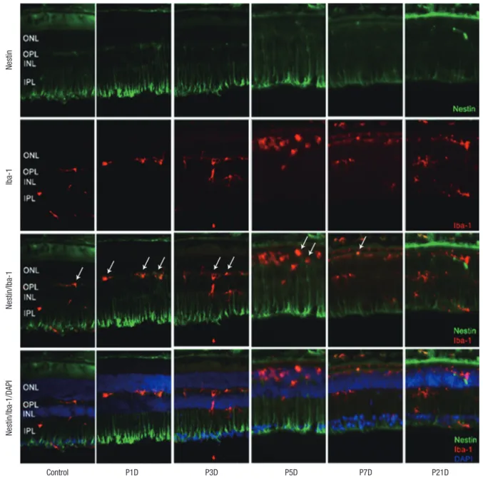
Fig. 1. Nestin and Iba-1 expression in control and degenerated adult mouse retina. Immunofluorescent labeling with nestin (green), Iba-1(red), and DAPI (blue) is shown. DAPI was used for nuclei staining to visualize the retinal layers. The decrease in thickness of the ONL due to selective loss of photoreceptor cells was becoming obvious by day 3 af-ter MNU injection. Progressive loss of photoreceptor cells ultimately led to a complete loss of the ONL at day 7 PI. Immunoreactivity of nestin was increased at day 1 PI, peaked at day 3, and declined gradually until day 21. MNU treatment resulted in migrating nestin+/Iba-1+ cells into the ONL. The number of nestin+/Iba-1+ cells peaked at day 3 PI. After that, the number of nestin+/Iba-1+ cells diminished progressively, particularly at day 7, and the cells were sparsely distributed by day 21. Arrows indicate nestin/Iba-1 co-immunolabeling.
Iba-1 = ionized calcium-binding adaptor molecule, DAPI = 4-6-diamino-2-phenylindole, ONL = outer nuclear layer, MNU = N-methyl-N-nitrosourea, PI = post injection, OPL = outer plexiform layer, INL = inner nuclear layer, IPL = inner plexiform layer.
Nestin
Iba-1
Nestin/Iba-1
Control P1D P3D P5D P7D P21D
of photoreceptor cells led to a nearly complete loss of ONL by day 7 PI (Fig. 1).
Nestin expression in resting retina
In the normal adult mouse retina, nestin was rarely expressed except for some vascular profiles in the outer plexiform layer (OPL) (Fig. 1).
Up-regulation of nestin expression after MNU treatment
Nestin expressing Iba-1+ microglia with amoeboid shape were
identified mainly in outer plexiform layer at 1 day after retinal degeneration induced by MNU injection (Fig. 1). Nestin express-ing microglia, previously located mainly in the OPL, had migrat-ed into ONL. The number of microglia showing nestin expres-sion was markedly increased at day 3 PI (Fig. 1). At day 5 PI, cell dimensions of nestin+ microglia were more extended (Fig. 1). At day 7 PI, nestin expressing microglia were significantly de-creased compared with day 5 PI, and the immunoreactivity of nestin in the whole retina was also significantly diminished (Fig. 1). By 21 days after MNU, most of the retinal neurons nuclei were
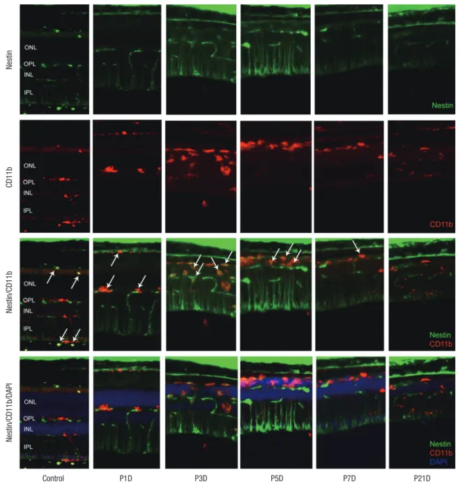
Fig. 2. Nestin and CD11bexpression in control and degenerated adult mouse retina. Immunofluorescent labeling with nestin (green), CD11b (red), and DAPI (blue) is shown. Ar-rows indicate nestin/CD11b co-immunolabeling. After a single injection of MNU, nestin+/CD11b+ cells migrated into the ONL at day 3 and showed a peak at day 5. The num-ber of nestin+/CD11b+ cells diminished progressively, particularly after 7 days, with sparse distribution by day 21.
DAPI = 4-6-diamino-2-phenylindole, ONL = outer nuclear layer, MNU = N-methyl-N-nitrosourea, OPL = outer plexiform layer, INL = inner nuclear layer, IPL = inner plexiform layer. Nestin CD11b Nestin/CD11b Control P1D P3D P5D P7D P21D Nestin/CD11b/DAPI
degraded and nestin expression had decreased to the baseline levels.
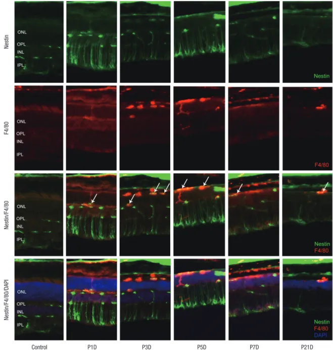
Expression of CD11b and F4/80 were rarely identified in the physiologic state prior to the MNU injection except for their ex-pression in the endothelium (Figs. 2 and 3). At day 1 after MNU injection, nestin+/CD11b+ cells were evident in the OPL. Nes-tin+/CD11b+ cells migrated into the ONL at day 3PI (Fig. 2), peak-ed at day 5 (Fig. 2), and decreaspeak-ed thereafter (Fig. 2). F4/80+ cells observed in the subretinal space revealed oval or spindle shaped cytoplasm without ramified processes at day 1 PI (Fig. 3). The
number of amoeboid nestin+/F4/80+ cells increased markedly from day 3 to day 5 PI and these cells were heavily confined with-in the subretwith-inal space and OPL (Fig. 3). Although the number of F4/80+ cells decreased thereafter, it was relatively preserved at 7 days after MNU injection (Fig. 3).
GFAP labeled Müller cells were distinctly increased at 1 day after MNU (Fig. 4). Nestin immunoreactivity of GFAP+ astro-cytes in the ganglion cell layer (GCL) and Müller cells was pro-gressively increased at day 3 PI (Fig. 4), and revealed peak activ-ity at day 5 (Fig. 4). After that, the number of nestin+/GFAP+
Mül-Fig. 3. Nestin and F4/80 expression in control and degenerated adult mouse retina. Immunofluorescent labeling with nestin (green), F4/80 (red), and DAPI (blue) is shown. Ar-rows indicate nestin and F4/80 co-immunolabeling. The number of amoeboid nestin+/F4/80+ cells increased markedly from day 3 to day 5 post injection and decreased there-after.
DAPI = 4-6-diamino-2-phenylindole, ONL = outer nuclear layer, OPL = outer plexiform layer, INL = inner nuclear layer, IPL = inner plexiform layer.
Nestin
F4/80
Nestin/F4/80
Control P1D P3D P5D P7D P21D
Fig. 4. Nestin and GFAP expression in control and degenerated adult mouse retina. Immunofluorescent labeling with nestin (green), GFAP (red), and DAPI (blue) is shown. Arrows indicate nestin/GFAP co-immunolabeling. The number of nestin+/GFAP+ Müller cells increased progressively at day 3 after MNU treatment, peaked at day 5, and decreased thereafter. GFAP-positive cells remained in an activated state at day 7 and day 21 post injection, despite the diminution in retinal thickness.
GFAP = glial fibrillary acidic protein, DAPI = 4-6-diamino-2-phenylindole, ONL = outer nuclear layer, OPL = outer plexiform layer, INL = inner nuclear layer, IPL = inner plexi-form layer. Nestin GF AP Nestin/GF AP Control P1D P3D P5D P7D P21D Nestin/GF AP/DAPI
ler cells was markedly reduced. However, GFAP expression re-mained in an activated state in the retinal tissue at day 7 (Fig. 4) and day 21 after MNU despite the diminution in retinal thick-ness (Fig. 4).
Western blot analysis
We used western blot analysis to quantify the expression of nes-tin and GFAP. Nesnes-tin showed increased immunoreactivation at day 1 PI. The expression of nestin peaked at day 3 PI and declin-ed at day 7 PI. At day 21 PI, the level declindeclin-ed to baseline when
compared with the control retinas (Fig. 5). GFAP expression ini-tially showed a gradual increase for the first 3 days after the MNU injection, and this level was maintained from day 3 to day 7 PI. GFAP expression decreased at day 21 PI; however, unlike nestin expression, GFAP expression was higher than that in the con-trol retina (Fig. 5).
Cell count
The number of nestin-expressing cells labeled with CD11b, Iba-1, and F4/80 was calculated in the retinal sections. After the MNU
Fig. 5. Western blot analyses of nestin and GFAP expression. (A) Significantly incre-ased immune-reactivity of nestin at day 1 and significantly decreincre-ased to mostly the level of control group at 21 days after MNU injection. (B) Immune-reactivity of GFAP showed gradually increasing for 3 days after MNU injection, and declined at 21 days. Nevertheless, GFAP remained in activated state compared to control group even after 21 days after MNU injection.
GFAP = glial fibrillary acidic protein, MNU = N-methyl-N-nitrosourea. Nestin Control P1D P3D P5D P7D P21D β-tubulin Nestin/ β-tubulin (% change)
Days after MMU
Control P1D P3D P5D P7D P21D 800 400 0 GFAP Control P1D P3D P5D P7D P21D β-tubulin GF AP/ β-tubulin (% change)
Days after MMU
Control P1D P3D P5D P7D P21D 120 80 40 0 A B
Fig. 6. Cell counts of Iba-1+, CD11b+, F4/80+, and nestin+ cells. (A) Iba-1+, CD11b+, F4/80+ cells were peaked at 5 days after injection. (B) Nestin/Iba-1 co-im-munolabelling cells and Nestin/F4/80 co-imco-im-munolabelling cells were peaked at 3 days after injection. Nestin/CD11b co-immunolabelling cells were peaked at 5 days after injection.
Iba-1 = ionized calcium-binding adaptor molecule, MNU = N-methyl-N-nitrosourea.
Number of cells
Days after MNU
0 5 10 15 20 25 500 400 300 200 100 0 CD11b F4/80 Iba-1 A Number of cells
Days after MNU
0 5 10 15 20 25 200 150 100 50 0 CD11b+Nestin F4/80+Nestin Iba-1+Nestin B
injection, the number of microglia/monocyte/macrophage mark-er-positive cells was increased at day 1 after MNU, peaked at day 5 (P < 0.001), and then decreased significantly at day 7 PI (P < 0.001) (Fig. 6). The number of nestin co-expressing microg-lia/ monocytes/macrophages was also increased at day 1 PI. The number of nestin+/Iba-1+ and nestin+/F4/80+ cells peaked at day 3 PI (P < 0.001). The number of nestin+/Iba-1+ cells decre-ased significantly at day 5 (P < 0.001), and the number of nes-tin+/F4/80+ cells was significantly reduced at day 7 (P < 0.001). The number of nestin+/CD11b+ cells peaked at day 5 PI (P < 0.001) and then decreased at day 7 (P < 0.001) (Fig. 6B).
DISCUSSION
Recently, nestin expression in the retina after acute injury has been widely studied (8-13). Previous studies have shown nestin expression in a degenerative retinal disease model. Wan et al. (11) investigated the possibility of photoreceptor regeneration from Müller glia after MNU induced retinal degeneration in the
adult rat retina. In that study, Müller glia underwent reactive gli-osis with the up-regulation of nestin and GFAP after MNU ad-ministration, and some Müller glia derived cells were induced to express rhodopsin exclusively. Valamanesh et al. (14) exam-ined the temporal pattern and cellular localization of nestin in the newborn rat retina in a neurodegenerative inherited disease model using genetically mutated rats. The study demonstrated that the level of nestin expression remained low after postnatal day 20 up to 1 year in normal animals but was up-regulated from postnatal day 30 in the dystrophic animals, and they concluded that nestin might be involved in mechanisms of migration, gen-eration of new neurons or glial cells and/or retinal modeling (14). In the present study, we assessed nestin expression in the adult mouse retina with MNU induced retinal degeneration model, and showed the up-regulation of nestin expression in microg-lia, monocytes/macrophages, Müller glia cells and astrocytes after MNU treatment. To the best of our knowledge, this is the first report of localized nestin expressing cells using microglia and monocyte/macrophage markers as well as a Müller cell mar-ker in the adult mouse degenerative retinal disease model.
Microglia and monocytes/macrophages showed differentia-tional changes in the adult mouse retina after MNU adminis-tration. Nestin up-regulation in the Iba-1 positive and CD11b/ F4/80 positive cells suggests that these cells may revert to a more developmentally immature state, since nestin serves as a cell cycle reentry marker (12). In the resting retina, microglia mark-er positive cells wmark-ere identified in the innmark-er plexiform laymark-er (IPL), the OPL, and the GCL. After MNU administration, these cells migrated toward the ONL and simultaneously showed increased nestin expression. Monocytes/macrophages marker positive cells were rarely recognized in the normal retina, but after MNU treatment, monocyte/macrophages were observed and also showed nestin co-expression.
The Müller cells also showed dedifferentiational changes af-ter MNU treatment. Previous studies have shown that nestin expression in differentiated Müller cell can be induced follow-ing retinal injury (7,10,12). Consistent with the results of previ-ous studies, the present study demonstrated reactive gliosis and nestin up-regulation of Müller cell after MNU injection. More-over, it is thought that, similar to GFAP, the nestin expression represent not only progenitor cells but also reactive gliosis in the pathologic retina (10,19,20). From this perspective, nestin might be used as an early stage marker of acute retinal injury when compared with the GFAP.
In conclusion, retinal microglia, macrophage, and Müller cells showed up-regulation of nestin expression in the adult mouse with pharmaceutically induced retinal degeneration model us-ing MNU, and the results suggest that these cells may revert to a more developmentally immature state. In addition, nestin ex-pression levels were sharply increased immediately after MNU administration and then returned to baseline levels. This find-ing implies that nestin might be used as an early-stage marker of acute retinal injury.
DISCLOSURE
The authors have no potential conflicts of interest to disclose.
AUTHOR CONTRIBUTION
Conceptualization: Moon CH, Cho H, Park TK. Data curation: Moon CH, Cho H, Kim YK. Funding acquisition: Park TK. In-vestigation: Moon CH, Cho H, Kim YK, Park TK. Supervision: Cho H, Park TK. Writing - original draft: Moon CH, Cho H, Park TK. Writing - review & editing: Moon CH, Cho H, Park TK.
ORCID
Chan Hee Moon http://orcid.org/0000-0003-1393-0242 Heeyoon Cho http://orcid.org/0000-0001-8744-4533 Yoon Kyung Kim http://orcid.org/0000-0002-2910-6904
Tae Kwann Park http://orcid.org/0000-0001-9689-4384
REFERENCES
1. Lendahl U, Zimmerman LB, McKay RD. CNS stem cells express a new class of intermediate filament protein. Cell 1990; 60: 585-95.
2. Reynolds BA, Weiss S. Generation of neurons and astrocytes from isolat-ed cells of the adult mammalian central nervous system. Science 1992;
255: 1707-10.
3. Suzuki S, Namiki J, Shibata S, Mastuzaki Y, Okano H. The neural stem/pro-genitor cell marker nestin is expressed in proliferative endothelial cells, but not in mature vasculature. J Histochem Cytochem 2010; 58: 721-30.
4. Dore-Duffy P, Katychev A, Wang X, Van Buren E. CNS microvascular peri-cytes exhibit multipotential stem cell activity. J Cereb Blood Flow Metab
2006; 26: 613-24.
5. Frisén J, Johansson CB, Török C, Risling M, Lendahl U. Rapid, widespread, and longlasting induction of nestin contributes to the generation of glial scar tissue after CNS injury. J Cell Biol 1995; 131: 453-64.
6. Clarke SR, Shetty AK, Bradley JL, Turner DA. Reactive astrocytes express the embryonic intermediate neurofilament nestin. Neuroreport 1994; 5:
1885-8.
7. Walcott JC, Provis JM. Müller cells express the neuronal progenitor cell marker nestin in both differentiated and undifferentiated human foetal retina. Clin Experiment Ophthalmol 2003; 31: 246-9.
8. Xue LP, Lu J, Cao Q, Hu S, Ding P, Ling EA. Müller glial cells express nestin coupled with glial fibrillary acidic protein in experimentally induced glau-coma in the rat retina. Neuroscience 2006; 139: 723-32.
9. Wohl SG, Schmeer CW, Kretz A, Witte OW, Isenmann S. Optic nerve le-sion increases cell proliferation and nestin expresle-sion in the adult mouse eye in vivo. Exp Neurol 2009; 219: 175-86.
10. Kohno H, Sakai T, Kitahara K. Induction of nestin, Ki-67, and cyclin D1 expression in Müller cells after laser injury in adult rat retina. Graefes Arch Clin Exp Ophthalmol 2006; 244: 90-5.
11. Wan J, Zheng H, Chen ZL, Xiao HL, Shen ZJ, Zhou GM. Preferential re-generation of photoreceptor from Müller glia after retinal dere-generation in adult rat. Vision Res 2008; 48: 223-34.
12. Luna G, Lewis GP, Banna CD, Skalli O, Fisher SK. Expression profiles of nestin and synemin in reactive astrocytes and Müller cells following reti-nal injury: a comparison with glial fibrillar acidic protein and vimentin.
Mol Vis 2010; 16: 2511-23.
13. Wohl SG, Schmeer CW, Friese T, Witte OW, Isenmann S. In situ dividing and phagocytosing retinal microglia express nestin, vimentin, and NG2 in vivo. PLoS One 2011; 6: e22408.
14. Valamanesh F, Monnin J, Morand-Villeneuve N, Michel G, Zaher M, Miloudi S, Chemouni D, Jeanny JC, Versaux-Botteri C. Nestin expression in the retina of rats with inherited retinal degeneration. Exp Eye Res 2013; 110:
26-34.
15. Ito D, Imai Y, Ohsawa K, Nakajima K, Fukuuchi Y, Kohsaka S. Microglia-specific localisation of a novel calcium binding protein, Iba1. Brain Res Mol Brain Res 1998; 57: 1-9.
16. London A, Itskovich E, Benhar I, Kalchenko V, Mack M, Jung S, Schwartz M. Neuroprotection and progenitor cell renewal in the injured adult mu-rine retina requires healing monocyte-derived macrophages. J Exp Med
17. Caicedo A, Espinosa-Heidmann DG, Piña Y, Hernandez EP, Cousins SW. Blood-derived macrophages infiltrate the retina and activate Muller glial cells under experimental choroidal neovascularization. Exp Eye Res 2005;
81: 38-47.
18. Erickson PA, Fisher SK, Guérin CJ, Anderson DH, Kaska DD. Glial fibril-lary acidic protein increases in Müller cells after retinal detachment. Exp
Eye Res 1987; 44: 37-48.
19. Xue LP, Lu J, Cao Q, Kaur C, Ling EA. Nestin expression in Müller glial cells in postnatal rat retina and its upregulation following optic nerve transec-tion. Neuroscience 2006; 143: 117-27.
20. Xue L, Ding P, Xiao L, Hu M, Hu Z. Nestin, a new marker, expressed in Mül-ler cells following retinal injury. Can J Neurol Sci 2010; 37: 643-9.