자궁경부상피내종양과 침윤성 편평상피암종의 혈관신생에서 비만세포와 혈관내피성장인자의 발현
원광보건대학 임상병리과1, 원광대학병원 병리과2
제갈승주1․이정아2․노종섭2
Mast Cells and Vascular Endothelial Growth Factor Expression in Neoangiogenesis of Cervical Intraepithelial Neoplasia and Invasive Squamous Cell Carcinomas of the Uterine Cervix
Seung-Joo Jekal
1, Jung-Ah Lee
2, and Jong-Sup Rho
2Department of Clinical Laboratory Science, Wonkwang Health Science College, Iksan 570-750, Korea
1Deparment of Pathology, Wonkwang University Hospital, Iksan 570-711, Korea
2To determine the correlation between mast cells(MCs) and neoangiogenesis in the growth and progression of cervical cancer, we investigated mast cell density(MCD), microvessel density(MVD) and the expression of vascular epithelial growth factor(VEGF) in cervical intraepithelial neoplasia and invasive suqamous cell carcinoma of the uterine cervix. Forty-five cervical intraepithelial neoplasia(CIN I, II and III), 15 microinvasive carcinomas, 15 invasive squamous cell carcinomas and 20 normal cervical epithelia were included in this study. MCs were stained with anti-c-Kit antibody and alcian blue, microvessels with anti-factor VIII antibody and VEGF with anti-VEGF antibody. The adjacent fields of both normal and neoplastic epithelium were used for counting MCs and microvessels. Computerized image analysis was used to evaluate MCD and MVD. MCD and MVD were the mean numbers per 1mm
2counted in 5-10 high and low power fields respectively. In both c-Kit and alcian blue stained sections, MCD progressively increased along the continuum from CIN I to invasive squamous cell carcinoma(p<0.001). MVD increased significantly with cervical neoplasia progression, from CIN to invasive squamous cell carcinoma (p<0.001). In double c-Kit and Factor VIII-stained sections, MCs were mainly present in the areas adjacent to newly formed blood vessels. However, there were no significant differences in MCD and MVD between normal epithelum and CIN I. A strong correlation was also observed between MCD and MVD. In double VEGF and alcian blue-stained sections, VEGF was expressed in only MCs. Strong VEGF-positive MCs were particularly abundant around the tumorous region. Our results suggest that MCs may upregulate neoangiogenesis by VGEF secretion in the development and progression of cervical neoplasia
Key Words : Mast cell, Neoangiogenesis, Vascular endothelial growth factor, Uterine cervix, Neoplasia
대한임상검사학회지 : 37권 제3호, 197-206, 20051)
교신저자 : 제갈승주, (우)570-750 전북 익산시 신룡동 344-2, 원광보건대학 임상병리과
Tel : 063-840-1215 E-mail : [email protected]
I. 서 론
혈관신생(neoangiogenesis)은 종양의 성장과 전이에 있 어 필요한 혈액 공급원으로서 중요한 역할을 하는 것으 로 알려져 있으며(Folkman, 1990; Weidner, 1993), 몇몇 종양의 정량적 연구에서는 종양 내 혈관신생이 종양의 전이와 환자의 예후를 예측할 수 있는 의미 있는 지표임 을 보여 주었다(Maeda 등, 1995; Saeki 등, 1997).
최근 비만세포는 주요 기능으로 알려진 알러지 및 염 증질환의 관련성 외에 종양을 대상으로 한 연구에서 종 양 간질부위나 그 주변부위에 많이 증가하며, 비만세포의 수적 증가가 암의 성장과 침윤, 전이 그리고 예후와 상관 성이 있는 것으로 보고되어 있다(Dabbous 등, 1986;
Jekal, 1999; Elpek 등, 2001; Acikalin 등, 2005). 아직 이 들 종양 주변부에 출현하는 비만세포가 종양의 기능적 관련성에 대해서는 정확히 밝혀져 있지 않으나 여러 연 구에서 비만세포가 혈관신생에 관여함으로써 종양의 진 행을 유도할 것이라는 증거들을 보여 주었고(Takanami 등, 2000; Toth 등, 2000; Ranier 등, 2003), 유방암과 전립 선암에서는 종양 주변의 혈관신생이 혈관내피성장인자 (vascular endothelial growth factor: VEGF)에 의해 촉진 될 것이라는 가능성을 시사하였다(Gasparini, 2000; Toth 등, 2000). 자궁경부암의 경우에 있어서도 비만세포는 자 궁경부상피내종양에서 침윤암종으로 진행하면서 종양상 피 주변에 비만세포밀도(mast cell index: MCD)가 증가하 고, 증가된 비만세포가 혈관신생과 밀접하게 연관되어 있 음을 보여 주었다(Benitez-Bribiesca 등, 2001; Cabanillas- Saez 등, 2002). 뿐만 아니라 자궁경부의 침윤전암종과 침 윤암종에 관한 연구에서는 미세혈관밀도(microvessel density: MVD)가 암의 진행 및 예후 사이에 상관성이 있 는 것으로 조사되었고(Ozalp 등, 2003), Sotiropoulou 등 (2004)도 미세혈관밀도가 상피내암종에 비해 미소침윤 암종에서 뚜렷한 증가를 보여 종양의 초기 침윤단계에서 혈관신생이 활발하게 이루어진다는 것을 암시하였으며, Lee 등(2003)은 자궁경부암의 진행과정에서 일어나는 혈 관신생이 종양세포로부터 유래한 VEGF에 의한 것으로 가정하였다. 이처럼 자궁경부암에서 비만세포의 증가와 혈관신생이 매우 밀접하게 관련되어 있다는 보고가 있음 에도 불구하고 자궁경부암의 발생과 진행과정에서 비만 세포가 어떻게 혈관신생을 촉진하는지에 대한 연구는 거 의 없는 실정이다.
따라서 본 연구는 자궁경부상피내종양과 침윤성 편평
상피암종의 신생혈관 형성과정에서의 비만세포와 VEGF 의 역할을 규명하기 위해 시도하였다.
II. 재료 및 방법
1. 재 료
본 실험에는 1999년부터 2004년까지 원광대학병원 병 리과에 의뢰된 자궁경부 생검조직 중 총 95건을 임의로 선별하여 사용하였다. 환자의 평균 연령은 47세(27-72세) 이었다. 선별된 조직은 다시 관찰하여 병변부위가 뚜렷하 게 보존된 조직만을 골라 형태의 특징에 따라 정상조직 (20건), 자궁경부상피내종양(cervical intraepithelial neo- plasia: CIN) I등급(13건), CIN II등급(15건). CIN III등급 (17건) 그리고 침윤성 편평상피암종을 다시 미소침윤암종 (icroinvasive carcinoma: MI)(15건)과 침윤암종(invasive carcinoma: IC)(15건)으로 나누어 총 6군으로 분류하였다.
모든 조직은 10% 중성완충포르말린에 고정된 것을 사용 하였다.
2. 방 법
1) 일반 병리조직학적 검사
10% 중성완충포르말린에 고정된 조직을 자동침투기를 사용하여 파라핀블록을 만든 후 박절기를 이용하여 4 ㎛ 두께로 박절하였다. 파라핀절편은 자일렌에서 탈파라핀 하여 하강계열 알코올단계의 함수과정을 거친 후 hema- toxylin-eosin(HE)염색을 하여 canada balsam으로 봉입하 였다.
2) 조직화학적 검사
비만세포의 분비과립 및 형태를 보기 위하여 Enerback
(1966)의 alcian blue/safranin 염색에서 대조염색을
safranin 대신 nuclear fast red로 바꾸어 사용하였다. 그
과정은 탈파라핀 후 함수한 절편을 1% alcian blue
8GX(0.7N HCl에 용해한 후 농염산으로 pH 0.3으로 조
정)에 옮겨 37℃에서 2시간 30분간 염색한 다음 0.7N
HCl에 잠시 헹군 후 nuclear fast red 용액으로 30분간 대
조염색하여 수돗물에 헹군 후 탈수 및 투명화하여 canada
balsam으로 봉입하였다. Alcian blue 8GX 및 nuclear fast
red는 모두 Sigma(Sigma Co., St. Louis, Mo, USA) 제품
을 사용하였다.
3) 면역조직화학적 염색
MicroProbe System(Fisher Scientific Co., USA)을 사용 하여 면역염색을 수행하였다. 비만세포 마커로 c-kit을, 내피세포 마커로 factor VIII, 그리고 신생혈관 형성 마커 로 VEGF를 택하여 각각 rabbit polyclonal anti-human c-kit(1:300, Dako, Glostrup, Denmark), mouse mono- clonal anti-human factor VIII-related Ag(1:50, Neo- Markers, Westinghouse Drive Fremont, USA) 및 mouse monoclonal anti-human VEGF(AB-5)(1:20, Oncogene, Cambridge, UK)를 사용하여 면역염색하였다. 그 과정을 간단히 요약하면 탈파라핀 후 함수한 절편을 항원부활을 위해 c-Kit은 pepsin에 넣어 45℃에서 4분간 두었으며, Factor VIII와 VEGF는 citrate buffer, pH6.0에 넣어 microwave oven에서 15분간 가열하였다. 그 다음 hydro- gen peroxidase block(Lab Vision, Westinghouse, USA)에 옮겨 45℃에서 7분간 방치하여 endogenous peroxidase를 차단한 후, 각각의 1차 항체에 넣어 냉장고에서 12시간 반응시켰다. 이어 2차 항체와 HRP polymer가 결합된 Super PicTure Polymer Detection Kit(Zymed, San Francisco, USA)를 사용하여 실온에서 30분간 반응시킨 다음 AEC(Zymed, San Francisco, USA)로 발색시켰다.
마지막으로 Mayer hematoxylin을 사용하여 대조염색한 후 Universal mount(Research genetics, Huntsville, USA) 로 봉입하였다. 각 단계마다 슬라이드는 Immuno/ DNA buffer(Invitrogen, Carlsbad, USA)를 사용하여 수세하였 다. 음성대조로는 1차항체를 사용하지 않았다.
4) Factor VIII-c-Kit 및 VEGF-alcian blue의 이중염색 비만세포와 미세혈관과의 조직학적 관련성을 보기 위 해 Factor VIII-c-Kit 이중염색을 하였으며, 비만세포에서 VEGF 발현을 확인하기 위해 VEGF-alcian blue 이중염색 을 하였다. Factor VIII-c-Kit 이중염색 과정은 탈파라핀 후 함수한 절편을 항원부활을 위해 pepsin으로 처리한 후 3% H
2O
2에 넣어 45℃에서 7분간 두어 endogenous peroxidase를 차단하였다. 이어 mouse monoclonal anti-human factor VIII-related Ag(1:50, NeoMarkers, Westinghouse Drive Fremont, USA)에 옮겨 냉장고에서 하루밤 반응시킨 후 Super PicTure Polymer Detection Kit(Zymed, San Francisco, USA)에 넣어 실온에서 30분 간 둔 다음 AEC를 사용하여 발색하였다. 그 다음
Immuno/DNA buffer로 수세한 후, citrate buffer, pH6.0에 넣어 microwave oven에서 20분간 가열하였고, 실온에서 10분간 식힌 후 enhancer(Zymed, San Francisco, USA)에 옮겨 45℃에서 30분간 두었다. 이어 rabbit polyclonal anti-human c-Kit(1:300, Dako, Glostrup, Denmark)에 옮 겨 45℃에서 30분간 반응시킨 후, Universal LSAB+AP kit Link(Dako, Glostrup, Denmark)에 옮겨 45℃에서 20분 간 반응시켰다. 그 다음 Universal LSAB+AP kit Strep- tavidin AP(Dako, Glostrup, Denmark)를 45℃ 20분간 반 응시킨 후 AP-blue substrate kit에 옮겨 실온에서 15분간 발색시킨 후 Universal mount로 봉입하였다. VEGF- alcian blue 이중염색과정은 탈파라핀 후 함수한 절편을 항원부활을 위해 citrate buffer, pH6.0에 넣어 microwave oven에서 15분간 가열한 다음 3% H
2O
2에 넣어 45℃에서 7분간 방치하여 endogenous peroxidase를 차단하였다. 그 다음 mouse monoclonal anti-human VEGF(AB-5)(1:20, Oncogene, Cambridge, UK)에 옮겨 냉장고에서 12시간 반응시킨 후 Super PicTure Polymer Detection Kit(
Zymed, San Francisco, USA)에 넣어 실온에서 30분간 둔 다음 AEC를 사용하여 발색시켰다. 이 슬라이드는 alcian blue, pH0.3을 사용하여 37℃에서 30분간 대조염색한 후 Universal mount(Research genetic, Huntsville, USA)를 사 용하여 봉입하였다.
5) 영상분석
단위면적당 MCD와 MVD를 구하기 위해 CCD 카메라 (Thosiba U-CMAD -2, Japan)가 부착된 현미경(Olymphus BX 50, Olymphus Optical Ltd,. Japan)을 사용하여 c-Kit 면역염색표본(배율 X200)과 alcian blue 염색표본(X200) 그리고 Factor VIII 면역염색표본(X100)으로부터 정상조 직과 CIN 병변의 경우는 상피 바로 밑 부분에서, 미소침 윤암종과 침윤암종의 경우는 종양세포 주변의 간질에서 5-10 시야를 선택하여 영상을 획득하여 컴퓨터에 저장하 였다. 저장된 영상은 분석 소프트웨어인 Image-Pro Plus ver 3.01(Media Cyberbetics Inc., USA)을 사용하여 1 mm
2당 비만세포와 미소혈관의 평균수를 세어 각각 MCD 와 MVD로 하였다.
6) 통계분석
각 군간 통계학적 차이는 일원분산분석(on-way
analysis of variance: ANOVA test)을 사용하여 조사하였
고 MCD와 MVD의 상관성을 보기 위해 Pearson's
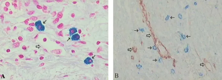
Fig. 2. Mast cells(arrow) stained with alcian blue(A) and Factor VIII/c-Kit(B). Mast cells were located in area adjacent to the
microvessel(open arrow) around the tumor foci. Original magnification X400(A), X200(B).Fig 1. Mast cells stained with alcian blue(A) and immunostained with c-Kit(B) in invasive squamous cell carcinoma of the uterine
cervix. Asterisk(✱) indicates tumoral epithelium; arrow indicates the mast cell. Original magnification X200.correlation coefficient를 적용하였다. p값이 0.05 이하이면 통계학적으로 유의한 것으로 판정하였다. 분석에는 SPSS ver 10.0 프로그램을 사용하였다.
III. 결 과
1. 비만세포의 분포와 밀도
자궁경부 조직 내의 비만세포 형태와 아형의 종류를 보기 위해 Enerback(1966)의 원법에 따라 alcian blue 염 색 후 safranin으로 대조염색한 결과 모두 청색으로 염색 되어 alcian blue-양성세포임을 확인하고 배경염색을 위해 safranin 대신 nuclear fast red를 사용하여 대조염색하였 다. 또한 c-kit 면역염색을 하여 alcian blue-양성 비만세포
와 c-kit-양성 비만세포 밀도를 비교하였다. 그 결과 자궁 경부 정상조직, 자궁경부상피내종양, 침윤암종 조직 내의 비만세포는 난원형, 다각형 및 방추형으로 상피 기저막 밑의 간질조직이나 종양세포 주변부의 혈관 가까이에 출 현하였으며 과립형과 탈과립형의 두 형태로 관찰되었다 (Fig. 1, 2).
Alcian blue 염색표본에서 1 ㎟ 평균 비만세포 밀도를 구한 결과 정상(26.2±9.4개)에 비해 CIN I 등급(34.6±10.8 개)에서 약간 증가하였으나, 통계학적으로 의의가 없었다 (p=0.149). 그러나 CIN I
등급에서 CINII
등급(55.0±9.2개)(p<0.001), CIN III
등급(65.4±6.0개)(p<0.001), 미소침윤암종(77.9±9.7개)(p<0.001), 침윤암종(89.5±11.8개) (p<
0.001)로 진행하면서 각 군간 모두 통계학적으로 유의하 게 증가하였다(Fig. 3). c-kit-양성 비만세포 역시 1 mm
2당 정상조직(44.8±7.2개)에 비해 CIN I등급(50.7±12.3개)
A B
A
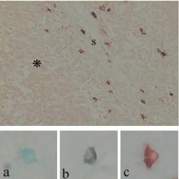
Fig. 5. Microvessels immunostained with anti-Factor VIII in section of both normal tissue(A) and invasive squamous cell
carcinoma(B) of the uterine cervix. Original magnification X200.Fig. 3. Mast cell density in normal, CIN and invasive squamous
cell carcinoma of the uterine cervix. Bar represent mean±SD of the six groups.N, normal tissue; CIN, cervical intraepithelial neoplasia; MI, microinvasive carcinoma, IC, invasive carcinoma.
NS, no significant compared with normal and MI respectively;
* p<0.05 compared with the data of CIN II, CIN III and MI respectively; **p<0.01 compared with the data of CIN I, CIN II, CIN III respectively.
은 약간 증가하였을 뿐 통계학적으로 유의한 차이가 없 었으나(p=0.839), CIN I 등급에서 CIN II등급(69.7±12.1 개)(p<0.01), CIN III 등급(84.0±10.1개) (p<0.05), 미소침 윤암종(99.4±19.8개)(p<0.05) 으로 진행하면서 각 군간 모 두 통계학적으로 유의한 증가를 나타내었다. 그러나 미소 침윤암종에 비해 침윤암종(106.0±18.7개)에서는 약간의 증가는 있었으나 통계학적으로 유의한 차이가 없었다 (p=0.782)(Fig. 3). 비만세포에 대한 alcian blue 염색과 c-Kit 면역염색을 비교한 결과 alcian blue-양성 비만세포 에 비해 c-Kit-양성 비만세포가 전 군에서 높게 출현하였 으나, 두 염색 방법 간에 상관관계를 조사한 결과 매우
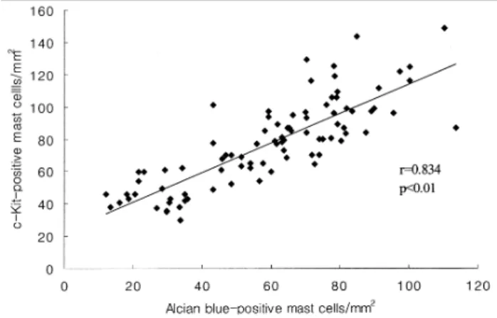
Fig 4. Correlation between c-kit-positive and alcian blue-
positive mast cells in normal, CIN and invasive squamous cell carcinoma of the uterine cervix. Linear correlation for two elements is highly significant.상관성이 높은 것으로 나타났다(Fig. 4).
2. 미세혈관의 밀도
Factor VIII을 사용하여 미세혈관을 면역염색한 결과
정상조직에 비해 자궁경부상피내종양 그리고 침윤암종으
로 진행하면서 미세혈관 수의 증가와 함께 염색강도가
점점 강하게 나타남을 관찰할 수 있었다(Fig. 5). 미세혈
관은 1 mm
2당 정상조직(11.2±3.1개)에 비해 CIN I 등급
(14.9±3.0개)에서 약간 증가하였으나 통계학적 유의성은
없었다(p=0.320). 그러나 CIN I등급에서 CIN II등급
Fig. 6. Microvessel density in normal, CIN and invasive
squamous cell carcinoma of the uterine cervix. Bar represent mean±SD of the six groups.NS, no significant compared with normal; * p<0.05 compared with the data of CIN I, CINII, CIN III and MI respectively