Cell Migratory Induction by Expression of Angiogenin and Vascular Endothelial Growth Factor in Resveratrol Treated HeLa Cells
I-Seul Joe
1,2, Sin-Gu Jeong
1,2and Goang-Won Cho
1,2*
1
Department of Biology, College of Natural Science, Chosun University, Gwangju 501-759, Korea
2
Department of Life Science, BK21-Plus Research Team for Bioactive Control Technology, Chosun University, Gwangju 501-759, Korea
Received January 13, 2014 /Revised February 26, 2014 /Accepted February 27, 2014Resveratrol (RSV), a natural polyphenolic compound, is a modulator for cell division and cell migra- tion, and has diverse beneficial properties. Angiogenin (ANG) and vascular endothelial growth factor (VEGF) are considered to be important mechanisms for cell proliferation, angiogenesis, the formation of tubular structures, and migration. In this study, we investigated whether RSV has a migratory ef- fect in HeLa cells. When cells were treated with 0~50 μM of RSV for 24 hr, the expression of ANG and VEGF was significantly increased in a dose dependent manner measured by real-time PCR.
Similarly, we performed time dependent experiments for 50 μM RSV treated cells and identified the optimized time at 24 hr. The increased expression in RSV treated cells was confirmed by Western blot analysis. To examine the toxic effects of RSV at the determined conditions, MTT assays were performed. The viabilities were unchanged for 0~50 μM RSV treated cells, while they decreased at 100 μM RSV. To examine the effect of migration in RSV treated cells, we performed a wound-healing assay. The migratory rates were significantly enhanced in the RSV treated group. In this study, we found that RSV induces an increase in the expression of migration factors ANG, VEGF, and enhances cell migration for the determined conditions.
Key words : Angiogenin, migration, resveratrol, vascular endothelial growth factor, wound-healing
*Corresponding author
Tel : +82-62-230-6641, Fax : +82-62-230-6650 E-mail : [email protected]
This is an Open-Access article distributed under the terms of the Creative Commons Attribution Non-Commercial License (http://creativecommons.org/licenses/by-nc/3.0) which permits unrestricted non-commercial use, distribution, and reproduction in any medium, provided the original work is properly cited.
Journal of Life Science 2014 Vol. 24. No. 4. 337~342 DOI : http://dx.doi.org/10.5352/JLS.2014.24.4.337
서 론
천연 폴리페놀계 화합물인 resveratrol (RSV, 3, 4, 5’–tri- hydroxy–trans - stillbene, C
14H
12O
3)은 포도, 땅콩, 산딸기류 열매 등 다양한 식물에서 발견되며 , 이러한 식물은 스트레스, 손상 , 햇볕 과잉 노출, 감염 등으로부터 방어하기 위해 RSV를 생산한다 [2, 20]. 많은 연구들을 통해 RSV는 항산화, 항염증, 항노화 등의 효과뿐 아니라 , sirtuins의 활성 자극, AMP-acti- vated Kinase (AMPK) 신호경로, 미토콘드리아의 생합성과 기능 , p53의 발현 등 다양한 작용기전과의 연관성이 보고 되 었다 [5, 12, 19].
RSV는 세포에서 pro-angiogenic effect와 anti-angiogenic effect으로 서로 상반된 성격을 지닌다고 알려져 있으나, 여러 연구들에서 사용한 pro-angiogenic effect와 anti-angiogenic effect를 유발하는 RSV 농도범위의 차이는 미미하다[3, 11, 21, 22]. 이것은 약물의 농도뿐 아니라, 세포주의 특성 및 세포가 처한 환경 등이 중요한 요인으로 작용함을 암시한다 .
Angiogenin (ANG)은 123개의 아미노산으로 구성된 단일 가닥 단백질로 , ribonuclease (RNase)라 불리는 유전자 fam- ily에 속해있다[14, 16, 17, 23]. ANG는 단백질 생산을 조절 하기 위해 ribonucleolytic activity, ribosomal RNA (rRNA) 전사를 향상시키는 등의 작용으로 세포이동 (cell migration), 신생혈관형성 (angiogenesis), 증식(proliferation), tubular structures의 형성 등을 포함한 다양한 생명현상에 관계한다 [6, 8, 10, 17]. ANG는 암세포의 발생에 밀접한 관계를 지닌 다고 알려져 있지만 [14], 한편으로는 신생혈관형성이 잘 형성 되지 않아 생기는 허혈성 뇌질환 , 허혈성 심장질환 또는 혈관 및 척수 손상 등에 ANG, Vascular endothelial growth factor (VEGF)가 치료적 효능을 갖는다[1, 9, 17]. 또한 amyotrophic lateral sclerosis (ALS)는 운동신경세포만 사멸하는 신경계 퇴 행성질환으로 , ANG mutation이 ALS질병에 있어서 중요한 역할을 하는 것으로 보고 되었다 [7, 13]. 뿐만 아니라, ANG는 신경세포에서 세포의 죽음을 유도하는 저산소증 (hypoxia)으 로부터 보호효과를 나타낸다 [4].
본 연구에서는 천연 폴리페놀계 화합물인 RSV에 의한 ANG와 VEGF의 발현을 확인하고, 그 역할을 확인하기 위해 세포의 이동효과를 비교 분석하였다 .
재료 및 방법
세포주와 세포배양조건
HeLa 세포는 자궁경부 상피 암으로부터 유래된 것으로 세
Table 1. Oligonucleotides used for real - time - PCR
Gene Forward primer (5’→3’) Reverse primer (5’→3’) Acc. No.
ANG VEGF
β-action
ATGGTGATGGGCCTGGGCGT AGAAAATCCCTGTGGGCCTT ATCCGCAAAGACCTGTACGC
CAGTCAATTTTCCGTCGTCCGTA GTCACATCTGCAAGTACGTTCG TCTTCATTGTGCTGGGTGCC
NM_001145 NM_001025368 NM_001101 Acc. No. indicates gene access number.
포주의 배지는 10% fetal Bovine Serum (FBS), 1% antibiotics (100 mg/l streptomycin, 100 U/ml penicillin)을 포함한 Dulbecco’s Modified Eagle Medium (DMEM, HyClone Laboratories, Inc., USA) 배지를 사용하여 37°C, 5% CO
2조건 에서 배양하였다 .
Materials
본 실험에서 사용된 resveratrol (Sigma Aldrich, R5010-100 mg)은 dimethyl sulfoxide (DMSO)에 녹인 뒤 50 mM로 희석 하여 사용 전까지 -20°C에 보관하였다.
Real-time PCR
HeLa 세포를 6-well에 1×10
5cell/well이 되도록 seeding하 고 안정된 다음 날 배지를 제거 한 후 phosphate-buffered sal- ine (PBS)로 세척하고 FBS 없는 배지로 overnight 배양하였 다 . 후에 RSV를 농도와 시간 별로 처리하였다. 전체 RNA는 RNAiso (TAKARA, Japan)을 이용하여 분리하였다. 추출한 RNA의 5 μg으로 cDNA synthesis kit (TAKARA, Japan)와 oligo-dT primer를 이용하여 제조사의 프로토콜에 따라 cDNA를 합성하였다. 합성된 cDNA는 SYBR Green PCR master mix (Applied Biosystems Inc., USA)와 함께 real-time PCR을 수행하였다. PCR에 사용된 primer는 human의 ANG, VEGF, β-actin을 사용하였다(Table 1). Real-time PCR cycling 은 95°C에서 10분 동안 반응시킨 후 40 cycles을 95°C에서 15 초 , 58°C에서 1분간 반응하였다.
Cell viability assay
RSV에 대한 세포의 독성평가, 생존능력 또는 증식능력을 보기 위한 실험으로 , HeLa 세포를 96-well plate에 seeding하 여 하룻동안 배양하였다 . 세포의 안정화 후에 PBS로 세척하 고 FBS가 없는 배지에 overnight 배양하였다. 다음날 FBS가 없는 배지에 RSV를 농도 별로 희석하여 24시간 또는 48시간 동안 처리하였다 . 배지를 제거한 후 5 mg/ml MTT solution (Sigma, USA)을 10:1 비율로 희석하여 2시간 동안 배양 후 DMSO로 녹여 ELISA microplate reader (Thermo Fisher Scientific Instruments co., Ltd.) 560 nm 흡광도에서 세포생 존능력 또는 세포증식능력을 평가하였다 . 약물에 대한 효과 는 약물을 처리하지 않은 대조 군에 대한 백분율 (%)로 나타내 었고 , 평균값과 표준오차는 Microsoft Excel program을 이용
하여 분석하였다 .
Western blot analysis
1×10
6cell/well에 protease 와 dephosphatase inhibitors가 포함된 50 μl RIPA buffer (Santa Cruz Biotechnology, USA) 를 처리해 4°C에 30분 동안 반응 시킨 후, 16,000× g 4°C에서 20분간 원심 분리하여 상층액에 있는 전체 단백질을 분리하 였다 . 50 μg의 sample을 만들어 sodium dodecyl sulphate (SDS) - polyacrylamide gel을 이용하여 전기영동을 한 뒤, ni- trocellulose membrane (Millipore, Germany)으로 transfer시 켰다 . 단백질이 전이된 membrane을 blocking buffer [5%
skim milk, 0.1% Tween-20 (pH 7.4)가 포함 된 TBS용액]에서 1시간 30분 동안 반응하였다. 1차 항체[1:500(Santa Cruz Biotechnology, USA)]는 blocking buffer 조건에 sodium azide 0.02%를 넣어 상온에서 2시간 또는 4°C 에 overnight 하였다 . 2차 항체는 TTBS에 1:20,000(Molecular probes®, USA)으로 희석해 1시간 30분 동안 상온에서 반응시켰다. 반 응 후 암실에서 ECL solution kit (Amersham Life Science, UK)를 사용하여 X-ray film에 노출시켜 확인하였다. 단백질 의 정량적 분석은 Image J 프로그램으로써 이루어졌다.
Wound-healing assay
세포 배양용 6 well plate에 HeLa 세포를 2×10
5cell/well 로 배양한 후 PBS로 세척하여 FBS 없는 배지로 overnight 배 양하였다 . 200 μl Yellow tip을 이용하여 단일 세포층을 긁은 후 PBS로 1번 세척해 주었다. 그 후 RSV를 처리하여 CO
2in- cubator에서 배양하였다. 세포이동성은 0, 24, 48시간마다 현 미경 (canon i-SOLUTION IMTcam3)을 통해 사진 촬영하였고 Image J software를 이용해 긁혀진 면적을 측정하였다. 이때, 0시간은 세포를 긁은 직후를 말한다. 면적 측정은 촬영 된 사 진의 긁혀진 면적을 구한 뒤 0시간의 면적에서 24시간의 면적 값을 빼 24시간 동안 움직인 거리를 계산하였고, 같은 방법으 로 48시간 동안의 이동 거리를 계산하였다. 계산된 값을 대조 군의 24시간에 대한 백분율(%)로 환산 하였다.
통계적 분석
실험 결과는 mean ± S.D.으로 표현되었다. 실험결과에서
student t-test를 사용하여 p<0.05 이하인 경우를 유의한 것으
로 판정하였다 .
A B
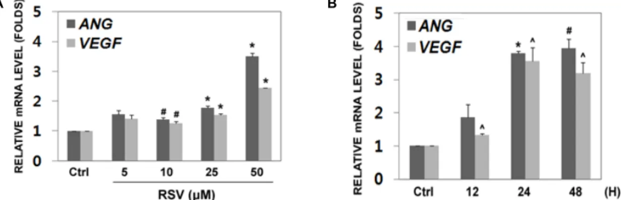
Fig. 1. Resveratrol induces the expression of
ANG
andVEGF
in HeLa cells. (A) Cells increased the expression ofANG
andVEGF
under treatment with 5~50 μM of RSV for 24 hr in compared with non-treated cells, measured by real-time PCR analysis (t
-test, *p
<0.005, #p
<0.05, mean ± S.D.,n
=4). (B) The expression ofANG
andVEGF
in the 50 μM RSV treated cells were increased in time dependent manner (0, 12, 24, and 48 hr), tested by real-time PCR (t
-test, *p
<0.001, #p
<0.01, ^p
<0.05 mean± S.D.,
n
=3).Fig. 2. Cellular toxicity in RSV treated cells. Cells were seeded at 96-well plates and then treated with 0, 5, 10, 25, 50 and 100 μM of RSV for 24 hr. Cell viabilities were de- termined by MTT assay (
t
-test, *p
<0.01, mean ± S.D.,n
=5).결과 및 고찰
Resveratrol (RSV)에 의한 ANG, VEGF 유전자의 발현 증가
HeLa 세포에서 파이토케미칼 RSV 처리 시, angiogenic factor 유전자의 발현을 확인 하기 위해 SYBR Green PCR master mix (Applied Biosystems Inc., USA)를 사용하여 re- al-time PCR을 수행하였다. 먼저, RSV의 최적 농도를 설정하 기 위해 RSV를 0~50 μM 24시간 동안 처리하였다. 대조군과 비교하여 농도가 증가함에 따라 Angiogenin (ANG), Vascular endothelial growth factor (VEGF) 유전자의 발현이 증가하였다.
50 μM 처리 시 ANG 유전자가 3.7배, VEGF 유전자가 2.4배 증가함을 관찰 하였다 (Fig. 1A). 시간에 따른 발현 변화를 확 인하기 위해 앞서 선정된 농도 (50 μM RSV)를 0~48시간까지 처리하여 분석하였다 . 그 결과 24시간에서 가장 높은 효과를 관찰하였다 (Fig. 1B). 이와 같이, HeLa 세포에 50 μM RSV를 24시간 동안 처리하였을 때 ANG와 VEGF가 가장 높게 발현 하여 , 이를 최적의 조건으로 선정하였다.
세포이동과 신생혈관형성을 촉진시키는데 ANG가 중요한 역할을 수행한다는 내용이 여러 선행연구를 통하여 확인되었 다 [4, 13]. 하지만, RSV에 의한 ANG 발현 변화에 대한 연구는 아직 보고된 바가 없다 . 본 연구에서는 HeLa 세포에 RSV를 처리하여 ANG, VEGF 유전자 발현이 증가함을 관찰하였다.
세포에서 Resveratrol 처리에 따른 세포독성 평가 RSV 처리에 따른 세포독성여부를 평가하기 위하여 MTT assay를 수행하였다. RSV를 24시간 동안 0~100 μM까지 처리 한 결과 , 0~50 μM 농도에서는 세포 독성이 관찰되지 않았으 나 , 100 μM에서 대조 군 대비 세포 생존율이 84.6%로 유의하 게 감소하였다 . 이와 같은 결과로, 앞서 선정된 조건(50 μM 24시간)이 세포의 생존에 영향을 미치지 않음을 확인하였다
(Fig. 2).
Resveratrol에 의한 ANG 단백질의 발현 증가
Fig. 1에서 RSV에 의한 ANG 유전자 발현이 증가함을 관찰 하였다 . 이 효과를 단백질 수준에서 관찰하기 위해 western blot을 수행하였다. 먼저, 세포에 RSV 50 μM을 24시간 동안 처리한 후 ANG 항체를 이용하여 발현 변화를 관찰하였다.
그 결과 , RSV를 처리한 그룹이 대조군에 비해 ANG 단백질이 19% 증가한 것을 확인하였다(Fig. 3A, Fig. 3B). 단백질의 발 현을 Image J 프로그램을 이용하여 분석하였고, 백분율로 환 산하여 그래프로 나타내었다 (Fig. 3B). 이를 통해, RSV가 ANG, VEGF 발현을 조절함을 확인하였다.
Resveratrol에 의한 세포의 이동 증가
여러 연구에서 ANG와 VEGF의 발현 증가에 따라 세포의
이동 능력이 향상됨을 보였다 [8, 20, 24]. 본 연구에서는 RSV
A
B
Fig. 3. ANG is increased in RSV treated cells. (A) Cells were treated with 50 μM of RSV for 24 hr and subjected to western blot analysis with antibodies against human ANG or β-actin. (B) ANG specific bands were quanti- fied using ImageJ software. Values represent the mean of three independent experiments (
t
-test, *p
<0.05, mean± S.D.,
n
=3).A
B
Fig. 4. Resveratrol treated cells were significantly enhanced the cell migration. (A) Cells were treated with/without 50 μM RSV in FBS free media, and subjected to wound- healing assay. The images were taken at the indicated time. RSV-treated cells present more rapidly recovered than non-treated cells. (B) The wound areas were calcu- lated by the Image J software. The migratory efficiency at the RSV-treated cells was compared with that of non-treated cells (
t
-test, *p
<0.005, #p
<0.01, mean ± S.D.,n
=9).Fig. 5. RSV uninfluenced cell growth and proliferations. Cells were treated with 50 μM RSV and incubated for 24~48 hr. The cell growths were then measured by MTT assay at the absorbance of 560 nm (
t
-test, *p
<0.01, mean ± S.D., n=3).가 ANG와 VEGF의 발현을 촉진시킴을 입증하였다. 이러한 효과가 세포 이동성에 영향을 미치는지 알아보기 위해 wound- healing assay를 수행하였다. 그 결과, RSV 50 μM을 처리한 그룹에서 24시간과 48시간에서 세포이동이 대조군과 비교하 여 유의성 있게 증가 함을 확인하였다 (Fig. 4A). 세포이동의 정도를 Image J 프로그램을 이용하여 백분율로 나타내었다 (Fig. 4B). 이와 같이, RSV가 HeLa 세포에서 ANG, VEGF를 증가시켰고 이에 따라 세포이동 능력이 향상됨을 입증하였다 .
선행 연구에 따르면 , HT-29 대장암세포에 고농도의 RSV 100, 200 μM 처리하면 wound-healing이 억제한다는 상반된 결과가 보고되었다 [15]. 반면에 다른 연구에서는 인간 관상동 맥의 소동맥 내피세포 (Human coronary arteriolar endothe- lial cells, HCAEC)에 RSV를 50 μM을 처리 했을 때 tubular structures의 형성이 대조 군과 비교하여 3배 가량 증가함을 보였다 [11]. RSV는 pro-angiogenic effect, anti-angiogenic ef- fect와 같이 상반된 효과를 나타낼 수 있으며, 이것은 RSV 처 리 농도와 시간뿐 아니라 이용된 세포 주에 따라 다름을 암시 한다 .
Resveratrol에 의한 세포증식 분석
세포이동 효과가 세포 증식에 의한 것 인지 검증하기 위하
여 Wound-healing assay와 동일한 조건인, RSV 50 μM을
24~48시간에서 세포의 증식 정도를 관찰하였다. Fig. 5에 나
타낸 것처럼 , 세포의 증식이 RSV에 의해 조절 되지 않음을
확인하였다 . 즉, RSV에 의한 세포 이동력 향상이 세포증식이
나 세포독성과 무관함을 암시한다 .
이상의 결과들을 살펴볼 때 , RSV는 앞서 설정된 조건(50 μ M, 24시간)에서 독성이 없으며, ANG와 VEGF의 발현을 증 가시켰고 , 그 결과 세포이동 능력을 향상시켰다. 본 연구결과 는 , RSV의 기전과 ANG, VEGF의 생리적 활성을 이해하는데 도움을 줄 것으로 기대된다 . 하지만, RSV에 의해 세포이동의 정확한 기전을 규명하기 위해서는 ANG, VEGF와 관련된 신 호전달경로를 확인해야 한다 .
감사의 글
이 논문은 2011학년도 조선대학교 학술연구비의 지원을 받아 연구되었음 .
References
1. Brudno, Y., Ennett-Shepard, A. B., Chen, R. R., Aizenberg, M. and Mooney, D. J. 2013. Enhancing microvascular for- mation and vessel maturation through temporal control over multiple pro-angiogenic and pro-maturation factors.
Biomaterials
34, 9201-9209.2. Carrizzo, A., Forte, M., Damato, A., Trimarco, V., Salzano, F., Bartolo, M., Maciag, A., Puca, A. A. and Vecchione, C.
2013. Antioxidant effects of resveratrol in cardiovascular, cerebral and metabolic diseases.
Food Chem Toxicol
61, 215-226.3. Chen, Y. and Tseng, S. H. 2007. Review. Pro- and anti-an- giogenesis effects of resveratrol.
In Vivo
21, 365-370.4. Cho, G. W., Kang, B. Y. and Kim, S. H. 2010. Human angio- genin presents neuroprotective and migration effects in neuroblastoma cells.
Mol Cell Biochem
340, 133-141.5. Csiszar, A., Labinskyy, N., Pinto, J. T., Ballabh, P., Zhang, H., Losonczy, G., Pearwon, K., de cabo, R., Pacher, P., Zhang, C. and Ungvari, Z. 2009. Resveratrol induces mi- tochondrial biogenesis in endothelial cells.
Am J Physiol Heart Circ Physiol
297, H13-20.6. Gao, X. and Xu, Z. 2008. Mechanisms of action of angiogenin.
Acta Biochim Biophys Sin (Shanghai)
40, 619-624.7. Greenway, M. J., Alexander, M. D., Ennis, S., Traynor, B.
J., Corr, B., Frost, E., Green, A. and Hardiman, O. 2004. A novel candidate region for ALS on chromosome 14q11.2.
Neurology
63, 1936-1938.8. Hu, G., Riordan, J. F. and Vallee, B. L. 1994. Angiogenin promotes invasiveness of cultured endothelial cells by stim- ulation of cell-associated proteolytic activities.
Proc Natl Acad Sci USA
91, 12096-12100.9. Huang, L., Huang, Y. and Guo, H. 2009. Dominant ex- pression of angiogenin in NeuN positive cells in the focal ischemic rat brain.
J Neurol Sci
285, 220-223.10. Jimi, S., Ito, K., Kohno, K., Ono, M., Kuwano, M., Itagaki, Y. and Ishikawa, H. 1995. Modulation by bovine angioge- nin of tubular morphogenesis and expression of plasmi- nogen activator in bovine endothelial cells.
Biochem Biophys Res Commun
211, 476-483.11. Kaga, S., Zhan, L., Matsumoto, M. and Maulik, N. 2005.
Resveratrol enhances neovascularization in the infarcted rat myocardium through the induction of thioredoxin-1, heme oxygenase-1 and vascular endothelial growth factor.
J Mol Cell Cardiol
39, 813-822.12. Khurana, S., Venkataraman, K., Hollingsworth, A., Piche, M. and Tai, T. C. 2013. Polyphenols: benefits to the car- diovascular system in health and in aging.
Nutrients
5, 3779-3827.13. Kieran, D., Sebastia, J., Greenway, M. J., King, M. A., Connaughton, D., Concannon, C. G., Fenner, B., Hardiman, O. and Prehn, J. H. 2008. Control of motoneuron survival by angiogenin.
J Neurosci
28, 14056-14061.14. Kishimoto, K., Liu, S., Tsuji, T., Olson, K. A. and Hu, G.
F. 2005. Endogenous angiogenin in endothelial cells is a general requirement for cell proliferation and angiogenesis.
Oncogene
24, 445-456.15. Lee, S. H., Park, S. Y., Kim, I. S., Park, O. J. and Kim, Y.
M. 2012. Effects of Resveratrol on Migration and Proliferation in HT-29 Colon Cancer Cells.
KSBB J
27, 289-294.16. Li, S. and Hu, G. F. 2012. Emerging role of angiogenin in stress response and cell survival under adverse conditions.
J Cell Physiol
227, 2822-2826.17. Ng, M. T., Stammers, A. T. and Kwon, B. K. 2011. Vascular disruption and the role of angiogenic proteins after spinal cord injury.
Transl Stroke Res
2, 474-491.18. Perrot-Applanat, M. and Di Benedetto, M. 2012. Autocrine functions of VEGF in breast tumor cells: adhesion, survival, migration and invasion.
Cell Adh Migr
6, 547-553.19. Price, N. L., Gomes, A. P., Ling, A. J., Duarte, F. V., Martin-Montalvo, A., North, B. J., Agarwal, B., Ye, L., Ramadori, G., Teodoro, J. S., Hubbard, B. P., Varela, A. T., Davis, J. G., Varamini, B., Hafner, A., Moaddel, R., Rolo, A. P., Coppari, R., Palmeira, C. M., de cabo, R., Baur, J. A.
and Sinclair, D. A. 2012. SIRT1 is required for AMPK acti- vation and the beneficial effects of resveratrol on mitochon- drial function.
Cell Metab
15, 675-690.20. Singh, C. K., Kumar, A., Lavoie, H. A., Dipette, D. J. and Singh, U. S. 2013. Diabetic complications in pregnancy: is resveratrol a solution?
Exp Biol Med (Maywood)
238, 482- 490.21. Simao, F., Pagnussat, A. S., Seo, J. H., Navaratna, D., Leung, W., Lok, J., Guo, S., Waeber, C., Salbego, C. G. and Lo, E. H. 2012. Pro-angiogenic effects of resveratrol in brain endothelial cells: nitric oxide-mediated regulation of vas- cular endothelial growth factor and metalloproteinases.
J Cereb Blood Flow Metab
32, 884-895.22. Srivastava, R. K., Unterman, T. G. and Shankar, S. 2010.
FOXO transcription factors and VEGF neutralizing anti- body enhance antiangiogenic effects of resveratrol.
Mol Cell Biochem
337, 201-212.23. Tsuji, T., Sun, Y., Kishimoto, K., Olson, K. A., Liu, S., Hirukawa, S. and Hu, G. F. 2005. Angiogenin is trans- located to the nucleus of HeLa cells and is involved in ribo- somal RNA transcription and cell proliferation.
Cancer Res
초록:Resveratrol 처리한 HeLa세포에서 angiogenin과 vascular endothelial growth factor 발현유도에 따른 세포이동촉진
조이슬
1,2․정신구
1,2․조광원
1,2*
(
1조선대학교 자연과학대학 생명과학과 ,
2조선대학교 자연과학대학 생명과학과 BK21 플러스 생리활성 제어 기술 인력양성 사업팀 )
Resveratrol (RSV)은 천연 폴리페놀계 화합물로 세포분열, 성장, 세포이동 등과 같은 다양한 효과가 보고되었 다 . Angiogenin (ANG)은 Vascular endothelial growth factor (VEGF)와 함께 세포의 증식, 신생혈관형성, tubular structures의 형성, 세포이동 등을 이끄는 중요한 단백질이다. 본 연구에서는 RSV에 의한 세포이동효과를 HeLa 세포에서 관찰하였다 . Real-time PCR을 통해 HeLa 세포에 RSV 0~50 μM을 24시간 동안 처리하였을 때, 농도에 따른 ANG, VEGF 유전자 발현이 의미 있게 증가 하였다. 같은 방법으로, RSV 50 μM을 시간에 따라(0~48시간) 처리하여 실험하였다 . 그 결과, RSV 50 μM을 24시간 동안 처리하였을 때 ANG, VEGF 유전자 발현이 가장 높게 증가하였고 , ANG 단백질 분석에서도 동일한 결과를 얻었다. 또한, MTT assay를 이용한 세포 독성연구에서, RSV 50 μM의 농도에서는 영향을 미치지 않음을 관찰하여, 이를 최적의 조건으로 결정하였다. RSV가 처리된 세포에서 세포이동효과를 조사하기 위해 wound-healing assay를 수행하였다. RSV가 처리된 그룹에서 세포이동이 의미 있 게 증가하였다 . 따라서, 본 연구에는 RSV에 의해 ANG, VEGF의 발현이 증가했고, 이에 따라 세포이동이 향상됨 을 관찰하였다 .
65, 1352-1360.
24. Wei, S., Gao, X., Du, J., Su, J. and Xu, Z. 2011. Angiogenin enhances cell migration by regulating stress fiber assembly and focal adhesion dynamics.