pISSN: 0378-6471 eISSN: 2092-9374
http://dx.doi.org/10.3341/jkos.2013.54.10.1534
= 증례보고 =
삼출성 나이관련황반변성 반대안의 새로운 신생혈관 발생 빈도
이정진⋅유영주⋅조성원⋅김종우 건양대학교 김안과병원 안과학교실 명곡안연구소
목적: 단안 나이관련황반변성에서 반대안의 새로운 삼출성 나이관련황반변성의 발생률을 알아보고자 하였다.
대상과 방법: 단안 삼출성 나이관련황반변성을 처음 진단받고 유리체내 베바시주맙이나 라니비주맙 주입을 받은 314명 환자에서 반대 안의 발생률을 후향 조사하였다.
결과: 2년 이상 추적관찰이 가능했던 314명중 1년 이내에 22명(7.0%), 2년 이내에 34명(10.8%), 2년 이상에서 43명(13.7%)에서 반대안 에 새로운 삼출성 나이관련황반변성이 발생하였다. 신생혈관의 유형별로 살펴봤을 때 전형맥락막신생혈관군, 잠복맥락막신생혈관군 및 결절맥락막신생혈관군의 세 유형 사이에서 발생률의 유의한 차이는 없었다. 치료 종류별로는 처음 12개월, 24개월까지는 라니비주 맙과 베바시주맙의 치료군 반대안 발생률의 차이는 없었으나, 24개월 이상에서는 베바시주맙 치료군이 반대안에 새로운 신생혈관 발 생률이 높았다.
결론: 24개월 이상의 경과관찰기간 동안 단안 삼출성 나이관련황반변성으로 치료받은 환자 중 13.7%에서 반대안에 새로운 삼출성황반 변성이 발생하였다.
<대한안과학회지 2013;54(10):1534-1539>
■Received: 2013. 2. 22. ■ Revised: 2013. 4. 18.
■Accepted: 2013. 8. 5.
■Address reprint requests to Young Ju Lew, MD, PhD Department of Ophthalmology, Kim’s Eye Hospital,
#136 Yeongsin-ro, Yeongdeungpo-gu, Seoul 150-034, Korea Tel: 82-1577-2639, Fax: 82-2-2671-6359
E-mail: [email protected]
* This study was presented as a narration at the 106th Annual Meeting of the Korean Ophthalmology Society 2011.
* The Association for Research in VIsion and Ophthalmology Annual Meeting, Fort Lauderdale, USA, 2012.
나이관련황반변성은 65세 이상의 고연령군에서 회복 불 가능한 실명의 주요 원인으로 알려졌다.1나이관련황반변성 의 유병률을 살펴보면, 40-79세의 인구 중 서양인을 대상 으로 한 연구를 분석한 결과 초기 황반변성은 8.8%, 후기 황반변성은 0.59%로 나타났으며, 동양인은 각각 6.8%, 0.56%
로 나타났다.2우리나라에서도 최근 식생활 등 생활형태가 서구화되고, 평균 수명이 늘어남에 따라 그 빈도가 과거에 비하여 늘었다는 의견이 있다.3Song et al4이 발표한 한 국 내병원에 건강검진에서 발견된 유병률을 보면 초기 황반변 성이 2.92%, 후기 황반변성이 0.19%로 보고되었다.
또한 나이관련황반변성에 대한 다른 역학연구를 살펴 보 면 단안 삼출성 황반변성 환자는 반대안에 새로운 신생혈 관이 발생할 잠재 위험이 있으며, 2년에 12-22%, 4년에 37%, 5년에 22-38.7% 정도의 발생률로 반대안에 새로운
신생혈관이 생기는 것을 보고하고 있다.5-8한편 MARINA와 ANCHOR 연구 결과 라니비주맙으로 치료받은 환자의 반대 안에 새로운 신생혈관이 생기는 발생률은 1년에 20.6%, 2년 에 32.1%로 보고하여 sham 또는 광역학치료 보다는 낮게 발생하지만 통계적으로 유의한 차이는 발견하지 못했다.9
삼출성 황반변성으로 치료받은 반대안의 새로운 발생빈 도에 대한 국내의 연구가 없어, 본 연구에서는 일차적으로 항혈관내피세포성장인자로 치료받은 환자의 반대안의 새로 운 삼출성 황반변성의 발생빈도를 알고 항혈관내피세포성 장인자의 종류와 삼출성 황반변성의 분류에 따른 발생빈도 의 차이가 있는지를 조사하였다.
대상과 방법
2009년 1월부터 2009년 8월까지 본원에서 삼출성 나이 관련황반변성을 새로 진단받고 항혈관내피세포성장인자 치 료를 받은 환자 중 반대안에 삼출성 나이관련황반변성의 징후가 없으며 24개월 이상 경과 관찰이 가능한 환자 314 명을 대상으로 후향 분석하였다.
반대안에 전형맥락막신생혈관(predominant classic choroidal neovascularization), 잠복맥락막신생혈관(occult/minimally classic choroidal neovascularization), 섬유혈관성 막막색 소상피박리(fibrovascular pigment epithelial detachment),
15
10
5
0
12 months 24 months Over 24 months (%)
Figure 1. Incidence of new neovascular AMD in the fellow eye.
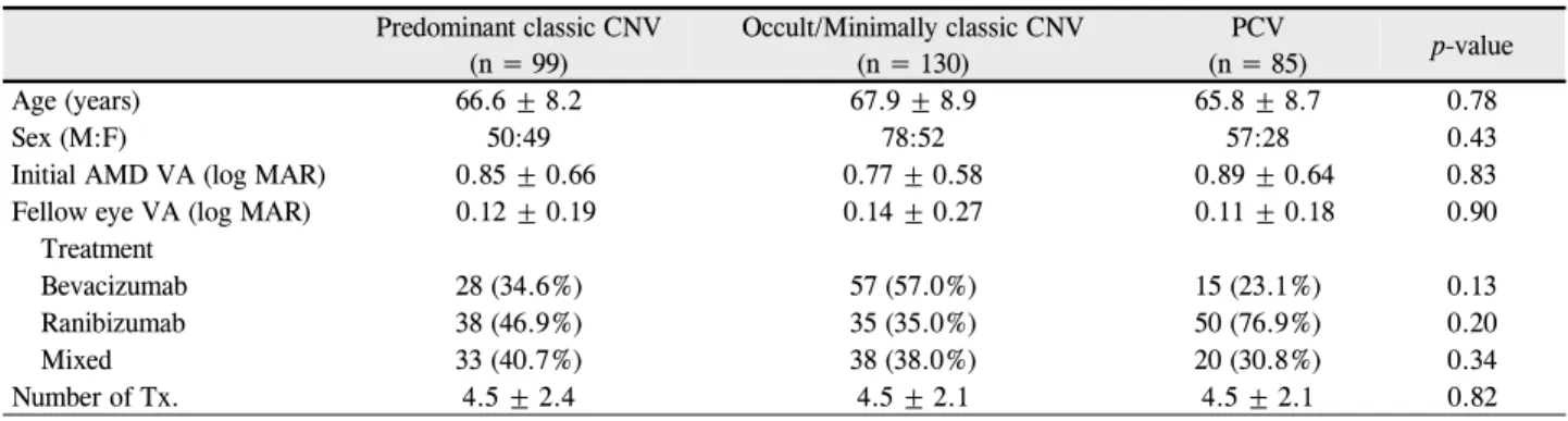
Table 1. Baseline characteristics of patients with unilateral exudative age-related macular degeneration (AMD) at baseline
Predominant classic CNV(n = 99)
Occult/Minimally classic CNV (n = 130)
PCV
(n = 85) p-value
Age (years) 66.6 ± 8.2 67.9 ± 8.9 65.8 ± 8.7 0.78
Sex (M:F) 50:49 78:52 57:28 0.43
Initial AMD VA (log MAR) 0.85 ± 0.66 0.77 ± 0.58 0.89 ± 0.64 0.83
Fellow eye VA (log MAR) 0.12 ± 0.19 0.14 ± 0.27 0.11 ± 0.18 0.90
Treatment
Bevacizumab 28 (34.6%) 57 (57.0%) 15 (23.1%) 0.13
Ranibizumab 38 (46.9%) 35 (35.0%) 50 (76.9%) 0.20
Mixed 33 (40.7%) 38 (38.0%) 20 (30.8%) 0.34
Number of Tx. 4.5 ± 2.4 4.5 ± 2.1 4.5 ± 2.1 0.82
Values are presented as mean ± SD.
NV = choroidal neovascularization; PCV = polypoidal choroidal vasculopathy.
Table 2. Number(%) of patients with fellow eyes exudative AMD conversion in each type of AMD
1 year 2 years Over 2 years
Predominant classic CNV (N = 99) 5 (5.1) 9 (9.1) 12 (12.1)
Occult / Minimally classic CNV (N = 130) 9 (6.9) 13 (10.0) 18 (13.8)
PCV (N = 85) 8 (9.4) 12 (14.1) 13 (15.3)
p-value 0.51 0.51 0.82 CNV = choroidal neovascularization; PCV = polypoidal choroidal vasculopathy.
망막출혈, 장액성 망막색소상피박리, 섬유성 조직, 장액성 감각망막박리. 레이저 반흔이 있는 경우는 삼출성 나이관련 황반변성 가능성이 있다고 보고 연구 대상에서 제외하였다.
대상환자의 반대안은 경과 관찰기간 동안 새로운 신생혈 관이 12개월 이내, 24개월 이내, 24개월 이상 지난 시점에 서 생기는 빈도로 구분하여 조사하였다.
모든 환자의 안저사진, 형광안저혈관조영사진, 인도시아 닌그린혈관조영사진과 빛간섭단층촬영을 취합하여 한 명의 망막전문의가 판독하여 나이관련황반변성의 유형을 전형맥 락막신생혈관, 잠복맥락막신생혈관, 결절맥락막신생혈관으 로 구분하였다. 또한 치료받은 항혈관내피세포성장인자의 종류를 조사하여 나이관련황반변성의 유형과 항혈관내피세 포성장인자의 종류에 따른 반대안의 신생혈관 발생빈도를 알아보았다. 통계학적 분석으로는 2 way ANOVA test를 사용하였으며 p-value가 0.05보다 작은 경우를 유의한 것 으로 간주하였다.
결 과
해당기간 동안 내원하여 처음 항혈관내피세포성장인자 의 치료를 받은 환자 중 24개월 이상 경과 관찰이 가능했던 환자는 총 419명이었다. 이 중 314명(74.9%)에서 단안에 삼출성 나이관련황반변성이 발견되었으며, 내원 당시 양안 에 삼출성 나이관련황반변성이 발견된 경우는 총 105명 (25.1%)이었다. 연구대상은 단안에만 발생한 314명을 대
상으로 하였으며 이중 99명(31.5%)이 전형맥락막신생혈 관, 130명(41.4%)이 잠복맥락막신생혈관, 그리고 85명 (27.1%)이 결절맥락막신생혈관으로 분류되었다. 내원 당 시 대상환자의 나이관련황반변성 유형에 따른 나이, 성별, 최 대교정시력과 치료방법은 유의한 차이가 없었다(Table 1).
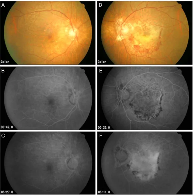
본 연구에서는 총 314명의 대상환자 중 12개월에 22명 (7.0%), 24개월에 34명(10.8%), 24개월 이상에서 43명 (13.7%)에서 반대안에 맥락막신생혈관이 발생하였으며 (Fig. 1) 이 중 2개월 후 반대안에 맥락막 신생혈관이 발 생한 한 예를 첨부하였다(Fig. 2, 3).
나이관련황반변성의 유형별로 본 반대안의 맥락막 신생 혈관의 발생률을 살펴 보면 12개월, 24개월과 24개월 이상 에서 전형맥락막신생혈관군, 잠복맥락막신생혈관군 및 결 절맥락막신생혈관군의 세 유형 사이에서 발생률의 유의한
A D
B E
C F
Figure 2. Fundus photograph and fluorescein angiograph of the right eye of a 64 year old female (A, B, C); left eye
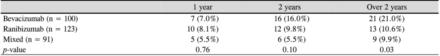
(D, E, F). (A, B, C) No specific abnormality (D) Choroidal neovascular membrane (CNVM) of the macula with ret- inal hemorrhage. (E) Predominant classic choroidal neovascularizaion with well demarcated lesion 23 seconds after injection. (F) Late phase shows staining implying old lesions. She was treated with intravitreal Ranibizumab in- jection 3 times.차이는 없었다(Table 2). 삼출성 나이관련황반변성의 치료 받은 항혈관내피성장인자의 종류별 발생률 비교에서는 12 개월과 24개월에서 치료법에 따른 발생률의 차이는 없었으 나, 24개월 이상에서 베바시주맙 단독은 21명(21%), 라니 비주맙 단독은 13명(10.6%), 베바시주맙과 라니비주맙을 모두 치료 받은 경우는 9명(9.9%)으로 통계적으로 유의한 차이를 보였다(p=0.03)(Table 3).
결론적으로 본원에 내원한 단안 삼출성 나이관련황반변 성 환자에서 반대안에 새로운 삼출성 황반변성이 발생하는 빈도는 1년에 7.0%, 2년에 10.8%, 2년 이상에서 13.7%였 으며 유형에 따른 반대안 맥락막신생혈관의 발생률의 차이
는 없었다. 치료종류별로 살펴보면 24개월까지는 유의한 차이가 없었지만, 24개월 이상에서 베바시주맙으로 치료받 은 경우가 반대안에 새로운 맥락막신생혈관의 발생률이 높 았다.
고 찰
삼출성 나이관련황반변성을 가진 경우 반대안에 새로운 황반변성이 생길 잠재 위험은 황반변성이 없는 사람보다 높을 것으로 여겨지고 있다. 여러 보고에서 발생빈도는 2년 에 12-22%, 4년에 37%, 5년에 22-38.7% 정도의 발생률
A D
B E
C F
Figure 3. Fundus photograph and fluorescein angiograph of same patient with figure 2 after 2 months of right visual
disturbance. Right eye (A, B, C) and left eye (D, E, F). (A) New hemorrhagic spot around macula (B, C) Class choroidal neovascularization which was absent 2 months prior (D) Retinal hemorrhage was decreased with fibrotic tissue changes. (E, F) Scar change with staining.Table 3. Number (%) of patients with fellow eyes exudative AMD conversion in each type of treatment
1 year 2 years Over 2 years
Bevacizumab (n = 100) 7 (7.0%) 16 (16.0%) 21 (21.0%)
Ranibizumab (n = 123) 10 (8.1%) 12 (9.8%) 13 (10.6%)
Mixed (n = 91) 5 (5.5%) 6 (5.5%) 9 (9.9%)
p-value 0.76 0.10 0.03
로 반대안에 새로운 신생혈관이 생기는 것을 보고하고 있 다.5-8
잠복맥락막신생혈관을 대상으로 한 MARINA 연구에서는
0.3 mg 또는 0.5 mg의 라니비주맙 치료를 받은 경우1년에 20.7%, 2년에 34.0%에서 새로운 맥락막신생혈관이 반대안 에 생겼으며, 전형맥락막신생혈관을 대상으로 한 ANCHOR
연구에서는 1년에 20.4%, 2년에 29.9%가 발생하였다. 따라 서 전체 삼출성 나이관련황반변성에서는 0.3 mg 또는 0.5 mg의 라니비주맙 치료를 받은 경우 1년에 20.6%, 2년에는 32.1%에서 반대안에 맥락막신생혈관이 발생하였다.9본 연 구에서는 라니비주맙 또는 베바시주맙으로 치료 받은 총 314명의 대상환자 중 12개월에 7.0%, 24개월에 10.8%, 24개월 이상에서 13.7%에서 반대안에 맥락막신생혈관이 발생하였다. 이는 이미 나이관련황반변성으로 초진 진료를 본 환자 중 양안에 삼출성 나이관련황반변성 양상을 가진 환자가 25.1%로 많은 수를 차지하고 있었으며, 또한 모든 대상 환자를 24개월 이상 경과관찰이 가능했던 경우로 제 한하였기 때문에 실제 진료실에서 보는 환자를 대상으로 하는 후향 연구의 한계로 인해 비교적 적은 발생률을 보였 을 것이라고 생각한다. 그리고 Ueta et al10이 일본인 대상 으로 한 연구를 살펴보면 첫 1년 발생률은 3.4%, 3년은 9.3%, 5년은 11.3%로 일본인의 경우 서양인에 비해 발생 률이 낮은 것으로 보고하고 있어 동양인에서 발생률이 낮 은 것도 원인으로 볼 수 있다.
삼출성 나이관련황반변의 유형별 분류를 살펴보면, 본 연구에서 단안 삼출성 나이관련황반변성 환자의 31.5%가 전형맥락막신생혈관, 41.4%가 잠복맥락막신생혈관이었으 며, 27.1%가 결절맥락막신생혈관으로 분류되었다. Park et al3의 국내 나이관련황반변성에 대한 기초 역학 연구는 전 형 또는 잠복맥락막신생혈관이 62.1%였으며 결절맥락막신 생혈관은 31.7%로 본 연구와 비슷하게 결절맥락막신생혈 관이 차지하는 비율이 비교적 높았다. 본 연구에서 반대안 에 새로운 신생혈관 발생률을 유형별로 관찰한 결과 2년 이 상 경과 관찰 동안 전형맥락막신생혈관 12.1%, 잠복맥락막 신생혈관 13.8%, 결절맥락막신생혈관에서 15.3%가 발생 하여 통계적 유의한 차이는 없었다(p=082). 따라서 본 연 구에서는 신생혈관의 유형별로는 반대안 발생률에는 큰 차 이가 없는 것으로 조사되었다. 국내 연구는 없으나 일본의 연구를 살펴보면, 반대안의 발생률은 전형 또는 잠복맥락막 신생혈관의 경우 1년, 3년, 5년 이상에 3.6%, 7.3%, 11.2%
였으며, 결절맥락막신생혈관은 3.2%, 11.1%, 11.1%로 역시 유형별로 유의한 차이는 없다고 보고하였다(p=0.876).10
단안 황반변성 환자에서 여러 치료가 반대안의 새로운 황반변성 발생에 미치는 영향에 대한 연구는 여러 가지가 진행되고 있다. 이전 AREDS연구에서 단안의 삼출성 나이 관련황반변성을 가진 환자들에서 고용량의 비타민이나 아 연 복용이 반대안의 삼출성 나이관련황반변성의 진행을 억 제하는 데는 제한적이라고 보고하였다.11그러나 최근 동물 연구에서는 베바시주맙 같은 항혈관내피세포성장인자의 주 입이 치료받지 않은 반대안에 대한 예방적인 효과의 가능
성에 대해 발표하였다.12-15 Bakri et al15은 토끼실험에서 유리체강내 베바시주맙 주입후에 반대안과 혈청내에 소량 의 베바시주맙이 검출되었으며, Avery et al13도 증식성당 뇨망막병증에서 단안에 유리체강내 베바시주맙 주입후에도 반대안의 망막 신생혈관에서의 누출이 감소하였음을 보고 하여 항혈관내피세포성장인자의 전신효과를 보고하였다.
하지만 항혈관내피세포성장인자의 전신효과에 대해서는 명 확하게 알려져 있지 않기 때문에 임상적 결과로 유추하게 된다.
MARINA연구와 ANCHOR연구에서 반대 안의 새로운 맥 락막신생혈관의 발생률은 라니비주맙을 치료 받은 군에서 sham군이나 PDT치료를 받은 군보다는 낮았으나, 통계적 으로는 의미가 없었다. 실제로 매달 라니비주맙을 주사받은 환자의 최고 혈청 농도가 0.3-2.36 ng/ml으로 측정되었으 며, 이것은 실제로 VEGF-A의 활동성을 50% 가량 억제할 수 있는 데 필요한 농도인 11-27 ng/ml 보다 지극히 낮은 것으로 in vitro 세포 증식 연구에서 보고되고 있다.16
따라서 본 연구에서 2년 이상이 지난 시점에서만 베바시 주맙에서 반대안의 맥락막신생혈관 발생률이 라니비주맙에 비해 유의하게 높게 나타난 것에 관하여, 항혈관내피세포억 제인자 종류에 따른 효과로 보는 것은 아직은 설득력 있는 이론적 뒷받침을 찾기 어려웠다. 또한 본 연구는 후향적인 연구로 통계적으로 차이는 없었으나 환자가 동일한 간격과 횟수의 베바시주맙 또는 라니비주맙 치료를 받지 않았기 때문에 두 군을 동일하게 비교하는데 한계점이 있다. 실제 기존의 여러 연구도 치료가 반대안의 발생률을 낮춘다는 것을 입증하지 못했고, 동물 실험과 in vitro 실험 등 여러 보고가 상이하므로 항혈관내피세포억제인자의 전신효과를 본 연구 결과로 추측하는 것은 어렵다고 볼 수 있다.
결론적으로 단안에 삼출성 나이관련황반변성을 가진 환 자에서 반대안의 신생혈관 발생률에 관한 국내 첫 보고로 향후 진단기술의 발전과 국민생활습관의 변화 등으로 발생 률은 달라질 수 있지만, 환자 진료에 참고가 될 수 있는 자 료로 의미가 크다. 그리고 나이관련황반변성의 유형에 따른 반대안의 신생혈관 발생률 차이는 없었으며 항혈관내피세 포인자의 종류에 따른 연구는 추가 연구가 필요하다고 여 겨진다.
REFERENCES
1) Friedman DS, O’Colmain BJ, Muñoz B, et al. Prevalence of age- related macular degeneration in the united states. Arch Ophthalmol 2004;122:556-72.
2) Kawasaki R, Yasuda M, Song JS, et al. The prevalance of age-re- lated macular degeneration in Asians: a systematic review and
=ABSTRACT=
Incidence of New Choroidal Neovascularization in Fellow Eyes of Patients Treated for Age-Related Macular Degeneration
Jung Jin Lee, MD, Young Ju Lew, MD, Sung Won Cho, MD, Jong Woo Kim, MD
Myung-Gok Eye Research Institute, Department of Ophthalmology, Kim’s Eye Hospital, Konyang University, Seoul, Korea
Purpose: To investigate the development of new choroidal neovascularization in fellow eyes of patients treated for unilat- eral exudative age-related macular degeneration (AMD).
Methods: Three hundred fourteen patients who were first diagnosed with unilateral exudative AMD and treated with intra- vitreal bevacizumab or ranibizumab were studied retrospectively.
Results: New exudative AMD developed in 7.0% of fellow eyes after 1 year, 10.8% after 2 years and 13.7% after more than 2 years. According to the subtype of exudative AMD, there were no significant differences between classic CNV, occult or minimally classic CNV, and PCV in the incidence of new exudative AMD. After 2 years, a higher conversion rate was found in the bevacizumab-treated group than the ranibizumab-treated group.
Conclusions: The cumulative incidence of involvement in fellow eyes with exudative AMD was 13.7% over 2 years.
J Korean Ophthalmol Soc 2013;54(10):1534-1539
Key Words: Bevacizumab, Exudative age-related macular degeneration, Incidence, Ranibizumab
Address reprint requests to Young Ju Lew, MD, PhD Department of Ophthalmology, Kim’s Eye Hospital
#136 Yeongsin-ro, Yeongdeungpo-gu, Seoul 150-034, Korea
Tel: 82-1577-2639, Fax: 82-2-2671-6359, E-mail: [email protected] meta-analysis. Ophthalmology 2010;117:921-7.
3) Park KH, Song SJ, Lee WK, et al. The results of nation-wide regis- try of age-related macular degeneration in Korea. J Korean Ophthalmol Soc 2010;51:516-23.
4) Song SJ, Youm DJ, Chang Y, Yu HG. Age-related macular degen- eration in a screened Korean population: prevalence, risk factors and subtypes. Ophthalmic Epidemiol 2009;16:304-10.
5) Klein R, Klein BE, Jensen SC, Meuer SM. The Five-year incidence and progression of age-related maculopahty: the Beaver Dam Eye Study. Ophthalmology 1997;104:7-21.
6) Klein R, Klein BE, Knudtson MD, et al. Fifteen-year cumulative incidence of age-related macular degeneration: the Beaver Dam Eye Study. Ophthalmology 2007;114:253-62.
7) Mukesh BN, Dimitrov PN, Leikin S, et al. Five-year incidence of age-related maculopathy: the Visual Impariment Project. Ophthalmology 2004;111:1176-82.
8) van Leeuwen R, Klaver CC, Vingerling JR, et al. The risk and natu- ral course of age-related maculopathy: follow-up at 6 1/2 years in the Rotterdam study. Arch Ophthalmol 2003;121:519-26.
9) Barbazetto IA, Saroj N, Shapiro H, et al. Incidence of new choroi- dal neovascularization in fellow eyes of patients treated in the
MARINA and ANCHOR trials. Am J Ophthalmol 2010;149:939-46.e1.
10) Ueta T, Iriyama A, Francis J, et al. Development of typical age-re- lated macular degeneration and polypoidal choroidal vasculopathy in fellow eyes of Japanese patients with exudative age-related mac- ular degeneration. Am J Ophthalmol 2008;146:96-101.
11) Klaver CC, Assink JJ, van Leeuwen R, et al. Incidence and pro- gression rate of age-related maculopathy: the Rotterdam study.
Invest Ophthalmol Vis Sci 2001;42:2237-41.
12) Neri P, Mariotti C, Mercanti L, Salvolini S, et al. Vitritis in the con- tralateral uninjected eye following intravitreal bevacizumab (Avastin). Int Ophthalmol 2008;28:425-7.
13) Avery RL, Pearlman J, Pieramici DJ, et al. Intravitreal bev- acizumab (Avastin) in the treatment of proliferative diabetic retinopathy. Ophthalmology 2006;113:1695.e1-15.
14) Sawada O, Kawamura H, Kakinoki M, Ohji M. Vascular endothe- lial growth factor in fellow eyes of eyes injected with intravitreal bevacizumab. Graefes Arch Clin Exp Ophthalmol 2008;246:1379-81.
15) Bakri SJ, Snyder MR, Reid JM, et al. Pharmacokinetics of intra- vitreal bevacizumab (Avastin). Ophthalmology 2007;114:855-9.
16) Bakri SJ, Snyder MR, Reid JM, et al. Pharmacokinetics of intra- vitreal ranibizumab (Lucentis). Ophthalmology 2007;114:2179-82.