Three Cases of Rare Anatomic Variations of the Long Head of Biceps Brachii
Sang-Ho Kwak, Seung-Jun Lee , Byung Wook Song1, Min-Soo Lee2, Kuen Tak Suh
Department of Orthopedic Surgery, Pusan National University Yangsan Hospital, Yangsan, 1Nalgae Hospital, Seoul, 2Hanseo Hospital, Busan, Korea
In general, the long head of the biceps brachii originates from the superior glenoid labrum and the supraglenoid tubercle, crosses the ro- tator cuff interval, and extends into the bicipital groove. However, rare anatomic variations of the origins of the long head have been re- ported in the past. In this report, we review the clinical history, radiologic findings, and arthroscopic identifications of 3 anatomic variants of the biceps tendon long head. As the detection of long head of biceps tendon pathology during preoperative radiologic assessment can be difficult without prior knowledge, surgeons should be aware of such possible anatomic variations.
(Clin Shoulder Elbow 2015;18(2):96-101)
Key Words: Shoulder; Anatomic variation; Long head of biceps brachii
Copyright © 2015 Korean Shoulder and Elbow Society. All Rights Reserved. pISSN 2383-8337
Clinics in Shoulder and Elbow Vol. 18, No. 2, June, 2015 http://dx.doi.org/10.5397/cise.2015.18.2.96
Received October 24, 2014. Revised January 26, 2015. Accepted March 12, 2015.
Correspondence to: Seung-Jun Lee
Department of Orthopedic Surgery, Pusan National University Yangsan Hospital, 20 Geumo-ro, Mulgeum-eup, Yangsan 626-770, Korea Tel: +82-55-360-2125, Fax: +82-55-360-2155, E-mail: [email protected]
Financial support: None. Conflict of interests: None.
In general, the long head of the biceps brachii originates from the superior glenoid labrum and the supraglenoid tubercle.
From its origin, the long head traverses along the rotator interval and the humeral head and extends into the bicipital groove.1) Cadaver studies by Vangsness et al.2) have shown that around 50% of long heads of the biceps brachii originate from the supe- rior labrum while the rest arises from the supraglenoid tubercle.
It is reported that most tendinous fibers of the long head of the biceps brachii merge with the labrum at the posterior superior labrum.
Anatomic variations in the origins of the long head of the biceps brachii are rare. Dierickx et al.3) have shown only 1.91%
of patients undergoing arthroscopic surgery show such anomaly.
Broadly, anatomic variations is classified into whether they have anomalous origins or there is a congenital absence of the long head itself.3,4) Most of these variations are only revealed coinci- dently during arthroscopic surgery, and the clinical significance of these anatomic variants remain unclear. Although there have been reports that congenital absence of the long head of biceps brachii contributes to the instability of the shoulder or to related congenital defects,5) there is no conclusive evidence as of yet to implicate these anatomic variations to shoulder pathology.
Until now, only a single case of an anatomic variation of the origin of the long head other than congenital absence has been reported in the Korean literature.6) In this report, we describe 3 cases of anatomic variations of the long head of the biceps bra- chii found during arthroscopic repairs of supraspinatus tendon tears.
Case Report
Case 1
A 40-year-old female was hospitalized with chronic pain on the right shoulder that began 3 weeks prior to the observation.
The patient had no job sequence and no exercise routines other than static yoga poses that she performed. The patient showed no obvious signs of trauma, but experienced severe pain with forward elevation of the arm and night pain. She had a visual analogue scale (VAS) for pain of 5. During physical examination, the patient presented with limited passive range of motion (ROM) of the shoulders; 150o of forward elevation and 150o of abduc- tion of the arm. Further, the patient showed signs of impinge- ment, a positive outcome for the empty can test, and tenderness at the supraspinatus attachment site. Speed’s and Yergason’s
tests, however, were negative and neither anterior nor posterior instability were detected.
Through plain radiographs, we found a subacromial arch on the right shoulder and, through magnetic resonance imag- ing (MRI), a supraspinatus tear and bony erosion at the greater tuberosity. During arthroscopy, we identified adhesive capsulitis and bifurcation at the origin of the long head of the biceps bra- chii (Fig. 1A).
For the shoulder pathology, the patient initially received con- servative management through ROM restorative exercises and administration of anti-inflammatory drugs. Despite an improved ROM, the VAS score of 5 failed to improve even after a year.
Thus, the patient underwent an arthroscopic rotator cuff repair, during which we were able to note a bifurcated long head.
The intra-articular thinner branch was attached to the labrum whereas the extra-articular thicker branch lay underneath the supraspinatus tendon. Since the movement of the supraspinatus tendon induced the movement of the intra-articular branch of the biceps brachii long head through its connection to the extra- articular branch, we excised the supraspinatus tendon before the rotator cuff repair. An unperturbed extra-articular branch of the biceps tendon was confirmed (Fig. 1B–D).
The patient was administered an abduction brace for 6 weeks postoperatively, after which passive and active ROM exercises were begun. By the 3rd postoperative month, the patient’s ROM level was improved to 170o of forward elevation and to 160o of abduction of the arm. By the 6th postoperative month, the VAS score decreased to 1 and the patient was capable of sustaining daily activities, even returning to yoga. Further, we did not detect any deterioration of biceps strength or a secondary lesion at the long head biceps tendon.
Case 2
A 46-year-old female who was injured as she fell on her right hand a month before presented with chronic pain on the right shoulder. The shoulder pain was not position-specific and the patient’s VAS score for pain was 4 points. Upon physical examination, the patient’s shoulder ROM was 180o of forward elevation and 170o of abduction of the arm. Results of the Neer, Hawkins, and the empty can tests came back positive, whereas those of the Speed’s and Yergason’s tests were negative. Instabil- ity of shoulder was not seen. By MRI, we found a tear of around 1 cm in size on the bursal-sided supraspinatus tendon (Fig. 2A).
As the patient was unresponsive to conservative management
A B
C D
SST
TLBT
GL
SST
BT
HH
TLBT
SST
TLBT
GL
SST
BT
HH
TLBT
SST
TLBT BT
HH SST
TLBT BT
HH
Fig. 1. (A) Coronal T2-weighted fat-sup- pressed magnetic resonance images of the shoulder of a 40-year-old female patient. The thick (white arrow) and thin limbs (white ar- rowhead) of the biceps tendon are visualized.
(B) The thin limb (black arrow) of the biceps tendon is visualized. (C, D) After identifying the thick limb (black arrowhead) of the biceps tendon, the thin limb (black arrow) was de- brided.
SST: supraspinatus tendon, TLBT: thin limb of biceps tendon, GL: glenoid fosssa, BT:
thick limb of biceps tendon, HH: humeral head articular surface.
of the supraspinatus tear, we carried out an arthroscopic repair on the 3rd month of trauma. Arthroscopically, we found that the attachment of the long head to the superior labrum was not in- tra-articular. Further, we saw that the lower supraspinatus tendon was positive for cord sign, which is reflective of the presence of capsular hypertrophy. This capsular hypertrophy began at the medial superior glenoid and ran pass the lower supraspinatus tendon into the bicipital groove. During the surgery, we could see through the supraspinatus tear that the long head traversed through the intra-articular capsule and ended up in an extra- articular location. Other than the supraspinatus repair, we did not otherwise disturb the long head (Fig. 2B–D). The patient was administered abduction shoulder braces for 6 weeks after the operation and by the 6th postoperative month, her VAS score decreased to 1 point. No symptoms indicative of abnormalities in the biceps muscles or the long head biceps brachii were de- tected postoperatively.
Case 3
A 55-year-old woman came into our clinic with complaints of pain on the left shoulder that began 3 months prior to the visit.
At the initial examination, the patient’s VAS score was 5 points,
and the passive ROM of the shoulder managed by the patient was 160o of forward elevation and 160o of abduction. The results for the Neer and Hawkins tests were negative whereas that of the empty can test was positive. However, Speed’s and Yergason’s test results were negative and there were no bicipital groove tenderness or instability of the shoulder. We detected a 1-cm large supraspinatus tear by MRI (Fig. 3A).
After 3 months of conservative management to no avail, we performed arthroscopic repair of the rotator cuff tear. Through arthroscopy, we found a type 1 superior labral anterior to poste- rior lesion and could confirm that the long head biceps tendon did not reside in an intra-articular location. Also, we found that the lower supraspinatus tendon was positive for Cord sign indi- cating the presence of capsular hypertrophy and that the long head originated from the medial superior glenoid and extended to the biceps brachii. Although we also detected synovitis along the hypertrophied region, since its indication was not strong enough we did not take immediate measures (Fig. 3B). At the 8 month follow-up of the supraspinatus repair, we found that the patient’s VAS score improved to 1 point. She felt no discomfiture during daily activities and there were no recurrent symptoms of the biceps brachii.
Fig. 2. (A) Coronal T2-weighted fat-sup- pressed magnetic resonance images of the shoulder of a 46-year-old female patient. The torn tendon of the rotator cuff and the long head of the biceps tendon are visualized. (B–D) A discrete cord-like thickening (black arrows) of the capsule underneath the supraspinatus tendon (SST) began just medial to the supe- rior glenoid at the supraglenoid tubercle and continued along the undersurface of the su- praspinatus into the biceps groove (BG; black arrowhead) within the rotator interval.
GL: glenoid fosssa, HH: humeral head articu- lar surface, BL: biceps long head, GT: greater tubercle.
A B
C D
SST
GL
BG
GT HH
BL SST
HH SST
SST
GL
BG
GT HH
BL SST
HH SST
Discussion
Although no standardized classification system exists to clas- sify anatomic variants of the long head of biceps tendon, in general, variants, except congenitally absent long heads, are classified in terms of their relationship, or extent of fusion, with the supraspinatus tendon. For instance, Wahl and MacGillivray4) arthroscopically classified the pathology into 4 types; incomplete proximal mesenteric, incomplete distal mesenteric, complete mesenteric, and congenitally absent (Table 1). Similarly, Dierickx et al.3) analyzed 2,976 shoulder pathologies and classified the 57 shoulders (1.91%) that had an anatomic variation into 4 types;
mesotenon, adherent, bifurcated, and congenitally absent, of which there were 29, 15, 11, and 2 of each, respectively (Table 2).
When we adopt these classification systems, we find that all our cases fall into the mesenteric type if using and Wahl and Mac- Gillivray’s classification system,4) whereas we find that shoulder pathology in Case 1 is categorized as an adherent bifurcated long head and those in Cases 2 and 3 as adherent if following Dierickx et al.’s classification.3)
The clinical significance of these anatomic variants of the long heads of the biceps brachii remains unknown. Especially, the
benefits of surgically treating the long heads aberrantly attached to the rotator cuffs or those passing beneath the inner supra- spinatus tendon are debated. For the support of surgical treat- ment, Ogawa and Naniwa7) reported of a long head that passed through the supraspinatus tendon whilst not passing the bicipital groove. Proposing this as one of the possible etiologies of the rotator cuff tear, they showed that when the long head of the biceps brachii was attached to the supraspinatus anterior aspect, good clinical outcome was achieved. Another study that sup- ports the surgical treatment of variations of the long had is that of Zhang et al.8) They reported of two cases of variations of the long head that originated from the anterior aspect of the supra- spinatus tendon, and suggested this as an etiological deformity that induced the tear of the supraspinatus through the input of excessive tension. In accordance to their hypothesis, tenodesis of the biceps tendon of these two patients, which reduces the excess tension on the tendons, gave good clinical outcomes.
Conversely, Kim et al.6) found that a conservative management of a long head of the biceps brachii adhered to the superior glenoid and the supraspinatus tendon was met with a good clinical outcome. Likewise, MacDonald9) reported that a conser- vative treatment of an adherent long head running underneath
Fig. 3. (A) Coronal T2-weighted fat-suppressed magnetic resonance images of the shoulder of a 55-year-old female patient. The torn rotator cuff tendon is shown and the long head of the biceps tendon with a superior labral anterior to posterior (SLAP) lesion is visualized. (B) A cord-like thickening of the capsule (black arrow) underneath the supraspinatus tendon (SST) and SLAP lesion (black arrowhead) are shown. Synovitis around the capsular layer overlying the long head of the biceps was evi- dent in this patient.
GL: glenoid fossa.
A B
SST
SLAP
GL SST
SLAP
GL
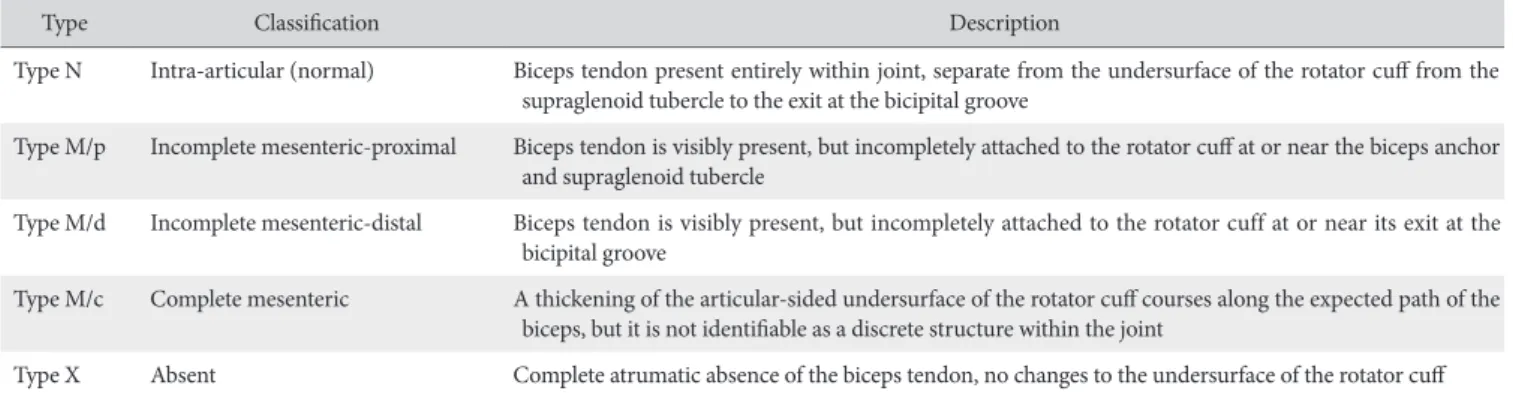
Table 1. Wahl and MacGillivray’s classification system of variants of intra-articular anatomy of the long head of the biceps tendon4)
Type Classification Description
Type N Intra-articular (normal) Biceps tendon present entirely within joint, separate from the undersurface of the rotator cuff from the supraglenoid tubercle to the exit at the bicipital groove
Type M/p Incomplete mesenteric-proximal Biceps tendon is visibly present, but incompletely attached to the rotator cuff at or near the biceps anchor and supraglenoid tubercle
Type M/d Incomplete mesenteric-distal Biceps tendon is visibly present, but incompletely attached to the rotator cuff at or near its exit at the bicipital groove
Type M/c Complete mesenteric A thickening of the articular-sided undersurface of the rotator cuff courses along the expected path of the biceps, but it is not identifiable as a discrete structure within the joint
Type X Absent Complete atrumatic absence of the biceps tendon, no changes to the undersurface of the rotator cuff Cited from the article of Wahl et al. (J Shoulder Elbow Surg. 2007;16:e25-30) with original copyright holder’s permission.4)
the supraspinatus tendon was sufficient. Similar to these latter findings, in this case report, we found that the outcomes of the conservative management of all variants attached to the rotator cuff except for the treatment of one intra-articular bifurcated long head were successful. Overall, these studies give a mixed message for the treatment of anatomic variations of the biceps brachii long head. However, when we consider that the rotator cuff tears of the cases described by Ogawa and Naniwa7) and Zhang et al.8) cannot solely be explained by anatomic variations of the long head, it is difficult to implicate anatomic variations as being the direct causative factor and erroneously conclude a benefit of surgical interventions. This is because Ogawa and Na- niwa7) sought to prove a clinical significance of disconnecting a long head using an already disconnected one, and Zhang et al.8) studied subjects, currently active young volleyball and baseball athletes, wherein it is difficult to preclude sports-related injuries as etiology for the rotator cuff tear. As surgical intervention has not yet been proven as a significantly better treatment method over conservative management, we believe that when there is
no clear arthroscopic indication of lesions of the long head in a physical examination or of mechanical stress, for example on supraspinatus tears, the merit of surgical treatment for long head of biceps tendons that are attached to the rotator cuff or run to inner supraspinatus tendon remains questionable. Further retro- spective studies are needed to elucidate the clinical and tangible benefit of surgical correction of anatomical variations.
Radiologic methods to detect anatomic variations of the long head of biceps brachii include ultrasounds, MRI, and magnetic resonance (MR) arthrograms. However, ultrasounds have limited capabilities to discern intra-articular lesions of the biceps brachii long head. Despite the high sensitivity and specificity offered by MRIs in terms of the detection of full-thickness tears of the biceps tendon, their usefulness as diagnostic tools for concurrent lesions of the shoulder is yet undetermined.5) MR arthrograms have similar limitations as MRIs in terms of diagnostic abili- ties.10) In the clinical setting, the majority of anatomic variants of the long head of the biceps brachii are not diagnosed during radiological screenings but during close scrutiny at the time of Table 2. Classification: 12 types of variations of the intra-articular portion of the long head of the biceps3)
Type Classification Description
MESO MESO-VI Fine string, providing vascularization to the tendon
MESO-SB Small, synovial band from medial to lateral, connecting the rotator cuff with the biceps. They are never on stress.
MESO-PU Pulley or hammock-like sling, whereby the biceps can move or slide freely up and down.
MESO-PA A hammock-like synovial sling in which the biceps tendon is able to move but not to glide.
MESO-CO The biceps tendon runs in a synovial sheath that is connected, loose woven but well vascularized, to the inferior surface of the capsule. No sliding is possible.
ADH ADH-PM A partial but strong medial adhesion runs cranial and medial to the inferior surface of the capsule. This fan-wise expansion to the articular side of the SSP tendon stops laterally and does not involve the cable. This type of adhesion becomes taut on the abduction maneuver and will usually give a downward and inferior traction on the rotator cuff.
ADH-PL The adhesion is laterally located and involves also the rotator cuff cable. This adhesion becomes taut on abduction. The medial portion of the LHB is free from the upper synovial layer and will relax on abduction. This gives an hourglass- type of impingement of the free medial portion of the LHB between humeral head and glenoid, well seen during dy- namic arthroscopic inspection.
ADH-CL No mesotenon is visible; instead a taut synovial covering runs in front of and behind the biceps tendon in continuity with the capsular synovium. The biceps tendon, fanning out to the upper labrum, appears no longer to be able to move up and down during the abduction maneuver in this extracapsular position.
ADH-CO A complete adherent course, without extension to the upper labrum, was only seen in a case with full-thickness SSP tear. The biceps tendon was located in the mass of the rotator cuff (SSP tendon).
SPL SPL-DO The biceps partially originates from the inferior surface of the SSP, partially from the glenoid, and joins before the bicipital groove. This extra band gets tight in abduction.
SPL-RE Besides the loose-woven mesotenon there is also a part that is clearly firmer and harder, running out from the biceps tendon, then laterally to the inferior surface of the capsule. This type of adhesion relaxes upon the abduction maneuver when the SSP tendon moves medially and the biceps glides laterally.
Absent biceps ABS (complete
absence of LHB) A complete absence of the LHB
Cited from the article of Dierickx et al. (J Shoulder Elbow Surg. 2009;18:556-65) with original copyright holder’s permission.3)
MESO: mesotenon, VI: vinculum, SB: small band, PU: pulley-like sling, PA: partial mesotenon, CO: complete mesotenon, ADH: adherent, ADH-PM: partially medially adherent to the supraspinatus (SSP), ADH-PL: partially laterally adherent to the SSP, LHB: long head of biceps, ADH-CL: complete adherent; attaching to the labrum, ADH-CO: complete adherent to SSP; not attaching to the labrum, SPL: split biceps, SPL-DO: split biceps double origin, SPL-RE: split biceps re- versed type, ABS: absent biceps.
arthroscopic viewing. Thus, surgeons should be aware at the time of arthroscopic surgery of possible unforeseen anatomic variations or of other lesions of the long head of biceps tendon.
Surgeons should keep a vigilant eye on these lesions to see whether they have an effect on surrounding structures such as the supraspinatus tendon.
Anatomic variations of the long head of biceps brachii can- not be easily detected through non-invasive radiological assess- ments. Surgeons’ stance on the benefit of correcting the ana- tomic variation on the outcome of the treatment of concurrent shoulder pathology remains controversial. The authors propose that arthroscopists should actively survey the possible influence of anatomically varied long heads of the biceps tendon on con- comitant shoulder lesions.
References
1. Zappia M, Reginelli A, Russo A, et al. Long head of the biceps tendon and rotator interval. Musculoskelet Surg. 2013;97 Suppl 2:S99-108.
2. Vangsness CT Jr, Jorgenson SS, Watson T, Johnson DL. The origin of the long head of the biceps from the scapula and glenoid labrum. An anatomical study of 100 shoulders. J Bone Joint Surg Br. 1994;76(6):951-4.
3. Dierickx C, Ceccarelli E, Conti M, Vanlommel J, Castagna A.
Variations of the intra-articular portion of the long head of the
biceps tendon: a classification of embryologically explained variations. J Shoulder Elbow Surg. 2009;18(4):556-65.
4. Wahl CJ, MacGillivray JD. Three congenital variations in the long head of the biceps tendon: a review of pathoanatomic considerations and case reports. J Shoulder Elbow Surg. 2007;
16(6):e25-30.
5. Franco JC, Knapp TP, Mandelbaum BR. Congenital absence of the long head of the biceps tendon. A case report. J Bone Joint Surg Am. 2005;87(7):1584-6.
6. Kim YJ, Jeong H, Ha JK, Lee KH, Lee WJ. Rare normal variation between biceps anchor and superior labrum: a case report. J Korean Shoulder Elbow Soc. 2009;12(2):245-9.
7. Ogawa K, Naniwa T. A rare variation of the biceps: a possible cause of degeneration of the rotator cuff. J Shoulder Elbow Surg. 1998;7(3):295-7.
8. Zhang AL, Gates CH, Link TM, Ma CB. Abnormal origins of the long head of the biceps tendon can lead to rota- tor cuff pathology: a report of two cases. Skeletal Radiol.
2014;43(11):1621-6.
9. MacDonald PB. Congenital anomaly of the biceps tendon and anatomy within the shoulder joint. Arthroscopy. 1998;14(7):
741-2.
10. Zanetti M, Weishaupt D, Gerber C, Hodler J. Tendinopathy and rupture of the tendon of the long head of the biceps brachii muscle: evaluation with MR arthrography. AJR Am J Roentgenol. 1998;170(6):1557-61.