Isokinetic Muscle Strength and Muscle Endurance by the Types and Size of Rotator Cuff Tear in Men
In Bo Kim , Do Keun Kim
Department of Orthopedic Surgery, Bumin Hospital, Busan, Korea
Background: Our study was to determine the effect on shoulder isokinetic muscle strength and muscle endurance in isolated full-thick- ness supraspinatus tendon tear and combined other rotator cuff tear.
Methods: Total of 81 male patients (mean age 57.8 ± 7.4 years) who were diagnosed as a full-thickness supraspinatus tendon tear were included. They were classified into isolated or combined tear. The isokinetic muscle strength and muscle endurance were measured using the Biodex multi-joint system PRO® (Biodex Medical Systems, Shirley, NY, USA) in following movements: shoulder abduction, ad- duction, flexion, extension, external rotation, and internal rotation. Then, the difference in muscle function according to the type of tears were assessed. Fifty-seven patients had isolated supraspinatus tendon (mean age 56.9 ± 7.3 years). They were classified into either an- teroposterior tear or modified mediolateral tear. The size were measured using T2-weighted magnetic resonance imaging scans in sagittal plane.
Results: Between subjects categorized into the type of tear, we found significant inter-categorical differences in isokinetic muscle strength during abduction, adduction, flexion, extension, and internal rotation, and in muscle endurance during flexion, extension, and internal rotation. Anteroposterior diameter tear, we did not show significant differences in either isokinetic muscle strength or muscle en- durance during any movements. However, with modified mediolateral diameter, we found significant differences with isokinetic muscle strength during adduction, and in muscle endurance the external rotation and internal rotation.
Conclusions: We found that a supraspinatus tendon tear associated with more numbers of rotator cuff tears has lower isokinetic muscle strength and muscle endurance than a tear found alone.
(Clin Shoulder Elbow 2014;17(4):166-174)
Key Words: Rotator cuff; Tear; Muscle strength; Isokinetic test
Copyright © 2014 Korean Shoulder and Elbow Society. All Rights Reserved. pISSN 2383-8337
Clinics in Shoulder and Elbow Vol. 17, No. 4, December, 2014 http://dx.doi.org/10.5397/cise.2014.17.4.166
Received July 14, 2014. Revised November 1, 2014. Accepted November 2, 2014.
Correspondence to: In Bo Kim
Department of Orthopedic Surgery, Bumin Hospital, 59 Mandeok-daero, Buk-gu, Busan 616-819, Korea Tel: +82-51-330-3082, Fax: +82-51-330-5041, E-mail: [email protected]
Financial support: None. Conflict of interests: None.
Introduction
Rotator cuff injuries are common causes of chronic shoulder pain in adults.1,2) A cuff tear-induced biomechanical imbalance of the shoulder can compromise the coordinated and synchro- nized action of the rotator cuffs. A dysfunctional rotator cuff can be the cause of pain and decreased muscle strength.3) Rotator cuff injuries are known to show a mismatch between the extent of deterioration of the cuffs and the extent of detectable symp- toms.4) Nevertheless, restoration of muscle strength after repair of rotator cuff tears is one of the key clinical markers of patient recovery.5-8) Therefore for clinicians the pre- and post-operative
assessments of the shoulder muscle strength make up an impor- tant part of the treatment and rehabilitative procedure. The peak torque, a parameter for muscle strength, is the maximum energy generated from a single muscle contraction. And a weakened peak torque in torn rotator cuffs is considered as one of the most prominent factors that contribute to the loss of initial torque and overall function of each individual muscle of the rotator cuffs. As well as a reduction in initial torque, the ability of each indepen- dent muscle to engage in contraction over a sustained period of time, which is called muscle endurance, is reduced in impaired rotator cuffs. Thus, a weakened muscle strength and reduced muscle endurance may lead to many clinical consequences.
Study Design
1) Methods of measurement
A Biodex multi-joint system PRO® (Biodex Medical Systems, Shirley, NY, USA) was used to measure the concentric isokinetic
Table 1. Goutellier Classification Stage
0 1 2 3 4
Supraspinatus 2 24 36 14 5
Infraspinatus 3 54 18 3 3
Subscapularis 5 51 22 0 3
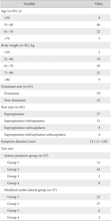
Table 2. Demographics, Tear Type, Tear Size and Tear Chronicity
Variable Value
Age (n=81), yr
≤50 8
51−60 46
61−70 22
>70 5
Body weight (n=81), kg
≤50 2
51−60 18
61−70 30
71−80 22
>80 9
Dominant arm (n=81)
Dominant 59
Non-dominant 22
Tear type (n=81)
Supraspinatus 57
Supraspinatus+infraspinatus 11
Supraspinatus+subscapularis 9
Supraspinatus+infraspinatus+subscapularis 4
Symptom duration (mo) 12.1 (1−120)
Tear size
Antero-posterior group (n=57)
Group 1 12
Group 2 43
Group 3 2
Group 4 0
Modified medio-lateral group (n=57)
Group 1 22
Group 2 27
Group 3 6
Group 4 2
Values are presented as number or median (range). Antero-posterior group (AP group): AP group 1<1 cm; 1 cm≤AP group 2< 3 cm; 3 cm≤ AP group 3<5 cm; AP group 4≥5 cm. Modified medio-lateral group 1: the torn end of the supraspinatus is within its insertion site of the greater tubercle of humer- us, group 2: the torn end of the supraspinatus is located medial to the greater tubercle and lateral to the center of the humeral head, group 3: the torn end of the supraspinatus is medial to the center of the humerus and lateral to the glenoid, and group 4: the torn end of the supraspinatus is found at or medial to the glenoid, which are illustrated in Fig. 2.
Muscle strength and muscle endurance can be improved by training in terms of both growth and cooperativeness. Individuals who have enhanced muscle strength and endurance of rotator cuffs show less sports-related degeneration of rotator cuff mus- cles and more protection against damages from accidents than those who do not.9) We postulated three hypotheses from these findings. First, we hypothesized that isokinetic muscle strength and endurance will become lower as rotator cuff tears are as- sociated with multiples tears. Second, we hypothesized that isokinetic muscle strength and endurance will become lower as the size of the all-thickness tears of the supraspinatus tendon becomes larger. Third, a closer correlation will be seen between muscle strength and endurance with the medio-lateral diameter than the antero-posterior diameter of the tears. In this study, we assessed how isokinetic muscle strength and muscle endurance correlated to the type and size of all-thickness tears of the rotator cuff.
Methods
Subjects of Study
To assess the effect of the type of rotator cuff tears, we first selected patients with all-thickness tears of the supraspinatus tendon of the shoulder, as diagnosed by magnetic resonance im- aging (MRI) from September 2012 and February 2014, including only male patients with tears that were classified as single all- thickness tears. Next, we selected patients with all-thickness tears of the supraspinatus tendon that were combined all-thickness tears of either the infraspinatus tendon and/or the subscapularis tendon. We categories all the patients into the type of tears they had but did not categorize the tears into horizontal lengths. In total, 81 patients were enrolled with an average age of 57.8 ± 7.4 years into the tear-type group. To assess the effect of the size of rotator cuff tears, we classified the patients with single all-thick- ness tears of the supraspinatus tendon into tear-size. In total, 57 patients with an average age of 56.9 ± 7.3 years were included into the tear-size group. In both groups, patients with bilateral tears of the rotator cuffs, previous history of shoulder surgery, partial tears of the rotator cuffs, a diagnosed frozen shoulder, chronic pseudoparalysis, or with neurological condition were excluded from the study subjects (Table 1, 2).
muscle strength (60o/s) and muscle endurance (180o/s) of the shoulder at flexion, extension, abduction, adduction, external rotation, and at internal rotation. Although the optimal veloc- ity for measuring the isokinetic strength is unknown, following past examples of use of low angular velocity to measure muscle strength, and high angular velocity to measure muscle endur- ance, in our study, we used 60o/s and 180o/s angular velocities for the measurement of isokinetic muscle strength and endur- ance, respectively.7,10) To prepare patients for optimal condition for measurement, the patients were asked to do 5 minutes of stretching before the examination. The measurement of muscle strength and endurance for the flexion and extension were taken with the arms starting from various positions; elbows at extension or the shoulder girdle at maximum flexion, whilst the patient sat with their antebrachium in neutral position. For ab- duction and adduction, the measurements were taken with the elbows from extension and the shoulder girdle from maximum adduction. For external and internal rotation, the measurements were taken with the shoulder from 90o of abduction and the elbow from 90o flexion. The elbows were fixed at 90o through- out the assessment. During the measurements, to ensure that patients generated force using only the rotator cuff muscles, the patients’ abdominal region and trunk were fastened using a belt. Further, the shoulder axis was matched to the center of the dynamometer by asking the patient to grab the handles comfort- ably on either side of the seat. In total, an average of the 5 mea- surements for isokinetic muscle strength and an average of 10 measurements of muscle endurance were taken after a practice run of 3 times. All measurements were taken by an experienced examiner with 11 years of experience. A resting period of 3 minutes between each measurement ensured initialization to normal resting conditions before the next measurement. For all patients, the contra-lateral side was measured first to compare with the affected arm and to give time for patients to get ac- custom to the dynamometer. Shklar and Dvir11) have shown that in healthy individuals, irrespective of laterality there is no differ- ence in the isokinetic muscle function between the two sides of the shoulders. Given that the range of motion was normal, there was no pain, and no shoulder abnormalities when assessed by physical examination, the unaffected arm of the affected arm was termed as the contra-lateral side. No radiological tests were performed on the contra-lateral arm.
2) Classification of tears and tear-size
Patients with rotator cuff tears were classified according to the type of, or the combination of, tears they had. Group 1 included those with a single all-thickness tear of the supraspinatus ten- don. Group 2 included those with a supraspinatus tendon tear that was combined with an infraspinatus tendon tear. Group 3 included those with a supraspinatus tendon tear that was com- bined with a subscapularis tendon tear. Lastly, group 4 included those with multiple tears of supraspinatus tendon, infraspinatus
tendon, and the subscapularis tendon.
We used a modified approach of DeOrio and Cofield’s ar- throscopic approach12) that measures the antero-posterior diam- eter of tears. However, instead of an arthroscopic approach we used a radiological approach to measure the antero-posterior and the medio-lateral diameters of cuff tears as our parameters for tear size. First, a T2-weighted MRI scan was taken at sagit- tal plane to measure the maximum antero-posterior diameter of the supraspinatus tendon tear (henceforth called the antero- posterior diameter) (Fig. 1). Next, a T2-weighted MRI scan at the coronal plane was taken to measure the maximum medio-lateral diameter of the supraspinatus tendon tear (henceforth called the medio-lateral diameter). For the medio-lateral diameter classifi- cation of the supraspinatus tendon size, an additional classifica- tion was made according to how the supraspinatus tears were positioned relative to the glenohumeral joint (henceforth called the modified medio-lateral diameter). First, if the tear resided within the root of the greater tuberosity of the supraspinatus ten- don, the patients were grouped into group 1. Second, if the tear was medial but was more lateral than the center of the humeral head then the patients were grouped into group 2. Third, if the tear was more medial than the center of the humeral head and lateral to the glenoid cavity, then they were grouped into group 3.
Lastly, if the tear was medial to or further than the glenoid cavity they were grouped into group 4 (Fig. 2).
3) Statistical analysis
Statistical analyses of the effect of the type of cuff tear on muscle strength and endurance were performed using ANOVA.
Statistical analyses of the effect of antero-posterior and modified medio-lateral diameters of the supraspinatus tear were also per- formed using ANOVA. Post hoc analyses were performed using a Dunnett T3 multiple comparison test. All statistical tests were run on IBM SPSS Statistics ver. 20.0 (IBM Co., Armonk, NY, USA).
Fig. 1. This figure shows the anterior to posterior diameter of the rotator cuff tear. AP: antero-posterior.
AP diameter
Posterior Anterior
Results
Effect of the Type of Rotator Cuff Tears on Isokinetic Muscle Strength and Muscle Endurance
Isokinetic muscle strength: The isokinetic strength was the largest in group 1 but the smallest in group 4 during abduction (p=0.038), flexion (p=0.011), extension (p=0.004), and inter- nal rotation (p=0.004). The isokinetic strength largest in group 1 but the smallest in group 2 during adduction (p=0.019). These differences in isokinetic strength between the groups were sig- nificant except during external rotation (Table 3).
Muscle endurance: Muscle endurance was the highest in group 1 but the lowest in group 2 during flexion (p=0.015), the highest in group 1 and the lowest in group 4 during extension (p=0.032) and internal rotation (p=0.018). These differences in muscle endurance between the groups were significant. No significant differences were seen in muscle endurance between different tear-types during abduction, adduction, or during ex- ternal rotation (Table 3).
Effect of the Supraspinatus Tendon Tear-size on Isokinetic Muscle Strength and Muscle Endurance
1) Effect of supraspinatus tendon tears classified accord- ing to the antero-posterior diameter
The different tear sizes classified by antero-posterior diameter showed no significant differences in the isokinetic strength and muscle endurance during all movements (Table 4).
2) Effect of supraspinatus tendon tears classified accord- ing to the medio-lateral diameter
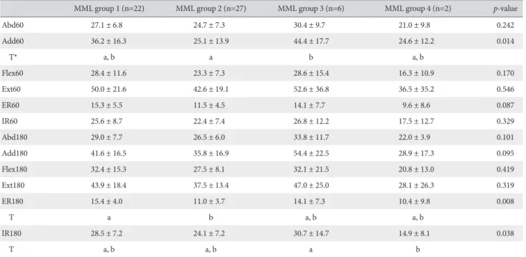
Isokinetic muscle strength: We found that group 3 showed the largest isokinetic muscle strength during abduction, adduc- tion, flexion, extension and at internal rotation, whereas, group 1 showed smallest value during internal rotation. Group 4 showed
the smallest isokinetic strength during all movements. However, between all movements only did we see a significant difference between the values of isokinetic strength during internal rotation between the different groups. We did not see any significant dif- ferences between the groups during abduction, flexion, exten- sion, external rotation or during internal rotation (Table 5).
Muscle endurance: We found that group 3 had the highest muscle endurance during abduction, adduction, extension, or at internal rotation, whereas group 1 had the highest during flexion and external rotation. Again, group 4 had the lowest value for muscle endurance during every position. However, a significant difference in the values of muscle endurance between all the groups were seen during external rotation (p=0.008) and inter- nal rotation (p=0.038), but not during abduction, adduction, flexion or extension (Table 5).
Discussion
In support of our first hypothesis, we found that isokinetic strength and muscle endurance was lower when the rotator cuffs were associated with more tears, especially if combined tears were found in the supraspinatus, infraspinatus, or the sub- scapularis tendons a complete imbalance in force coupling led to a prominent decrease in these strength parameters. We found that the decrease in isokinetic muscle strength varied in terms of the type of rotator cuff tears associated. The isokinetic muscle strength was the largest during all the movements that were tested in patients with only single tears of the supraspinatus ten- don, and the lowest, except during adduction, in patients with combined triple tears of the supraspinatus, infraspinatus, and the subscapularis tendons. Similarly, the muscle endurance was the highest during all movements in patients with single tears of the supraspinatus tendon and lowest in patients with a combination Fig. 2. Modified medio-lateral group. (A) The torn end of the supraspinatus is within its insertion site of the greater tubercle of humerus. (B) The torn end of the supraspinatus is located medial to the greater tubercle and lateral to the center of the humeral head. (C) The torn end of the supraspinatus is medial to the center of the humerus and lateral to the glenoid. (D) The torn end of the supraspinatus is found at or medial to the glenoid.
A B C D
of triple tears of the supraspinatus, infraspinatus, and the sub- scapularis tendons.
Rejecting our second hypothesis, we found that muscle strength and endurance did not decrease with the increase in the size of the single all-thickness tear of the supraspinatus ten- don. For patients classified according to antero-posterior diame- ter, we found that the isokinetic strength was largest in all move- ments except during flexion in patients with a tear size of an antero-posterior diameter of 1 to 3 cm and smallest in all move- ments in the 3 to 5 cm group. We found that muscle endurance was highest in all movements except during flexion and external rotation in the 1 to 3 cm group. For patients classified according to the modified medio-lateral diameter, the isokinetic strength was the largest during all movement except during external rota- tion in group 3, and the smallest during all movements in group 4. We found that the muscle endurance was highest during all
movements except during flexion and external rotation in group 3, and the lowest during all movements in group 4.
Lastly, we cautiously conclude, in support of our third hy- pothesis, that there is a closer correlation between medio-lateral diameter than the antero-posterior diameter with isokinetic strength and muscle endurance in patients with torn supraspi- natus tendons. We found that within patients classified by either measures of tear size, those in the classification just below the largest sized tears showed the highest isokinetic strength and muscle endurance for most measurements whereas those of the largest sized tears showed the lowest isokinetic strength and muscle endurance. Although we cannot say that tear size com- pletely reflects tear chronicity and that a supraspinatus tendon tear can become a chronic tear before it becomes a massive tear, it requires further study to reveal whether the results are an artifact of a compensatory growth of muscles other than the Table 3. Differences according to the Type of Torn Cuffs
Type group 1 (n=57) Type group 2 (n=11) Type group 3 (n=9) Type group 4 (n=4) p-value
Abd60 26.1 ± 7.5 20.2 ± 8.2 23.8 ± 6.8 18.6 ± 3.1 0.038
T* a a, b a, b b
Add60 31.4 ± 16.4 17.1 ± 8.3 28.0 ± 13.0 17.4 ± 10.7 0.019
T a B a, b a, b
Flex60 25.6 ± 10.4 16.7 ± 7.7 19.5 ± 7.6 15.5 ± 9.1 0.011
T a a, b a, b b
Ext60 46.3 ± 22.5 27.1 ± 18.2 37.0 ± 17.7 15.9 ± 7.9 0.004
T a B a, b b
ER60 13.1 ± 5.6 10.0 ± 4.0 12.0 ± 2.8 9.7 ± 5.6 0.189
T a B a, b a, b
IR60 24.0 ± 8.7 19.0 ± 7.8 19.0 ± 4.8 10.2 ± 3.5 0.004
T a a, b a, b b
Abd180 28.1 ± 7.6 24.0 ± 10.4 27.5 ± 9.2 19.0 ± 5.0 0.099
Add180 39.8 ± 17.9 29.0 ± 19.0 37.3 ± 20.5 24.7 ± 16.9 0.167
T a a a a
Flex180 29.6 ± 13.1 18.0 ± 9.6 25.3 ± 11.0 17.1 ± 12.1 0.015
T a b a, b a, b
Ext180 40.7 ± 17.3 28.4 ± 18.3 34.0 ± 15.1 20.2 ± 18.6 0.032
T a a, b a, b b
ER180 13.0 ± 4.9 12.0 ± 6.0 12.2 ± 3.4 7.9 ± 2.8 0.221
T a a a a
IR180 26.2 ± 8.7 20.5 ± 9.3 21.9 ± 6.7 14.8 ± 6.4 0.018
T a a, b a, b b
Values are presented as mean ± standard deviation.
Type group 1: supraspinatus, Type group 2: supraspinatus+infraspinatus, Type group 3: supraspinatus+subscapularis, Type group 4: supraspinatus+infraspinatus +subscapularis, Abd: abduction, Add: adduction, Flex: flexion, Ext: extension, ER: external rotation, IR: internal rotation.
*The same letters indicate insignificant difference between groups based on Dunnett T3 multiple comparison test.
Table 4. Differences between Groups Classified by the Antero-posterior Diameter of the Supraspinatus Tendon Tear
AP group 1 (n=12) AP group 2 (n=43) AP group 3 (n=2) AP group 4 (n=0) p-value
Abd60 24.9 ± 5.8 26.7 ± 7.9 21.0 ± 9.8 0.478
Add60 29.8 ± 17.0 32.1 ± 16.6 24.6 ± 12.2 0.766
Flex60 29.2 ± 13.1 25.0 ± 9.4 16.3 ± 10.9 0.206
Ext60 41.8 ± 22.4 48.0 ± 22.4 36.5 ± 35.2 0.582
ER60 13.3 ± 5.0 13.3 ± 5.7 9.6 ± 8.6 0.662
IR60 22.9 ± 6.7 24.5 ± 9.0 17.5 ± 12.7 0.488
Abd180 26.7 ± 7.9 28.7 ± 7.6 22.0 ± 3.9 0.373
Add180 34.7 ± 18.3 41.7 ± 17.8 28.9 ± 17.3 0.339
Flex180 34.1 ± 18.2 28.8 ± 11.3 20.8 ± 13.0 0.297
Ext180 36.4 ± 19.7 42.4 ± 16.3 28.1 ± 26.3 0.335
ER180 13.6 ± 3.7 13.0 ± 3.7 10.4 ± 9.8 0.687
IR180 26.0 ± 3.6 26.8 ± 9.4 14.9 ± 8.1 0.165
T* a a a
Values are presented as mean ± standard deviation. Antero-posterior group (AP group 1)<1 cm, 1 cm≤AP group 2<3 cm, 3 cm≤AP group 3<5 cm, AP group 4≥5 cm.Abd: abduction, Add: adduction, Flex: flexion, Ext: extension, ER: external rotation, IR: internal rotation.
*The same letters indicate insignificant difference between groups based on Dunnett T3 multiple comparison test.
Table 5. Differences between Groups Classified by the Modified Mediolateral Diameter of the Supraspinatus Tendon Tear
MML group 1 (n=22) MML group 2 (n=27) MML group 3 (n=6) MML group 4 (n=2) p-value
Abd60 27.1 ± 6.8 24.7 ± 7.3 30.4 ± 9.7 21.0 ± 9.8 0.242
Add60 36.2 ± 16.3 25.1 ± 13.9 44.4 ± 17.7 24.6 ± 12.2 0.014
T* a, b a b a, b
Flex60 28.4 ± 11.6 23.3 ± 7.3 28.6 ± 15.4 16.3 ± 10.9 0.170
Ext60 50.0 ± 21.6 42.6 ± 19.1 52.6 ± 36.8 36.5 ± 35.2 0.546
ER60 15.3 ± 5.5 11.5 ± 4.5 14.1 ± 7.7 9.6 ± 8.6 0.087
IR60 25.6 ± 8.7 22.4 ± 7.4 26.8 ± 12.2 17.5 ± 12.7 0.329
Abd180 29.0 ± 7.7 26.5 ± 6.0 33.8 ± 11.7 22.0 ± 3.9 0.101
Add180 41.6 ± 16.5 35.8 ± 16.9 54.4 ± 22.5 28.9 ± 17.3 0.095
Flex180 32.4 ± 15.3 27.5 ± 8.1 32.1 ± 21.5 20.8 ± 13.0 0.419
Ext180 43.9 ± 18.4 37.5 ± 13.4 47.0 ± 25.0 28.1 ± 26.3 0.319
ER180 15.4 ± 4.0 11.0 ± 3.7 14.1 ± 7.3 10.4 ± 9.8 0.008
T a b a, b a, b
IR180 28.5 ± 7.2 24.1 ± 7.2 30.7 ± 14.7 14.9 ± 8.1 0.038
T a, b a, b a b
Values are presented as mean ± standard deviation. Modified medio-lateral (MML group): modified mediolateral group 1: the torn end of the supraspinatus is within its insertion site of the greater tubercle of humerus, group 2: the torn end of the supraspinatus is located medial to the greater tubercle and lateral to the center of the humeral head, group 3: the torn end of the supraspinatus is medial to the center of the humerus and lateral to the glenoid, and group 4: the torn end of the supraspinatus is found at or medial to the glenoid, which are illustrated in Fig. 2.
Abd: abduction, Add: adduction, Flex: flexion, Ext: extension, ER: external rotation, IR: internal rotation.
*The same letters indicate insignificant difference between groups based on Dunnett T3 multiple comparison test.
supraspinatus tendon, such as rotator cuff and the subscapularis muscles, that compensates for the loss in muscle strength in rota- tor cuffs with small-sized tears but not by massive tears.
The isotonic, isometric, and isokinetic strength of the shoul- der can be used to manually assess shoulder muscle strength.
As these measurements can be taken quickly and easily, these manual tests are often used to pre- and post-operatively assess the shoulder muscle strength in rotator cuff tear patients.12) Al- though as manual tests they are open to influence of subjective examination, especially for the isometric strength tests, mean- ing that reliability decreases,12,13) the isokinetic strength test has shown to be effective to measure relatively objectively despite the subnormal force in muscle contraction in injured arms. For isokinetic strength testing, it is however important that the pa- tient is still able to generate the set angular velocity and length- tension function of the muscle is unimpaired. The isokinetic muscle strength has an added advantage of overcoming the problem of spasticity7) and that it objectively measures dynamic rotator strengths using functional muscle movements.13) How- ever, isokinetic strength tests are not always useful in measuring the shoulder muscle strength in rotator cuff conditions. For ex- ample, if patients cannot attain the set angular velocity then the isokinetic strength measured becomes 0% of the contra-lateral side. Also, if patients are in too much pain, cannot maintain the required position whilst testing, is limited in their range of mo- tion they will not be able to generate the required angular veloc- ity. In this study, 3 patients were excluded midway as they could not maintain the required position for testing, and thus did not reach the required angular velocity.
Previous studies have shown that a loss in strength transfer in torn rotator cuffs leads to decrease in isokinetic muscle strength of the affected arm by around 37% to 70% of the contra-lateral unaffected arm.7,14,15) In case of small-sized tears, the force is partially transmitted through the center of the tear,16) but as the tear size increases not only does the supraspinatus tendon lose muscle strength, the infraspinatus tendon and the subscapularis tendon are affected too.17-19) The muscles involved in shoulder flexion are the anterior deltoid muscle, clavicular pectoralis ma- jor muscle, biceps brachii and the coracobrachial muscle. The muscles involved in shoulder extension are posterior deltoid muscle, teres major, clavicular pectoralis major muscle, latissi- mus dorsi muscle and the biceps brachii long head. The muscles involved in abduction are central deltoid muscle, supraspinatus muscle and the biceps brachii long head. The muscles involved in adduction are pectoralis major muscle, latissimus dorsi mus- cle, teres major, triceps brachii long head and the coracobrachial muscle. The muscles for external rotation are posterior deltoid muscle, infraspinatus muscle and teres minor. Lastly, the muscles for internal rotation are pectoralis major muscle, teres major, latissimus dorsi muscle, anterior deltoid muscle, and the sub-
scapularis muscle.11,20-22) The external rotators and internal rota- tors are directly influenced by the actions of rotator cuffs. How- ever, in our study, we found that the isokinetic strength during external rotation was the lowest in only two groups classified by the type of rotator cuff tears. We believe that this is because the actual shoulder motion is brought about the cooperation of rota- tor cuffs along with several other muscles, and we did not assess functional strength ratio, which has recently been highlighted as an important indicator of muscle strength. Further, although this was not considered in our study, the chronicity of tears is expect- ed to have a large influence on muscle strength and endurance.
Although most people believe that contraction of the muscle is concentric, eccentric muscle contraction, which balance high levels of force without much input of energy, is considered as a far more useful type of contraction than concentric contraction in sports training and in the rehabilitation setting.23) Recently, the conventional strength ratio, which is the ratio of the external rotator to internal rotator strength in concentric action, and the functional strength ratio, which is the ratio of eccentric external rotator strength to concentric internal rotator strength, over the simple peak torques have been highlighted as useful indicators of strength. The functional strength ratio is especially useful in that it takes into consideration the various antagonistic muscles of eccentric contraction involved in the dynamic stability of the glenohumeral joints.24)
The authors compared the difference in muscle strength and muscle endurance between sex, age, and weight. When we compared the data according to the type of rotator cuff tears, males showed a significant difference in terms of the muscle strength during abduction, adduction, flexion, extension, and internal rotation, and in terms of the muscle endurance during flexion, extension, and the internal rotation. However, females did not show any significance difference in muscle strength and endurance during all movement, thus they were not included in this study. In female patients who showed some extent of sig- nificant difference between movements in terms of the kinetic muscle strength and endurance, may have been influenced by shoulder girdle muscles rather than the rotator cuff tendons themselves. We found that the isokinetic strength and endur- ance of both the affected and the contra-lateral arms increased with age, but this trend was more prominent for males than for females. The isokinetic muscle strength and the muscle endur- ance did not show a significant relationship with weight of either the affected or the contra-lateral arm in both males and females.
For the experimental approach to assess the isokinetic muscle strength and muscle endurance in terms of the size of the rotator cuff tears, we modified DeOrio and Cofield’s method of clas- sifying rotator cuffs by their antero-posterior diameter using ar- throscopy by taking the measurements from MRI scans instead.
As well as classifying the patients into different types of tears by
their antero-posterior diameter, we also classified patients in terms of the position of the tears relative to the humeral head and glenoid cavity (we called this the modified medio-lateral diameter) and assessed these factors. There were several reasons behind the modification. First, DeOrio and Cofield’s classifica- tion could only be made at the time of surgical intervention- our attempt was to make a preoperative decision on the clas- sification through MRI. Second, a clinician tends to be more familiarized with the medio-lateral diameter of rotator cuff tears than the preoperative antero-posterior diameter, thus we deter- mined to see whether there was a difference between the two diameters. Third, we predicted that the anatomical size of the humeral head of an Asian background would be smaller than of a Caucasian background, and we reasoned that the same clas- sification system may not be appropriate for Korean patients. So, if DeOrio and Cofield’s classification is followed, a preoperative MRI assessment that rules out a massive tear of the rotator cuffs may sometimes actually be difficult to repair surgically. Using our modified approach to classify tear-size, we did not find a signifi- cant correlation between the antero-posterior diameter, which were subcategorized into tear size of 1 cm, 3 cm, and 5 cm, and our parameters of strength to support our second hypothesis.
However, we found a significant correlation between the antero- posterior diameter that was subcategorized into their position relative to the humeral head and glenoid cavity and muscle strength. We suggest from these results that at least for the as- sessment of muscle strength of shoulders of Korean males a new classification of rotator cuff tear sizes may be needed.
There are several limitations to our study. First, we did not factor out individual pain levels at the time of isokinetic muscle strength measurement. An individual with pain would have underachieved in the preoperative rotator cuff muscle strength test, thus potentially giving an underestimated value. As the pain resolved postoperatively, a greater improvement may be seen in the muscle strength than if the pain had been relieved momen- tarily at the preoperative assessment.15) However, giving patients subacromial localized injection of anesthesia could unwittingly enhance muscle strength in individuals with rotator cuff tears.25) Nevertheless, we cannot rule out that not having controlled the pain before a preoperative rotator cuff muscle strength test may have influenced our results. Second, patients with asymptom- atic rotator cuff tears could be present in the contra-lateral side.
Although we questioned patients of the absence or presence of pain, their previous medical history of the contra-lateral shoul- der, and we excluded all individuals who were found to have a bilateral tear through MRI or ultrasound in our study, since pro- spective studies using MRI or ultrasounds show that all-thickness tears are in 28% of individuals over 60s, 65% in over 70s,26) a high prevalence of asymptomatic tears makes it likely our sub- ject of studies may have included bilateral patients. Later studies where objectively assessed controls are included and a proper
diagnosis of the contra-lateral side to eliminate any possibility of bilateral tears should be performed.
Conclusion
We found that rotator cuff tears that are combined with ad- ditional tears are more likely to show lower muscle strength and endurance. However, in case of tears of the supraspinatus ten- don, we found that how large the size of the single all-thickness tears of the supraspinatus tendon did not influence the isokinetic strength nor the endurance of the muscles, i.e. a large tear of the supraspinatus tendon was not associated with a lower isokinetic strength or muscle endurance any more than a small tear. Fur- thermore, we found that our modified medio-lateral diameter was more correlated with isokinetic strength and muscle endur- ance than the antero-posterior diameter. Currently, no studies exist to objectively inform clinicians as to changes the in muscle strength and endurance according to the type and size of rota- tor cuff tears. Our study gives methods to measure parameters of muscle strength and endurance so that they may be used as objective markers to assess the extent of preoperative condition and postoperative recovery of muscle function.
References
1. Cameron BD, Iannotti JP. Clinical evaluation of the painful shoulder. In: Zlatkin MB, ed. MRI of the shoulder. 2nd ed.
Philadelphia: Lippincott Williams & Wilkins; 2003. 47-84.
2. Bigliani LU, Morrison DS, April EW. The morphology of the ac- romion and its relationship to rotator cuff tears. Orthop Trans.
1986;10:228-35.
3. Soslowsky LJ, Carpenter JE, Bucchieri JS, Flatow EL. Bio- mechanics of the rotator cuff. Orthop Clin North Am.
1997;28(1):17-30.
4. Dunn WR, Kuhn JE, Sanders R, et al. Symptoms of pain do not correlate with rotator cuff tear severity: a cross-sectional study of 393 patients with a symptomatic atraumatic full-thickness rotator cuff tear. J Bone Joint Surg Am. 2014;96(10):793-800.
5. Ellman H, Hanker G, Bayer M. Repair of the rotator cuff. End- result study of factors influencing reconstruction. J Bone Joint Surg Am. 1986;68(8):1136-44.
6. Gore DR, Murray MP, Sepic SB, Gardner GM. Shoulder- muscle strength and range of motion following surgical re- pair of full-thickness rotator-cuff tears. J Bone Joint Surg Am.
1986;68(2):266-72.
7. Rabin SI, Post M. A comparative study of clinical muscle test- ing and Cybex evaluation after shoulder operations. Clin Or- thop Relat Res. 1990;(258):147-56.
8. Walker SW, Couch WH, Boester GA, Sprowl DW. Isokinetic strength of the shoulder after repair of a torn rotator cuff. J Bone Joint Surg Am. 1987;69(7):1041-4.
9. Kibler WB, Chandler TJ, Stracener ES. Musculoskeletal adap- tations and injuries due to overtraining. Exerc Sport Sci Rev.
1992;20:99-126.
10. Forthomme B, Croisier JL, Ciccarone G, Crielaard JM, Cloes M.
Factors correlated with volleyball spike velocity. Am J Sports Med. 2005;33(10):1513-9.
11. Shklar A, Dvir Z. Isokinetic strength relationships in shoulder muscles. Clin Biomech (Bristol, Avon). 1995;10(7):369-73.
12. Marino M, Nicholas JA, Gleim GW, Rosenthal P, Nicholas SJ.
The efficacy of manual assessment of muscle strength using a new device. Am J Sports Med. 1982;10(6):360-4.
13. Davies GJ, Ellenbecker TS, Wilk KE. Isokinetic testing and reha- bilitation of the shoulder complex. In: Wilk KE, Reinold MM, Andrews JR, eds. The Athlete’s shoulder. 2nd ed. Philadelphia:
Churchill Livingstone; 2009. 719-47.
14. Basmajian JV. Therapeutic exercise. 4th ed. Baltimore: Wil- liams and Wilkins; 1984. 88.
15. Ivey FM Jr, Calhoun JH, Rusche K, Bierschenk J. Isokinetic test- ing of shoulder strength: normal values. Arch Phys Med Reha- bil. 1985;66(6):384-6.
16. Mccardle WD, Katch FI, Katch VL. Exercise physiology. Energy, nutrition and human performance. Philadelphia: Lea & Fe- biger; 1981. 288-94.
17. Kirschenbaum D, Coyle MP Jr, Leddy JP, Katsaros P, Tan F Jr, Cody RP. Shoulder strength with rotator cuff tears. Pre- and postoperative analysis. Clin Orthop Relat Res. 1993;(288):174- 8.
18. Ben-Yishay A, Zuckerman JD, Gallagher M, Cuomo F. Pain inhibition of shoulder strength in patients with impingement syndrome. Orthopedics. 1994;17(8):685-8.
19. Burkhart SS. Reconciling the paradox of rotator cuff repair versus debridement: a unified biomechanical rationale for the treatment of rotator cuff tears. Arthroscopy. 1994;10(1):4-19.
20. Tae SK, Kim JY, Park JS. MRI follow-up study after arthroscopic repair of multiple rotator cuff tendons. J Korean Shoulder El- bow Soc. 2008;11(2):96-103.
21. Clark JM, Harryman DT 2nd. Tendons, ligaments, and capsule of the rotator cuff. Gross and microscopic anatomy. J Bone Joint Surg Am. 1992;74(5):713-25.
22. Romanes GJ. Cunningham’s textbook of anatomy. 12th ed.
London; New York: Oxford University Press; 1981. 373-7.
23. Miller MD, Thompson SR. DeLee & Drez’s orthopaedic sports medicine: principles and practice. 4th ed. Philadelphia: Else- vier; 2014. 442-4.
24. Andrade Mdos S, de Lira CA, Vancini RL, de Almeida AA, Benedito-Silva AA, da Silva AC. Profiling the isokinetic shoul- der rotator muscle strength in 13- to 36-year-old male and female handball players. Phys Ther Sport. 2013;14(4):246-52.
25. Minagawa H, Itoi E, Konno N, et al. Humeral attachment of the supraspinatus and infraspinatus tendons: an anatomic study. Arthroscopy. 1998;14(3):302-6.
26. Watson MS, Surgical disorders of the shoulder. New York:
Churchill Livingstone; 1991.