Dynamic Stabilization of the Scapula for Serratus Anterior Dysfunction:
A Retrospective Study of Functional Outcome and Results
Soo Tai Chung* , Jon J. P. Warner1,*
Department of Orthopaedic Surgery, Catholic Kwandong University International St. Mary’s Hospital, Incheon, Korea, 1Harvard Shoulder Service, Massachusetts General Hospital, Boston, MA, USA
Background: Twenty-six patients (12 male and 14 female) with symptomatic scapular winging caused by serratus anterior dysfunction were managed by split pectoralis major tendon transfer (sternal head) with autogenous hamstring tendon augmentation from 1998 to 2006.
Methods: Twenty-five patients showed positive results upon long thoracic nerve palsy on electromyography. The mean duration of symptoms until surgery was 48 months (range, 12–120 months). Four patients had non-traumatic etiologies and 22 patients had trau- matic etiologies. On follow-up assessment for functional improvement, a Constant-Murley score was used. Twenty-one patients were completely evaluated, while five patients who had less than 12 months of follow-up were excluded.
Results: Pain relief was achieved in 19 of the 21 patients, with 20 patients showing functional improvement. The pain scores improved from 6.0 preoperatively to 1.8 postoperatively. The mean active forward elevation improved from 108o (range, 20o–165o) preoperatively to 151o (range, 125o–170o) postoperatively. The mean Constant-Murley score improved from 57.7 (range, 21–86) preoperatively to 86.9 (range, 42–98) postoperatively. A recurrence developed in one patient. Of the 21 patients, ten had excellent results, six had good results, four had fair results, and one had poor results.
Conclusions: Most patients with severe symptomatic scapular winging showed functional improvement and pain relief with resolution of scapular winging.
(Clin Shoulder Elbow 2015;18(4):229-236)
Key Words: Split pectoralis major tendon transfer; Serratus anterior dysfunction; Scapular winging; Long thoracic nerve palsy
Copyright © 2015 Korean Shoulder and Elbow Society. All Rights Reserved. pISSN 2383-8337
Clinics in Shoulder and Elbow Vol. 18, No. 4, December, 2015 http://dx.doi.org/10.5397/cise.2015.18.4.229
Received September 30, 2015. Revised November 11, 2015. Accepted November 30, 2015.
*These authors contributed equally to this paper as co-first authors.
Correspondence to: Soo Tai Chung
Department of Orthopaedic Surgery, Catholic Kwandong University International St. Mary’s Hospital, 25 Simgok-ro 100beon-gil, Seo-gu, Incheon 22711, Korea
Tel: +82-32-290-2925, Fax: +82-32-290-3879, E-mail: [email protected] Financial support: None. Conflict of interests: None.
Introduction
When the balance of periscapular muscles is broken, ab- normal scapulothoracic movement can develop. The most symptomatic abnormal scapulothoracic movement is scapular winging.1) The causes for scapular winging can be divided into three groups; serratus anterior dysfunction, trapezius palsy and facioscapulohumeral muscular dystrophy. Scapular winging caused by serratus anterior dysfunction is the most common disorder. Since the serratus anterior muscle has only one inner- vation via the long thoracic nerve, incidents such as trauma, viral
infection, brachial neuritis, and iatrogenic injuries can lead to its dysfunction.2-7)
The serratus anterior muscle originates from the anterolateral surface of the first 8 or 9 ribs, and inserts at the anterior surface of the medial border of the scapula. This muscle functions in the protraction and lateral rotation of the scapula, and stabilizes the scapula while elevating the arm. The long thoracic nerve that innervates the serratus anterior muscle is long in shape and lo- cated subcutaneously over the ribs of the thorax ; therefore, it is very vulnerable to both direct and indirect injury.8)
Although most patients with serratus anterior palsy can be
Table 1. Patients Data (Causes & Results) Case No. Sex/Age (yr)Injured sideCauses of injuryPrior surgeryApprehension test/ instability symptomElectromyographic dataScapula stabilization testPostop complicationPostop wingingPain scoreActive flexion (preop/postop)Constant- Murley scoreFinal result 1F/30R (D)Ski injuryNoNoLTN palsy+NoNo 5/0 110/170 64/98E 2M/45LIdiopathicC6-7 laminectomyNoLTN palsy+NoNo 4/0 90/150 61/93E 3M/31L (D)IdiopathicLong thoracic nerve decompressionYesLTN & DSN palsy+NoMild 8/2 165/165 76/86G 4M/21LTractionNoNoLTN palsy+-- 7/- 80/- 58/-- 5M/33R (D)IdiopathicNoNoLTN palsy-NoNo 7/2 90/160 64/88G 6F/38LMVANoNoLTN palsy- (no correction)NoNo 2/0 140/150 77/98E 7N/51LWork-relatedNoNoLTN palsy+Pectoralis muscle spasmNo 10/5 100/160 54/91E 8F/25R (D)MVANoNoLTN palsy+NoNo 10/2 80/155 40/93E 9F/33R (D)MVABankart/Putti-PlattNo/secondaryLTN palsy+RecurrenceSevere 8/8 90/80 44/42P 10F/46R (D)TractionAnterior capsular shiftTrace/secondary(?)Normal+NoNo 5/0 90/150 48/85G 11F/34LWork-relatedNoNoLTN palsy+NoNo 5/2 90/140 52/78F 12F/35R (D)MVANoNoLTN palsy+NoNo 5/2 90/150 45/88G 13M/33LWork-relatedCervical discectomyNo/secondaryLTN palsy+Scapulothoracic bursitisNo 10/5 160/160 55/75F 14F/16R (D)Soccer injuryNoNoLTN palsy+NoNo 5/0 130/160 72/98E 15F/30R (D)MVANoYesLTN palsy+NoNo 5/2 80/160 58/88G 16M/38R (D)Work relatedNoNo/secondaryLTN palsy+Idiopathic pain syndromeNo 5/2 150/160 77/79F 17F/18R (D)Lacrosse injuryNoNo/secondaryLTN palsy+NoNo 2/0 160/170 86/98E 18F/31R (D)Work-relatedNoNoLTN palsy+NoMild 5/2 150/150 71/86G 19M/50L (D)MVANoNoLTN palsy+-- 5/- 110/- 56/-- 20M/33R (D)IdiopathicNoNo/secondaryLTN palsy+NoNo 5/0 140/150 64/96E 21M/38L (D)Work-relatedAcromioplastyNoLTN palsy- (no correction)-- 8/- 80/- 42/-- 22F/21LWork-relatedNoNoLTN palsy+NoNo 5/0 90/170 54/98E 23F/29LMVAPosterior capsulorraphyNo/secondaryLTN palsy+NoMild 7/2 20/130 21/76F 24M/48R (D)TractionNoNoLTN palsy- (no correction)NoNo 8/2 60/125 28/91E 25F/38R (D)Roller skating injuryNoNoLTN palsy+-- 7/- 80/- -/-- 26F/39R (D)Work-relatedCervical discectomyNoLTN palsy+-- 5/- 150/- -/-- F: female, M: male, R: right, L: left, D: dominant, MVA: motor vehicle accident, preop: preoperative, postop: postoperative, LTN: long thoracic nerve, DSN: dorsal scapula nerve, E: excellent, G: good, P: poor, F: fair.
treated conservatively, prolonged scapular winging after 12 to 24 months of non-operative management is an indication of sur- gery.3,9) Different techniques have been introduced for the surgi- cal stabilization of winged scapula.10-16) Today, the split pectoralis major tendon transfer is widely used for surgical management of scapular winging.13,15,17-21) However, owing to their being few examples and short follow-up periods, there is little information available regarding this technique for large groups and complete functional outcomes. This study was conducted to evaluate the functional outcome and clinical results in a relatively large group of patients with scapular winging caused by long thoracic nerve injuries.
Methods
Patient Selection
Between 1998 and 2006, 26 patients with serratus anterior dysfunction caused by long thoracic nerve palsy were treated with a split pectoralis major tendon transfer (sternal part) with autogenous hamstring tendon augmentation. All clinical and electromyographic data were reviewed retrospectively. For the final follow-up evaluation, five patients who had less than 12 months follow-up were excluded from the 26 patients due to too short follow-up period.
There were twelve men and fourteen women. The average age of the patients at the time of surgery was 34 years (range, 16–51 years). The dominant shoulder was involved in 18 of the 26 patients. Long thoracic nerve palsy resulted from idiopathic non-traumatic causes in four patients, and traumatic causes in 22 patients. Of the twenty-two patients who had a traumatic serratus anterior dysfunction, seven patients were involved in motor vehicle accidents, eight patients had work related injuries, four patients had sports injuries and three patients had minor traction injuries (Table 1). All patients had painful dysfunction of the shoulder, specifically posterior periscapular pain, scapular
winging and weakness or loss of forward flexion of the arm.
In 25 of the 26 patients, long thoracic nerve injuries were documented in the electromyographic data. All patients were initially treated conservatively for at least 12 months: Most cases had the neuroplaxia-like paresis of long thoracic nerve, therefore we recommended rest and inhibition of lifting activity above the level of the scapula, which usually leads to complete recovery.
The Costant-Murley score was used for postoperative functional evaluation.22) The final results of the 21 patients were graded as excellent, good, fair and poor. Patients were scored as excellent when they were satisfied with their results, exhibited painless, full use of the arm for daily living activity, and had a full range of motion in the shoulder without scapular winging (Fig. 1). Patients received a score of good when they were satisfied with their results but had one or two of the following symptoms, including mild pain when using the arm for daily living activity, mild limita- tion of motion in the shoulder and mild scapular winging. Pa- tients were scored as fair when they were not fully satisfied with their results, reporting mild pain when using the arm for daily liv- ing activities, and mild limitation of motion in the shoulder with or without mild scapular winging. Patients were scored as poor when they were not satisfied with their results, and noted recur- rences of painful scapular winging.
Surgical Procedure
All procedures concerning the split pectoralis major tendon transfer with hamstring tendons augmentation for serratus an- terior dysfunction have been explained in detail in previous ar- ticles.15,23,24)
A brief description of the surgical procedure is as follows (Fig. 2).
Postoperative Rehabilitation
A shoulder immobilizer with an abduction brace was ap- plied in the operating room and worn for the first six weeks.
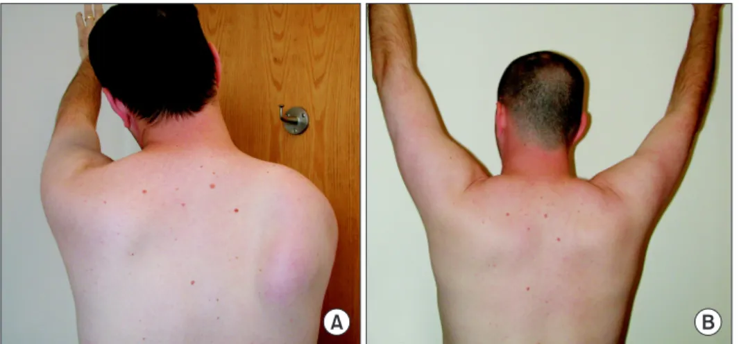
Fig. 1. (A) Scapular winging in patient with serratus anterior dysfunction. (B) Postop- erative image showing scapular stabilization one year after surgery.
A B
During this period, pendulum exercises and passive shoulder motion were encouraged to ensure the smooth gliding of the transferred tendon and graft, and to avoid scar formation. The sling was worn for the entire six weeks and taken off only for therapy. After six weeks, the sling was discontinued and active assisted range of motion as well as a home pulley unit was used.
Water therapy was used if available. At three months, isometric strengthening exercises with elastic bands were started. A bio- feedback program using Myotrac® (Thought Technology Ltd., Montreal West, QC, Canada) was started under the supervision of a well trained physical therapist. At six months, heavy labor or lifting was permitted. It was recommended that contact sports be avoided for one year after surgery.
Results
Twenty-six patients that underwent the procedure were eval-
uated clinically and electromyographically. The average follow up period was 42 months (range, 12–92 months).
All but one patient showed chronic denervation of the long thoracic nerve during electromyographic evaluation. The one exception was a patient who had normal electromyographic data despite typical scapular winging due to serratus anterior dysfunction. One of the twenty-five patients who had abnormal electromyographic data in the long thoracic nerve had a com- bined nerve injury in the dorsal scapular nerve, which might have developed via an idiopathic cause or a viral infection. Trau- matic long thoracic nerve palsy was observed in 22 of the 26 patients as a result of accidents (7 patients), sports and minor in- juries (7 patients), and work-related (mainly heavy lifting) injuries (8 patients) (Table 1).
The mean period from the beginning of symptomatic scapu- lar winging to surgery was 48 months (range, 12–120 months).
Seven patients had previous operations including one cervical Fig. 2. Surgical procedure. (A) Hamstring autograft harvesting and preparation. (B) Split pectoralis major tendon dissection.
(C) Preparation of pectoralis major tendon with hamstring autograft for transfer. (D) Preparation for recipient site of scapula and transfer path. (E) Securing of graft to infe- rior pole of scapula.
A
B
C
D
E
laminectomy, one cervical discectomy, one long thoracic nerve decompression, one anterior capsular shift, one posterior capsu- lar shift, one Putty-Platt procedure, and two acromioplasties in one patient. A scapular stabilization test improved the forward elevation of the arm in 22 patients, but could not stabilize the scapula in four patients (Table 1).
Instability was a concern in nine patients. Two patients had positive apprehension tests, while the other seven felt instability symptoms without apprehension signs on physical examination, despite three of these seven patients having prior instability op- erations (two had open surgery for anterior instability and one had arthroscopic posterior capsulorrhaphy). Instability symptoms were relieved in seven patients after surgical dynamic stabiliza- tion of the scapula. Two patients showed glenohumeral instabil- ity with scapular winging, and their positive apprehension signs remained after surgical correction for scapular winging. These patients were advised to get surgery for glenohumeral instability.
Twenty of the patients experienced pain relief from the pro- cedure, with eight patients experiencing no pain and twelve patients experiencing mild pain. The pain scores of patients im- proved from an average of 6.0 out of 10 preoperatively to an av- erage of 1.8 out of 10 postoperatively. The mean active forward elevation improved from 108o (range, 20o–165o) preoperatively to 151o (range, 125o–170o) postoperatively. The mean Constant- Murley score improved from 57.7 (range, 21–86) preoperatively to 86.9 (range, 42–98) postoperatively (Table 2).
Preoperatively, severe scapular winging was observed in all patients. After performing a split pectoralis major tendon transfer with autogenous semitendinous and gracilis augmentation, the winging was completely corrected in 18 of the 21 patients, mild winging remained in two patients, and severe scapular winging was recurrent in one patient. Although one patient had scapular winging caused by multiple nerve injuries, involving both the long thoracic nerve and dorsal scapular nerve, he had an excel- lent outcome.
Postoperative complications developed in 4 of the 21 pa- tients (19.0%). A recurrence of scapular winging 11 months after surgical dynamic stabilization occurred in one patient (4.8%), which was managed with a scapulothoracic fusion. A pectoralis major muscle spasm developed in one patient, but improved with physical therapy. A scapulothoracic bursitis developed in one patient and was treated with injection therapy into the bursa. Idiopathic upper extremity pain syndrome developed in one patient and was managed in the pain clinic department.
No other complicated symptoms were developed on the donor sites for harvesting of the semitendinous and gracilis tendons. All complications developed in trauma-related patients with one failure in a motor vehicle accident group, and the other three complications in the work-related injuries group.
In the final follow-up, ten of the twenty-one patients (47.6%)
had excellent results, six patients (28.6%) had good results, four Table 2.
Analysis of Final Results by the Causes of Injury VariableNon-traumatic; idiopathic, viral (4 shoulders)Motor vehicle accident (6 shoulders)Sport & minor trauma (5 shoulders)Work-related (6 shoulders)Total (21 shoulders) PreoperativePostoperativePreoperativePostoperativePreoperativePostoperativePreoperativePostoperativePreoperativePostoperative Active flexion (°)*121 (90–165)156 (150–165)83 (20–140)138 (80–160)110 (60–160)155 (125–170)123 (90–150)157 (140–170)108 (20–165)151 (80–170) Constant-Murley score (0–100)*66.3 (61–76)90.8 (86–96)47.5 (21–77)80.8 (42–98)59.6 (28–86)94.0 (85–98)60.5 (42–77)84.5 (75–98)57.7 (21–86)86.9 (42–98) Pain score (0–10)*6.0 (4–8)1.0 (0–2)6.2 (2–10)2.7 (2–8) 5.0 (2–8) 0.4 (0–2)6.7 (5–10)2.7 (0–5)6.0 (2–10)1.8 (0–8) Postoperative complicationNoneRecurrence (1)†NoneMuscle spasm (1)† Scapulothoracic bursitis (1)† Idiopathic upper extremity pain (1)†Complication rate: 4/21 (19.0)‡ Recurrence rate: 1/21 (4.8)‡ Final patient satisfactionS: 4/4†S: 4/6† I: 1/6† N:1/6†
S: 4/5† I: 1/5†S: 2/6† I: 3/6† N: 1/6†
S: 15/21 (71)‡ I: 4/21 (19)‡ N: 2/21 (10)‡ S: completely satisfied, I: improved but not completely satisfied, N: non-satisfied. *Values are presented as median (range). † Values are presented as number only. ‡ Values are presented as number/total number (%).
patients (19.0%) had fair results, and one patient (4.8%) had poor results.
Discussion
Although the majority of cases of serratus anterior dysfunc- tion due to long thoracic nerve palsy recover spontaneously with conservative management, a 26% mean failure rate for conser- vative treatment was reported.2) If symptomatic serratus anterior dysfunction persists for more than 16 months under conserva- tive treatment, then surgical management is indicated.15) Nerve transfer using the thoracodorsal nerve for acute long thoracic nerve palsy has been reported,25) but this procedure has lower success rates and is not effective on patients with nerve palsy lasting more than a year.26)
For patients with chronic long thoracic nerve palsy, several dif- ferent surgical techniques (scapulothoracic fusion, scapulopexy, tendon transfer) were introduced to stabilize the scapula10,11,14) Dynamic stabilization of the scapula using the pectoralis major tendon transfer enables return of almost normal symptomatic and functional improvement; therefore, it is widely used for the surgical management of symptomatic refractory scapular winging by serratus anterior dysfunction.4,13,15,17,21) Although many differ- ent muscles, such as the pectoralis minor, rhomboid, and levator scapula are used for tendon transfers,27,28) the pectoralis major muscle is most useful because of its similar excursion and power to the serratus anterior muscle.2,29)
Split pectoralis major tendon transfer resolved the problem of cosmetic concerns around the breast, and augmentation with twisted, multilayered fascia lata9,13) or hamstring autograft15) was introduced to decrease the failure rate on the attached site of the transferred tendon. Direct contact of the transferred split pectoralis major tendon to the inferior angle of the scapula was recommended for improving tendon to bone healing.17)
Long thoracic nerve palsy can be developed by non-traumat- ic and/or traumatic episodes. Vastamäki and Kauppila6) inves- tigated etiologic factors in the isolated paralysis of the serratus anterior muscle in 197 patients, and found that non-traumatic episodes (22.8%), such as idiopathic or viral infections, were less common than traumatic episodes (77.2%), such as traction or compression injuries. In our series, all patients with an irrevers- ible long thoracic nerve injury had surgical dynamic stabilization of the scapula, and this was caused by minor or major trauma in 20 of the 26 patients (83%).
Warner and Navarro15) reported that long thoracic nerve palsy could not be confirmed in many patients with scapular winging by electromyographic findings, and proposed the possibility of an inadequate electromyographic study or direct injury to the serratus anterior as its cause. However, in this series, we were able to obtain positive electromyographic findings for long tho- racic nerve palsy in 23 of the 24 patients. These findings indicate
that the accuracy of electromyographic diagnosis has recently improved because of increased concerns of long thoracic nerve injuries.
The scapular stabilization test15) helped confirm loss of scapu- lar stabilization, but there is no relationship between the success of scapular stabilization surgery and positive test results. This was demonstrated when we achieved excellent results after split pectoralis major tendon transfer with hamstring augmentation in three patients, whose scapular winging could not be corrected with the scapular stabilization test owing to the fixed stiffness of the scapula.
As previously described,13) there is a relationship between scapular winging and glenohumeral instability, in that scapular winging can make patients feel instability symptoms. However, actual apprehension tests can be negative in patients that devel- op instability symptoms secondarily via the scapular dyskinesis if we attempt physical examination in the supine position with a well-stabilized scapula.
In our series, nine patients were concerned with instability symptoms. In seven patients instability symptoms disappeared after surgical dynamic stabilization of the scapula with split pec- toralis major tendon transfer (Table 1). However two patients with positive apprehension tests still had glenohumeral instabil- ity, despite correcting their scapular winging by surgical dynamic stabilization, and an additional surgery for glenohumeral instabil- ity was recommended.
In most of the patients, we were able to achieve improve- ments in range of motion, correction of scapular winging, and significant pain relief in the shoulder.
Specifically, scapular winging was completely corrected by surgery in 17 of the 21 patients, while mild winging remained in 3 of the 21 patients.
Overly intensive rehabilitation17) or premature return to heavy lifting18) were noted as a cause of failure, as these could cause stretching or tearing of the graft extension or augmentation. By using hamstring augmentation, we can obtain a very strong con- nection between the transferred pectoralis major tendon and the scapula. It is well known that hamstring tendons are much stronger than the fascia lata, as shown in biomechanical stud- ies.30)
In our series we experienced one failure 11 months after surgery. This was a late development of recurrence compared to other failure reports for pectoralis major tendon transfer with fascia lata augmentation.17,18) This indicates that the cause of failure in our series was not the weak strength of the connection between the transferred tendon and the augmented autograft, but rather a problem in bone healing of the transferred tendon.
In other words, the hamstring autograft was strong enough for augmentation of the transferred tendon in dynamic scapular stabilization, but the transferred tendon to bone healing is an- other important factor that must be considered to ensure good
outcomes. Therefore, as Connor et al.17) recommended, obtain- ing direct contact between the transferred split pectoralis major tendon and the inferior angle of the scapula is important to in- ducing the tendon to heal to the bone.
Complication rates after a split pectoralis major transfer for serratus anterior dysfunction are variable in the literature.15,17,18,21)
Although many different kinds of complications have been reported, recurrence was the worst among the following com- plications: graft failure, adhesive capsulitis, infection, herniation of thigh muscle, and seroma on the thigh.15,17-19,21) All reported failures usually developed as graft failures within a few months of surgery due to aggressive physical therapy or heavy manual labor.17,20) However, changing the graft augmentation technique to use twisted, multilayered fascia lata or hamstring tendons, de- creases the earlier reported failure rates.13,15,17)
Conclusion
Split pectoralis major transfer with hamstring augmentation can stabilize the winged scapula by long thoracic nerve palsy, and result in good functional outcome and pain relief. Augment- ed hamstring tendon grafts provide a strong enough connection between the transferred pectoralis major tendon and the scap- ula. Although failures have been reported in previous series, as well as our own, these all developed within 12 months. There- fore at least one year of protection is needed to inhibit chronic stretching or tearing of the grafted tendon.17,18) Accordingly, it is important to create a supportive environment around the trans- ferred tendon for tendon to bone healing to decrease the failure rate.
References
1. Kuhn JE, Plancher KD, Hawkins RJ. Scapular winging. J Am Acad Orthop Surg. 1995;3(6):319-25.
2. Fery A. Results of treatment of anterior serratus paralysis. In:
Post M, Morey BF, Hawkins RJ, eds. Surgery of the shoulder.
St. Louis: Mosby-Year; 1990. 325-29.
3. Foo CL, Swann M. Isolated paralysis of the serratus anterior. A report of 20 cases. J Bone Joint Surg Br. 1983;65(5):552-6.
4. Gozna ER, Harris WR. Traumatic winging of the scapula. J Bone Joint Surg Am. 1979;61(8):1230-3.
5. Misamore GW, Lehman DE. Parsonage-Turner syndrome (acute brachial neuritis). J Bone Joint Surg Am. 1996;78(9):1405-8.
6. Vastamäki M, Kauppila LI. Etiologic factors in isolated paralysis of the serratus anterior muscle: a report of 197 cases. J Shoul- der Elbow Surg. 1993;2(5):240-3.
7. Wood VE, Frykman GK. Winging of the scapula as a compli- cation of first rib resection: a report of six cases. Clin Orthop Relat Res. 1980;(149):160-3.
8. Jobe C. Gross anatomy of the shoulder. In: Rockwood C, Mat-
sen F, eds. The shoulder. Philadelphia: W.B. Saunders Com- pany; 1998. 34-97.
9. Wiater JM, Flatow EL. Long thoracic nerve injury. Clin Orthop Relat Res. 1999;(368):17-27.
10. Dickson F. Fascial transplants in paralytic and other conditions.
J Bone Joint Surg Am. 1937;19(2):405-12.
11. Hawkins R, Willis R, Litchfield R. Scapulothoracic arthrodesis for scapular winging. In: Post M, Morey BF, Hawkins RJ, eds.
Surgery of the shoulder. St. Louis: Mosby-Year; 1990. 356-9.
12. Lindstrom N, Danielsson L. Muscle transposition in serratus anterior paralysis. Acta Orthop Scand. 1962;32:369-73.
13. Post M. Pectoralis major transfer for winging of the scapula. J Shoulder Elbow Surg. 1995;4(1 Pt 1):1-9.
14. Tubby A. A case illustrating the operative treatment of paralysis of the serratus magnus by muscle grafting. British Med J. 1904;
2:1159-60.
15. Warner JJ, Navarro RA. Serratus anterior dysfunction. Recogni- tion and treatment. Clin Orthop Relat Res. 1998;(349):139-48.
16. Whitman A. Congenital elevation of scapula and paralysis of serratus magnus muscle. J Am Med Asso. 1932;99(16):1332-4.
17. Connor PM, Yamaguchi K, Manifold SG, Pollock RG, Flatow EL, Bigliani LU. Split pectoralis major transfer for serratus ante- rior palsy. Clin Orthop Relat Res. 1997;(341):134-42.
18. Iceton J, Harris WR. Treatment of winged scapula by pectoralis major transfer. J Bone Joint Surg Br. 1987;69(1):108-10.
19. Noerdlinger MA, Cole BJ, Stewart M, Post M. Results of pecto- ralis major transfer with fascia lata autograft augmentation for scapula winging. J Shoulder Elbow Surg. 2002;11(4):345-50.
20. Perlmutter GS, Leffert RD. Results of transfer of the pectoralis major tendon to treat paralysis of the serratus anterior muscle.
J Bone Joint Surg Am. 1999;81(3):377-84.
21. Steinmann SP, Wood MB. Pectoralis major transfer for serratus anterior paralysis. J Shoulder Elbow Surg. 2003;12(6):555-60.
22. Constant CR, Murley AH. A clinical method of functional as- sessment of the shoulder. Clin Orthop Relat Res. 1987;(214):
160-4.
23. Clavert P, Warner JJ. Scapular winging caused by serratus an- terior dysfunction: recognition and treatment. In: Warner JJ, Iannotti JP, Flatow EL, eds. Complex and revision problems in shoulder surgery. Philadelphia: Lippincott Williams & Wilkins;
2005. 583-97.
24. Simovitch RW, Lavery KP, Chung ST, Warner JJ. Pectoralis major transfer for scapular winging. Tech Should Elbow Surg.
2006;7(4):191-9.
25. Novak CB, Mackinnon SE. Surgical treatment of a long thoracic nerve palsy. Ann Thorac Surg. 2002;73(5):1643-5.
26. Narakas AO, Hentz VR. Neurotization in brachial plexus inju- ries. Indication and results. Clin Orthop Relat Res. 1988;(237):
43-56.
27. Chaves JP. Pectoralis minor transplant for paralysis of the ser- ratus anterior. J Bone Joint Surg Br. 1951;33(2):228-30.
28. Herzmark MH. Traumatic paralysis of the serratus anterior re- lieved by transplantation of the rhomboidei. J Bone Joint Surg Am. 1951;33(1):235-8.
29. Povacz P, Resch H. Dynamic stabilization of winging scapula by direct split pectoralis major transfer: a technical note. J
Shoulder Elbow Surg. 2000;9(1):76-8.
30. Noyes FR, Butler DL, Grood ES, Zernicke RF, Hefzy MS. Bio- mechanical analysis of human ligament grafts used in knee- ligament repairs and reconstructions. J Bone Joint Surg Am.
1984;66(3):344-52.