Characteristics of Magnetic Resonance Arthrography Findings in Traumatic Posterosuperior Rotator Cuff Tears
Yung-Min Cho1, Sung-Jae Kim, Jin-Cheol Oh, Yong-Min Chun
Department of Orthopaedic Surgery, Arthroscopy and Joint Research Institute, Severance Hospital, Yonsei University College of Medicine, Seoul, 1Headquarter Company, Capital Corps Headquarters, Republic of Korea Army, Anyang, Korea
Background: Few studies have investigated magnetic resonance (MR) characteristics of traumatic posterosuperior rotator cuff tears involving the supraspinatus and infraspinatus. We hypothesized that traumatic rotator cuff tears may have MR characteristics distinguish- able from those of non-traumatic tears.
Methods: Preoperative MR arthrography and intraoperative tear size measurements were compared in 302 patients who underwent MR arthrography and subsequent arthroscopic rotator cuff repairs for traumatic (group T, 61 patients) or non-traumatic (group NT, 241 patients) tears. The inclusion criteria for both groups were posterosuperior full-thickness rotator cuff tear and age between 40 and 60 years. For group T, traumas were limited to accidental falls or slips, or sports injuries, motor vehicle accidents; injuries were associated with acute onset of pain followed by functional shoulder impairment; and time between injury and magnetic resonance imaging (MRI) was 6 weeks or less.
Results: In group T, 72.1% of shoulders (44 patients) had tendon tears with blunt edges while 27.9% of shoulders (17 patients) had tears with tapering edges. In contrast, 21.2% of patients in group NT (51 patients) had blunt-edge tears, while 78.8% (190 patients) of tears had tapering edges. These results were statistically significant (p<0.001) and estimated odds ratio was 9.6. The size of tear did not vary significantly between groups.
Conclusions: We found no exclusive MR characteristic to define traumatic tears. However, oblique coronal MRI of traumatic tears showed a significant tendency for abrupt and rough torn tendon edges and relatively consistent tendon thicknesses (without lateral taper- ing) compared to non-traumatic cuff tears.
(Clin Shoulder Elbow 2015;18(4):211-216)
Key Words: Rotator cuff tear; Trauma; Arthroscopy; Shoulder; Magnetic resonance arthrography
Clinics in Shoulder and Elbow Clinics in Shoulder and Elbow Vol. 18, No. 4, December, 2015
http://dx.doi.org/10.5397/cise.2015.18.4.211
Received November 2, 2014. Revised August 3, 2015. Accepted September 11, 2015.
Correspondence to: Yong-Min Chun
Department of Orthopaedic Surgery, Arthroscopy and Joint Research Institute, Severance Hospital, Yonsei University College of Medicine, 50-1 Yonsei-ro, Seodaemun-gu, Seoul 03722, Korea
Tel: +82-2-2228-5679, Fax: +82-2-363-6248, E-mail: [email protected] Financial support: None. Conflict of interests: None.
Introduction
The authors have often been queried with regard to whether it is possible to determine if a particular rotator cuff injury result- ed from trauma. These questions infer that this type of informa- tion could be interpreted from a patient’s magnetic resonance (MR) scan. Indeed, we have often been solicited for advice or opinions from courts or health insurance companies attempting to determine whether a rotator cuff injury resulted from trauma.
In general, most rotator cuff tears are considered to be as-
sociated with degenerative tendon changes.1-3) According to early reports, the incidence of acute traumatic rotator cuff tears was only 8% of 510 patients referred to the Mayo clinic for cuff repairs,4) while recent studies have reported up to 17%.5,6) Al- though several studies have reported on traumatic rotator cuff injuries, these have primarily focused on the subscapular tendon or on anterosuperior rotator cuff tears.5,7-9)
Several radiological studies regarding the distinction be- tween traumatic and non-traumatic rotator cuff tears have been reported, however these studies primarily investigated ultra-
sonographic findings.10,11) Although ultra-sonography has shown relatively high sensitivity and specificity ranging from 80% to 100%,10-14) the accuracy depends largely on the examiner’s ex- pertise. In contrast, magnetic resonance imaging (MRI) or MR arthrography (MRA) are now considered the most accurate diag- nostic tool for rotator cuff tears. Nevertheless, only a few studies regarding the traumatic rotator cuff tear have been reported, mainly on traumatic anterosuperior rotator cuff tear or isolated subscapularis tear.5,7,8) Previous studies on traumatic anterosu- perior rotator cuff tear only addressed functional or radiological outcomes after open or arthroscopic repair, and did not describe the distinguishable characteristic MR findings of the traumatic rotator cuff tear. Also, no study has been published that explore the radiological findings of traumatic posterosuperior rotator cuff tears by MRA. Thus, we attempted to determine some consis- tency or unique distinctions of traumatic posterosuperior rotator cuff tears, as shown by MRA.
The purpose of this study is to compare traumatic and non- traumatic posterosuperior rotator cuff tears using preoperative MRA findings, which were re-evaluated by arthroscopic surgery.
We hypothesized that traumatic posterosuperior rotator cuff tears would present characteristic MRA findings distinguishable from non-traumatic posterosuperior rotator cuff tears.
Methods
Study Populations
This study is a retrospective comparative study consisting of 302 patients who had undergone an MRA in our institute with our routine shoulder MR protocol, and had subsequently under- gone arthroscopic rotator cuff repairs for either traumatic (group T, 61 patients) or non-traumatic posterosuperior rotator cuff tears (group NT, 241 patients) between March 2008 and August 2012. Patients included in the study met the criteria described below. Despite the high accuracy of MRI for detection of rotator cuff tear, MRA can delineate anatomic structures and demon- strate subtle abnormalities by contrast solution.15) Thus, to outline the shape of the torn tendon of the rotator cuff, we thought that the MRA is more appropriate than conventional MRI. The com- mon inclusion criteria for both groups were (1) a posterosuperior full-thickness rotator cuff tear involving the supraspinatus or greater; (2) patients between the ages of 40 to 60 years, exclud- ing patients with asymptomatic pre-existing degenerative rotator cuff tears, which are common in elderly patients.16-18) Medical records including radiological images were reviewed retrospec- tively. Yonsei University College of Medicine, Severance Hospital Institutional Review Board approval was obtained by a waiver of informed consent.
For the trauma group, (1) traumas were limited to accidental falls or slips, sports injuries, or motor vehicle accidents; (2) inju- ries were associated with acute onset of pain, followed by func-
tional shoulder impairment; and (3) the time from injury to MR imaging was 6 weeks or less, excluding any chronic insidious de- generative changes at the edge of the torn cuff. For the non-trau- ma groups, the inclusion criteria were (1) patient who did not have accidents or injuries which were followed by acute onset of pain and dysfunction. The common exclusion criteria were (1) subscapularis-involving rotator cuff tears requiring repair;
(2) tears of only partial thickness; (3) greater tuberosity avulsion fractures; (4) prior shoulder surgery on the affected shoulder;
(5) rotator cuff arthropathy; (6) and patients who have claimed workers’ compensation. For each patient, the mechanism and degree of injury was evaluated in detail during examination. For all patients in the trauma group, pre-existing pain in the affected shoulder before trauma necessitated exclusion from the study. In the non-trauma group, patients with history of previous trauma in the affected shoulder were excluded.
Radiological Assessment
MRAs were taken of all shoulders. In our institution, MR ex- amination was performed using a 3.0T MR imager (Magnetom Trio Tim; Siemens, Erlangen, Germany) or a 3.0T MR imager system (Achieva®; Philips Healthcare, Best, Netherlands) fitted with a dedicated shoulder coil. Using an anterior approach un- der fluoroscopic guidance, direct arthrography was performed by intra-articular injection.
For evaluation of distinguishable characteristics in traumatic posterosuperior rotator cuff tear in MRI, we focused on the sta- tus of the edge of the torn tendon and the existence of the ten- don stump left on the greater tuberosity in the oblique coronal image. These characteristics were assessed in the image showing the longest retraction of the torn tendon, which was either T1- weighted fat suppression oblique coronal images or T2-weighted TSE oblique coronal images based on the PACS system (Centricity PACS; GE Medical System Information Technologies, Milwau- kee, WI, USA). The longest retraction on the oblique coronal image was defined as maximum medial to lateral length which was described by Davidson et al.19)
According to the shape of the torn edge of the tendon, it was classified as either blunt or tapering. A blunt edge was defined by an abrupt and rough edge of the torn tendon and character- ized by relatively consistent tendon thickness (without tapering laterally) on the oblique coronal image with arthroscopic refer- ence (Fig. 1). In contrast, tapering edges were defined by a later- ally tapered and smooth edge of the torn tendon (Fig. 2). Two independent observers reviewed the MRA images and classified the shape of the torn edge of the tendon. In cases of an ambigu- ous edge or disagreement between the observers, consensus was achieved based on whether the tendon had consistent thickness which was classified as a blunt edge, and if not, as a tapering edge. At the time of arthroscopic surgery, the tendon stump re- maining on the greater tuberosity was classified as either a true
tendon stump or merely fibrous scar tissue (Fig. 3).
Statistical Analysis
For comparison of the shapes of the torn edges and whether a tendon stump remained on the greater tuberosity between groups, chi-square tests were performed using the PASW Statis- tics software ver. 18.0 (IBM Co., Armonk, NY, USA). A critical p- value of 0.05 was considered as significant for all analyses.
Results
Patients’ Demographics
Group T included 36 males and 25 females, and group NT included 90 males and 151 females. The mean age of patients in group T was 55 years (range, 41 to 59 years old) and 56.2 years (range, 41 to 59 years old) in group NT. For group T, the mean time between the injury and MRA was 4.1 weeks (range, 0.5 to 6 weeks), and the mean period between the injury and surgery was 2.7 months (range, 1.5 to 4 months). Of the 61 patients, 30 patients were injured from falls, 25 were injured during sports activities, and six were injured in motorcycle accidents. In group NT, the mean period between symptom onset and MRA was 8.2 months (range, 3 to 15 months), and the mean time from symp- tom onset to surgery was 12.2 months (range, 6 to 22 months).
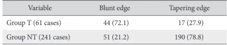
Of the 61 shoulders in group T, 44 shoulders (72.1%) had torn tendons with a blunt edge and 17 shoulders (27.9%) had tapering edges. The interclass correlation coefficient for reliabil- ity was 0.837 (p<0.001). In the 241 shoulders of group NT, 51 shoulders (21.2%) had torn tendons with a blunt edge, and 190 shoulders (78.8%) had tapering edges. The interclass correla- tion coefficient for reliability in this group was 0.829 (p<0.001).
Significant differences were observed between the groups (p<0.001) using a chi-square analysis. The estimated odds ratio between blunt shaped edges and group T was 9.6 (95% confi- dence interval: 5.1, 18.3; Table 1).
The incidence of tendon stumps remaining on the greater tuberosity was 26.2% in group T and 16.2% in group NT. There was no significant difference between the groups (p=0.055) (Table 2). However, a significant difference was observed be- tween blunt and tapering edges (p=0.037), and the estimated Fig. 1. A 53-year-old male with a traumatic rotator cuff tear. Torn tendon
with a blunt edge (white arrow) on the T2-weighted oblique coronal image.
Fig. 2. A 56-year-old female with non-traumatic rotator cuff tear. Torn tendon with a tapering edge (white arrow) on the fat-saturated T1-weighted oblique coronal image.
Fig. 3. A 51-year-old male with a traumatic rotator cuff tear. Tendon stump left on the greater tuberosity (white arrow) on the fat-saturated T1-weighted oblique coronal image.
odds ratio between the existence of a stump and a tendon tear with a blunt edge was 1.9 (95% confidence interval: 1.1, 3.5;
Table 2).
The mean tear size on oblique sagittal image was 19.7 ± 7.1 mm for patients in group T, and 19.5 ± 10.2 mm for patients in group NT. No significant difference in tear size was observed be- tween the groups. However, in both groups, tapering-edged torn tendons were associated with a larger tear size than those with blunt edges, and these results were significant (p<0.001; Table 3). In comparison of the tear size between patients with blunt- edged tears, group T patients showed a significantly larger tear size (p=0.035). However, no significant difference was observed between the tear sizes of patients with tapering edges, regardless of the study group.
Discussion
Our study found that the traumatic cuff tears had an abrupt and rough cuff-off edge with relatively consistent tendon thick- ness (without tapering laterally), compared to non-traumatic cuff tears with statistical significance. However, contrary to our expectation, this difference did not provide a clear distinction between traumatic and non-traumatic rotator cuff tears. In as- sessing the shape of the tendon tear, we employed MRA rather than MRI because we thought that MRA would be superior for
delineating the shape of the torn edge of the tendon, etc. How- ever, the hematoma around the cuff tear might become unclear by injecting a contrast into the joint.
For evaluation of this hypothesis, we excluded asymptom- atic degenerative rotator cuff tears from the traumatic cuff tear group. These types of rotator cuff tears have been reported in more than 20% of individuals older than 60 years of age.9,18) Thus, only patients between the ages of 40 and 60 years were included in this study. However, we acknowledge that we could not completely exclude asymptomatic tears from the traumatic cuff tear group because we did not have images showing an intact cuff before the trauma, and Kim et al.18) reported occur- rence of asymptomatic degenerative rotator cuff tears in as many as 10% of individuals younger than 60 years of age. Patients with an MRA more than 6 weeks after trauma were excluded to con- trol for time-sensitive changes in the shape of the torn rotator cuff.4)
Although we were unable to determine a method for the un- questionable definition of traumatic rotator cuff tears, we found significant differences in the distribution of tear shapes between traumatic and non-traumatic tears. In group T, 72.1% of tendons had a blunt edge and 27.9% had a tapering edge. In contrast, only 21.2% of tendons had a blunt edge and 78.8% had a ta- pering edge in group NT. Although the blunt edge proportion in group T was not as high as expected and the proportion in group NT was not as low as expected, the estimated odds ratio of blunt shaped edges and group T was 9.6, indicating a strong tendency for traumatic posterosuperior rotator cuff tears to have blunt shaped edges on oblique coronal images. There might be a degenerative change preceding the traumatic event. However, it is beyond the scope of this study to address this issue.
In addition to the tendon edge, we focused on the presence of a tendon stump remaining on the greater tuberosity. Although we found no significant differences between groups T and NT for this parameter, a significant difference was observed between Table 1. Distribution of Blunt and Tapering Edges of Torn Tendons
Variable Blunt edge Tapering edge
Group T (61 cases) 44 (72.1) 17 (27.9)
Group NT (241 cases) 51 (21.2) 190 (78.8)
Values are presented as number (%). Group T: traumatic rotator cuff tears, Group NT: non-traumatic rotator cuff tears, Blunt edge: abrupt and rough edge of the torn tendon with relatively consistent tendon thickness (without lateral tapering) on the oblique coronal image, Tapering edge: the relative lateral tapering and smooth edges of the torn tendon on the oblique coronal image.
Table 2. Distribution of the Tendon Stump Left on the Greater Tuberosity ac- cording to the Group and the Edge Type
Variable With stump Without stump p-value
Group T (61 cases) 16 (26.2) 45 (73.8) 0.055
Group NT (241 cases) 39 (16.2) 202 (83.8)
Blunt edge (95 cases) 24 (25.3) 71 (74.7) 0.037 Tapering edge (207 cases) 31 (15.0) 176 (85.0)
Values are presented as number (%). Group T: traumatic rotator cuff tears, Group NT: non-traumatic rotator cuff tears, Blunt edge: abrupt and rough edge of the torn tendon with relatively consistent tendon thickness (without lateral tapering) on the oblique coronal image, Tapering edge: the relative lat- eral tapering and smooth edge of the torn tendon on the oblique coronal im- age, With stump: tendon stump left on the greater tuberosity, Without stump:
tendon stump was not left on the greater tuberosity.
Table 3. Mean Tear Sizes in Groups T and NT
Variable Tear size (mm) p-value
Group T 19.7 ± 7.1 <0.001
Blunt edge 16.9 ± 6.2
Tapering edge 23.2 ± 6.7
Group NT 19.5 ± 10.2 <0.001
Blunt edge 14.0 ± 3.4
Tapering edge 21.4 ± 11.0
Values are presented as the mean ± standard deviation. Group T: traumatic rotator cuff tears, Group NT: non-traumatic rotator cuff tears, Blunt edge:
abrupt and rough edge of the torn tendon with relatively consistent tendon thickness (without lateral tapering) on the oblique coronal image, Tapering edge: the relative lateral tapering and smooth edge of the torn tendon on the oblique coronal image.
blunt and tapering edges, and the estimated odds ratio between the existence of a stump and having a tendon with a blunt edge was approximately 2. Thus, a stump remaining on the greater tuberosity appears to show association with shape of the torn tendon edge, but not with trauma.
Zanetti et al.20) suggested that occult greater tuberosity frac- tures were relatively common (38%) in patients younger than 40 years old with clinically suspected traumatic rotator cuff, indicat- ing that the greater tuberosity should be examined for fractures.
Importantly, assessment of tendon tear shapes on MRI was not performed in this group. However, although the patients in the current study had an MRI within 6 weeks of trauma, we found no occult fractures and only two cases of greater tuberosity frac- tures were excluded.
The current study has several limitations. As mentioned previ- ously, due to the lack of pre-trauma imaging we were unable to assess whether the patients in group T had pristine tendons and relevant tissues before the trauma. A subset of patients in group T may have had asymptomatic insidious degenerative tears, which could influence our current analysis of the results. Indeed, such asymptomatic degenerative tears may predispose individu- als to traumatic rotator cuff tears. Although we only included patients younger than 60 years of age, other investigators have reported asymptomatic cuff tears of 10% to 13% in people aged 50 to 59 years.9,18) Thus, this study would be weightier if we included only patients younger than 50 years of age. However, we required larger numbers of patients than those exclusion cri- teria would have afforded us. In addition, although we tried to exclude patients with a tendency for compensation, it would be impossible to rule them out completely, which might affect the results of this study.
Conclusion
We did not discover absolute differences in the shape of the edge of the torn tendon between traumatic and non-traumatic posterosuperior rotator cuff tears. However, we observed a sig- nificantly strong tendency for traumatic rotator cuff tears to have tendons tears with abrupt and rough edges and relatively consis- tent tendon thickness (without tapering laterally) on the oblique coronal MRI.
Acknowledgements
We thank Young-Jun Cho, our research assistant, for assis- tance with the figures.
References
1. Brewer BJ. Aging of the rotator cuff. Am J Sports Med. 1979;
7(2):102-10.
2. Neer CS 2nd. Impingement lesions. Clin Orthop Relat Res.
1983;(173):70-7.
3. McLaughlin HL, Asherman EG. Lesions of the musculotendi- nous cuff of the shoulder. IV. Some observations based upon the results of surgical repair. J Bone Joint Surg Am. 1951;33(1):
76-86.
4. Bassett RW, Cofield RH. Acute tears of the rotator cuff. The timing of surgical repair. Clin Orthop Relat Res. 1983;(175):18- 24.
5. Ide J, Tokiyoshi A, Hirose J, Mizuta H. Arthroscopic repair of traumatic combined rotator cuff tears involving the subscapu- laris tendon. J Bone Joint Surg Am. 2007;89(11):2378-88.
6. Hantes ME, Karidakis GK, Vlychou M, Varitimidis S, Dailiana Z, Malizos KN. A comparison of early versus delayed repair of trau- matic rotator cuff tears. Knee Surg Sports Traumatol Arthrosc.
2011;19(10):1766-70.
7. Namdari S, Henn RF 3rd, Green A. Traumatic anterosuperior rotator cuff tears: the outcome of open surgical repair. J Bone Joint Surg Am. 2008;90(9):1906-13.
8. Maier D, Jaeger M, Suedkamp NP, Koestler W. Stabilization of the long head of the biceps tendon in the context of early re- pair of traumatic subscapularis tendon tears. J Bone Joint Surg Am. 2007;89:1763-9.
9. Deutsch A, Altchek DW, Veltri DM, Potter HG, Warren RF.
Traumatic tears of the subscapularis tendon. Clinical diagnosis, magnetic resonance imaging findings, and operative treat- ment. Am J Sports Med. 1997;25(1):13-22.
10. Farin PU, Jaroma H. Acute traumatic tears of the rotator cuff:
value of sonography. Radiology. 1995;197(1):269-73.
11. Teefey SA, Middleton WD, Bauer GS, Hildebolt CF, Yama- guchi K. Sonographic differences in the appearance of acute and chronic full-thickness rotator cuff tears. J Ultrasound Med.
2000;19(6):377-8; quiz 383.
12. Dinnes J, Loveman E, McIntyre L, Waugh N. The effectiveness of diagnostic tests for the assessment of shoulder pain due to soft tissue disorders: a systematic review. Health Technol As- sess. 2003;7(29):iii, 1-166.
13. Rutten MJ, Jager GJ, Kiemeney LA. Ultrasound detection of rotator cuff tears: observer agreement related to increasing ex- perience. AJR Am J Roentgenol. 2010;195(6):W440-6.
14. Sipola P, Niemitukia L, Kröger H, Höfling I, Väätäinen U. De- tection and quantification of rotator cuff tears with ultrasonog- raphy and magnetic resonance imaging: a prospective study in 77 consecutive patients with a surgical reference. Ultrasound Med Biol. 2010;36(12):1981-9.
15. Steinbach LS, Palmer WE, Schweitzer ME. Special focus ses- sion. MR arthrography. Radiographics. 2002;22(5):1223-46.
16. Sher JS, Uribe JW, Posada A, Murphy BJ, Zlatkin MB. Abnor- mal findings on magnetic resonance images of asymptomatic shoulders. J Bone Joint Surg Am. 1995;77(1):10-5.
17. Tempelhof S, Rupp S, Seil R. Age-related prevalence of rotator
cuff tears in asymptomatic shoulders. J Shoulder Elbow Surg.
1999;8(4):296-9.
18. Kim HM, Teefey SA, Zelig A, Galatz LM, Keener JD, Yama- guchi K. Shoulder strength in asymptomatic individuals with intact compared with torn rotator cuffs. J Bone Joint Surg Am.
2009;91(2):289-96.
19. Davidson JF, Burkhart SS, Richards DP, Campbell SE. Use of
preoperative magnetic resonance imaging to predict rotator cuff tear pattern and method of repair. Arthroscopy. 2005;
21(12):1428.
20. Zanetti M, Weishaupt D, Jost B, Gerber C, Hodler J. MR imag- ing for traumatic tears of the rotator cuff: high prevalence of greater tuberosity fractures and subscapularis tendon tears. AJR Am J Roentgenol. 1999;172(2):463-7.