Clinical and Radiological Outcomes of Acute Acromioclavicular Joint Dislocation: Comparison of Hook Plate Fixation with Single Tight Rope Technique
Sung Hyun Lee, Jeong Woo Kim , Seng Hwan Kook
Institute of Wonkwang Medical Science and Department of Orthopedic Surgery, Wonkwang University Hospital, Iksan, Korea
Background: This study was conducted to compare the clinical and radiological outcomes of the locking hook plate fixation (HP) tech- nique and the single tight rope (TR) technique applied for acute high-grade acromioclavicular (AC) joint separations.
Methods: Between 2009 and 2014, 135 consecutive patients with acute AC joint separation Rockwood types III, IV, and V were sub- jected to surgical reconstruction. One hundred fourteen patients (84.4%) were available for retrospective evaluation. Of them, 62 and 52 were treated using the single TR group and clavicular HP group techniques, respectively. The visual analogue scale, Constant, Ameri- can Shoulder and Elbow Surgeons (ASES), and Taft scores were used for clinical assessment. Postoperative shoulder range of motion was also assessed. An anteroposterior radiograph of the coracoclavicular distance (CCD) was obtained to evaluate the radiographic signs of recurrence.
Results: The TR group patients had better Constant, ASES, and Taft scores than the HP group patients. The loss of reduction in terms of the CCD did not differ between groups. Subacromial osteolysis was observed in 34.6% of the cases in the HP group. However, there were no significant differences in the clinical outcomes between the patients with and without osteolysis in the HP group. Subcoracoid osteolysis, drill tunnel widening, and metal displacement were observed in 3.2%, 22.6%, and 4.8% of the cases in the TR group, respec- tively.
Conclusions: The single TR technique was relatively more effective at treating acute high-grade AC joint injuries than the HP fixation technique (level of evidence: therapeutic; retrospective comparative study, Level III).
(Clin Shoulder Elbow 2017;20(3):153-161)
Key Words: Acromioclavicular joint; Acute separation; Arthroscopic stabilization; Tight rope technique; Clavicular hook plate Clinics in Shoulder and Elbow Vol. 20, No. 3, September, 2017
https://doi.org/10.5397/cise.2017.20.3.153
Received July 6, 2017. Revised August 4, 2017. Accepted August 13, 2017.
Correspondence to: Jeong Woo Kim
Department of Orthopedic Surgery, Wonkwang University Hospital, 895 Muwang-ro, Iksan 54538, Korea Tel: +82-63-831-4546, Fax: +82-63-852-9329, E-mail: [email protected]
IRB approval (No. WKUHIRB 201611-HR-122).
Financial support: This paper was supported by Wonkwang Institute of Clinical Medicine in 2017. Conflict of interests: None.
Introduction
The optimal treatment of acromioclavicular (AC) dislocations is still a matter of discussion.1) However, some consensus has been reached regarding low- and high-energy trauma.2) Low- energy trauma injuries, which are classified as type I or type II injuries, are treated conservatively using either a harness or a sling,2,3) while high-energy trauma injuries, such as type IV, V, and VI injuries, are treated surgically4) and the treatment for type III injuries is still a matter of debate.5,6) Many operative techniques
have been developed over the years. However, the optimal operative treatments among these options is still unclear. The choice of surgical method is made based on multiple factors, in- cluding patient’s medical condition and activity level, surgeon’s experience, cost effectiveness, and insurance policy.
The use of cerclages for coracoclavicular (CC) augmentation is an alternative to pin fixation or hook plate procedures, which do not require an implant removal.4,7,8) The main problems as- sociated with CC loop augmentations are the highly invasive approach to the coracoid base and the anterior subluxation of
the clavicle, which leads to a mal-reduction of the AC joint in the postoperative course.4) Furthermore, a sawing effect of syn- thetic material owing to clavicle rotation has been reported.4) This highly invasive approach is also associated with increased postoperative morbidity.9) To overcome these disadvantages, minimally invasive techniques for CC augmentation using flip buttons have been developed (Fig. 1).10-12) These surgeries can be conducted in a minimally invasive or arthroscopically assisted manner. In fact, biomechanical clinical studies report high fixa- tion strength and good to excellent functional results following these types of surgeries.3,10,12-14)
Another well-established method for AC joint reconstruction is hook plate fixation (HP) (Fig. 2).15-17) Unlike the old Wolter plate, the HP method has been designed to include an exten- sion hook under the acromion to provide more stable fixation.18) The use of the HP method has increased since advent of the locking plate with locking screws, and good results following its use have been reported.19,20) However, there are known disad- vantages associated with this technique, including a mandatory second surgery for removal of the plate. Moreover, transarticular fixation using an HP may induce bony osteolysis, shoulder im- pingement, and rotator cuff damage, leading to poor functional results.15,21) Considering the advantages and disadvantages of these two surgical methods, there is little radiologic and clinical outcome evidence regarding the management of acute high- grade AC joint injuries using HP vs. the tight rope (TR) method.
Therefore, we compared the clinical and radiologic outcomes of the locking HP fixation technique with those of the single TR
technique for treatment of acute high-grade AC joint separa- tions. Our hypothesis was that the HP fixation would have equal functional and radiographic results to the single TR technique.
Methods
Patient Selection
Between 2009 and 2014, a consecutive series of 135 pa- tients with traumatic AC joint separation were surgically treated in Wonkwang University Hospital Trauma Center. The inclu- sion criteria of the patients were as follows: (1) radiographically confirmed closed Rockwood type III (with horizontal instability observed in manual/overhead workers or athletes) or higher AC dislocation; (2) surgery within 2 weeks of trauma (it is known that after this period, CC ligaments lack healing potential22));
(3) primary treatment using a clavicular HP (Synthes, Bettlach, Switzerland) or TR (Arthrex, Naples, FL, USA) devices; (4) age of
≥18 years; (5) mono trauma; and (6) minimum follow-up of 24 months after surgery. The exclusion criteria were as follows: (1) polytrauma, including associated nerve and/or vessel injury; (2) previous injury to the injured shoulder; and (3) disease processes that would preclude accurate evaluation of the outcomes (e.g., neuromuscular, rheumatic, or significant psychiatric or metabolic disorders).
Between 2009 and 2011, the single TR technique was per- formed in 61 patients. Between 2012 and 2014, clavicular HPs were implanted in 74 patients. HP surgeries were selected at later dates because of the termination of payments for TR sur-
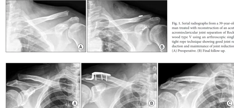
A B
Fig. 1. Serial radiographs from a 39-year-old man treated with reconstruction of an acute acromioclavicular joint separation of Rock- wood type V using an arthroscopic single tight rope technique showing good joint re- duction and maintenance of joint reduction.
(A) Preoperative. (B) Final follow-up.
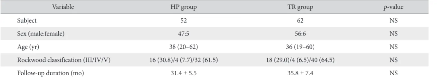
A B C
Fig. 2. Serial radiographs from a 35-year-old man treated with a hook plate fixation, showing good maintenance of joint reduction after plate removal. (A) Preop- erative. (B) Postoperative. (C) Last follow-up after plate removal.
geries by the national insurance policy of South Korea. Patients were divided into two groups according to the surgical method used (TR group and HP group). In the TR group, 52 of the initial 61 patients were evaluated, five patients were not available for follow-up, and four refused participation. In the HP group, 62 of the initial 74 patients were evaluated (nine patients were un- available; three patients refused participation). Generally, follow- up was available for 114 (84.4%) of these 135 patients.
Clinical Evaluation
Postoperative clinical evaluations were performed regu- larly on an outpatient basis (at 4 weeks, 6 weeks, 3 months, 6 months, and 12 months following surgery and at the last follow- up). The results of the evaluations at the last follow-up were ana- lyzed. At the time of follow-up, all patients were evaluated using the visual analogue scale (VAS) to assess subjective pain, and the Constant, ASES, and Taft23) scores were used for the clinical assessments. Postoperative shoulder ranges of motion (ROMs), including forward flexion, external rotation at the side, internal rotation to the back, and abduction were assessed. ROM of the injured shoulder was compared to that of the contralateral unaf- fected shoulder to assess ROM limitations.
Radiological Assessment
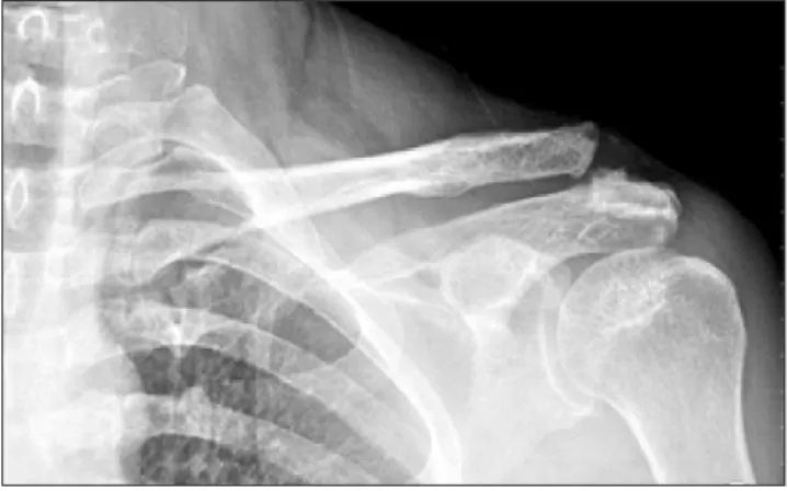
To evaluate reduction of the AC joint after surgery, we ana- lyzed the anteroposterior radiographs. The CC distance (CCD),24) which was defined as the perpendicular distance between the upper border of the coracoid process and the inferior cortex of the clavicle, was analyzed (Fig. 3). CCD was measured by three orthopedic surgeons who were blinded to the patient informa- tion. Interobserver reliability was assessed based on the kappa value, which was found to be 0.91. The CCDs of the contralat-
eral unaffected side and the affected side were compared at the last follow-up to determine if the anatomical reduction was ap- propriate. We considered increases in the CCD of 0% to 50%, 50% to 100%, and >100% with respect to the contralateral side assessed using the follow-up radiographs as intact, subluxation, and re-dislocation, respectively (Fig, 4).15) At the final follow- up, subacromial osteolysis, subcoracoid osteolysis, drill tunnel widening, and metal displacement were evaluated using plain radiographs.
Surgical Technique
All patients were placed in the beach-chair position under general anesthesia. All surgeries were performed by one sur- geon.
1) HP group
An approximately 5-cm-long skin incision was made in line with the lateral clavicle and the AC joint. The AC joint was ex- posed, and the articular disc was excised in case of disruption.
Following AC joint reduction, the clavicular HP was positioned with the hook placed dorsally under the acromion and fixed onto the clavicle with screws. If possible, the AC joint capsule was reconstructed. The deltoidotrapezoid fascia was then closed using resorbable sutures, and the wound was closed in layers.
2) TR group
Two standard arthroscopic portals were created (posterior and anteroinferior). The posterior portal was the initial viewing portal. After diagnostic glenohumeral arthroscopy and possible therapeutic interventions, the anteroinferior working portal was created. This portal was established in line with the upper subscapularis margin. The coracoid undersurface was prepared using a radio-frequency device. A skin incision approximately 1 cm long was then made in line with the lateral clavicle ~3.5 cm medial to the AC joint, after which the surface of this inci- sion was prepared. An anterior cruciate ligament drill guide with a thrust block (Arthrex) was subsequently introduced via the anteroinferior portal and positioned below the base of the cora- coid while the drill sleeve was vertically placed on the clavicular surface ~4 cm medial to the AC joint line. The bone tunnel was then overdrilled using a 4 mm drill bit, and the TR device was inserted via a shuttle-wire under arthroscopic control. The TR was secured using six alternating half hitches. Finally, the wound was closed in layers over the clavicular buttons.
Postoperative Management
Rehabilitation involved wearing a sling for 3 to 4 weeks. The patients were initially allowed to move fully and actively at the elbow, wrist, and hand. Patients in the HP group were allowed to elevate the shoulder passively by no more than 90° in the plane of the scapula after the first week. Patients in the TR group were allowed to perform this movement three weeks after sur- gery. Pendulum exercises began at the first week after the injury Fig. 3. Radiological analysis of coracoclavicular distance.
in both groups. The active ROM progressively advanced from the sixth week after surgery in both groups. Exercises to regain strength were initiated once the patient had full and pain-free passive and active ROMs. The exercises were primarily directed toward scapular stabilization. In the HP group, patients were ad- vised that the implant should be removed 16 to 20 weeks after surgery. Return to work without restrictions was allowed after 16 to 20 weeks, and contact sports or major efforts were avoided for 4 to 6 months in both groups.
Statistical Analysis
Statistical analyses were performed using SPSS ver. 12.0 (SPSS Inc., Chicago, IL, USA) Data were reported as medians (ranges, minimum–maximum) or means ± standard deviations. All data were tested for normal distribution using the Kolmogorov- Smirnov test. Subsequently, the data of the two groups were compared using the nonparametric Mann-Whitney U-test for independent samples and the Student’s t-test for dependent samples. The level of significance was defined as p<0.05 for all tests.
Ethical Approval
All procedures performed in studies were in accordance with the ethical standards of the institution or practice at which the studies were conducted. This retrospective comparative study was approved by our institutional review board (WKUHIRB 201611-HR-122).
And informed consent was obtained from all individual par- ticipants included in the study.
Results
Demographics
Age, sex, follow-up period, and Rockwood classification were evaluated as demographic factors. No significant differences were found in the demographic characteristics between the two groups (Table 1).
Clinical and Radiological Outcomes
The clinical and radiological outcomes are presented in Table 2. The VAS, Constant, ASES, and Taft scores and ROM were evaluated as clinical factors at the last follow-up. The functional scores differed significantly between two groups. Specifically, the mean Constant score was 77.2 ± 8.5 in the HP group and 89.5
± 7.4 in the TR group (p=0.018), while the ASES score was 80.3 ± 10.2 in the HP group and 92.1 ± 8.7 in the TR group (p=0.023) and the Taft score was 8.3 ± 2.7 in the HP group and 10.5 ± 2.3 in the TR group (p=0.015). No significant differ- ences in the VAS score or ROM were found between groups at the last follow-up.
The radiographic outcomes indicated that the CCDs de- creased in both groups from 210.7% ± 46.8% preoperatively to 107.2% ± 10.5% at the final follow-up in the HP group and from 224.5% ± 50.2% preoperatively to 109.1% ± 12.1% at
A
B Fig. 4. (A) Serial radiographs from a 20-year-old man treated with a tight rope fixation showing a subluxation of joint reduction at last follow-up. (B) Serial ra- diographs from a 53-year-old man treated with a hook plate fixation showing a redislocation after plate removal.
the final follow-up in the TR group. No significant differences were found between groups.
Six cases of recurrent subluxation and two cases of re- dislocation were observed in the HP group, while 10 cases of subluxation and two cases of re-dislocation were observed in the TR group at the final follow-up. These differences were also not statistically significant.
Complications and Revisions
Subacromial osteolysis was observed in 34.6% of the cases (18/52) in the HP group (Fig. 5). Of these, 66.7% (12/18) were considered to be mild osteolysis, while 33.3% (6/18) were con- sidered to be severe osteolysis.
There were no significant differences in the VAS, Constant, ASES, or Taft scores at the final follow-up between the patients
with osteolysis (n=18) and those without osteolysis (n=4) in the HP group (Table 3). The radiological findings indicated that there were no differences between the initial and final CCDs (Table 3).
Two patients (3.8% in HP group, 3.2% in TR group) required revision surgery in each group because of recurrent vertical and horizontal instability with pain. In all cases, the AC and CC liga- ments were reconstructed.
In 14 patients (22.6%) in the TR group, trapezoid-like tunnel widening in the clavicle bone by a mean of 7.3 ± 0.7 mm was observed when compared with that in the direct postoperative radiographs. Implant migration of the TR buttons was observed in three cases (4.8%) and subcoracoid osteolysis in two cases (3.2%).
Table 1. Demographics of the Two Groups
Variable HP group TR group p-value
Subject 52 62 NS
Sex (male:female) 47:5 56:6 NS
Age (yr) 38 (20–62) 36 (19–60) NS
Rockwood classification (III/IV/V) 16 (30.8)/4 (7.7)/32 (61.5) 18 (29.0)/4 (6.5)/40 (64.5) NS
Follow-up duration (mo) 31.4 ± 5.5 35.8 ± 7.4 NS
Values are presented as number only, median (range), number (%), or mean ± standard deviation.
HP: hook plate, TR: tight rope, NS: not significant.
Table 2. Comparison of Clinical and Radiological Outcomes between Groups
Variable HP group (n=52) TR group (n=62) p-value
VAS* 1.5 ± 1.5 1.8 ± 2.1 NS
Constant score* 77.2 ± 8.5 89.5 ± 7.4 0.018
ASES score* 80.3 ± 10.2 92.1 ± 8.7 0.023
Taft score* 8.3 ± 2.7 10.5 ± 2.3 0.015
Range of motion (°) NS
Active FE† -8.5 ± 11.2 -7.8 ± 10.8 NS
ER† -4.6 ± 7.4 -4.2 ± 6.7 NS
IR† -3.5 ± 5.1 -2.9 ± 4.5 NS
Abduction† -9.5 ± 13.7 -8.9 ± 14.4 NS
Initial CCD (%) 210.7 ± 46.8 224.5 ± 50.2 NS
Final CCD (%) 107.2 ± 10.5 109.1 ± 12.1 NS
Recurrence (intact/subluxation/re-dislocation) 44/6/2 50/10/2 NS
Postoperative complications (subacromial osteolysis/
subcoracoid osteolysis/drill tunnel widening/metal displacement)
18/52
(18 [34.6]/0 [0]/0 [0]/0 [0]) 19/62
(0 [0]/2 [3.2]/14 [22.6]/3 [4.8]) NS Values are presented as mean ± standard deviation, number only, or number (%).
VAS: visual analogue scale, ASES: American Shoulder and Elbow Surgeons, FE: forward elevation, ER: external rotation, IR: internal rotation, CCD: coracocla- vicular distance, HP: hook plate, TR: tight tope, NS: not significant.
*The results at the last follow-up were used for analysis. †Difference between affected side and contralateral unaffected side.
Discussion
The most important finding of this study is that the patients with acute high-grade AC joint injuries managed using the single TR technique showed better clinical outcomes as assessed using the Constant, ASES, and Taft scores than those managed using the HP. In addition, HP fixation induced accompanying compli- cations and required a second surgery for implant removal in all patients. However, the radiographic outcomes indicated that the CCDs decreased in both groups with no significant difference.
Accordingly, our hypothesis that the HP fixation would have equal functional and radiographic outcomes with those of the single TR technique was partly rejected.
There are a large number of surgical approaches used to treat acute high-grade AC joint injuries reported in the literature.25-28) It has been reported that coracoacromial ligament transfer, HP fixation, AC K-wire fixation, and CC screw fixation are biome- chanically insufficient.25) In vivo analysis of AC joint motions after HP fixation indicates that the clavicular motion and AC joint bio- mechanics change significantly after HP.26) The technique select- ed to treat an AC joint injury may have a strong influence on the outcomes, especially if open procedures or fixation using metal hardware are performed. Patients in the TR group in this study had better Constant, ASES, and Taft scores at the last follow-up than those in the HP group. The results of our study agree with those published by Andreani et al.,27) who found that patients treated using the TR technique had a mean Constant score of 90, while those managed using an HP had a mean Constant score of 75. In addition, Boström Windhamre et al.28) reported that patients managed using HP fixation have significantly more pain than those managed using a CC non-rigid fixation. They argued that this may be because of the presence of chronic irri- tation in the subacromial space, which then leads to a persistent pain syndrome.28)
We considered the following reasons as contributors to the
inferior functional outcomes. A portion of the hook may enter the posterior aspect of the AC ligaments, which may then com- promise the proper healing of these structures. Similarly, HPs must be removed, and the period during which the plate is im- planted might be characterized by the development of serious complications, such as the upward cutting of the hook through the acromion,29) subacromial osteolysis,30) fracture,31) AC joint osteoarthritis, subacromial impingement, rotator cuff tears,5) and plate bending.24) Even when there is timely removal of the plate, the patient may be subjected to an increased risk of fracture of the distal clavicle after low-energy trauma.32) Further, the HP is a stiff plate that does not allow normal rotation of the clavicle, which may account for some of the late complications and ar- thritis.26) In this study, 34.6% (18/52) of the patients had mild or severe subacromial osteolysis. Although there were no differenc- es in the clinical and radiologic outcomes based on the presence of subacromial osteolysis, the relatively frequent occurrence of this complication is a considerable disadvantage of the HP fixa- tion. Lin et al.21) reported that, among a group of patients with AC joint injuries and distal third clavicle fractures managed using HPs, 37.5% (15/40) developed subacromial impingement syn- drome. The functional scores of these patients were lower than those of patients without impingements. Among these patients, 40.0% (6/15) had a rotator cuff tear diagnosed using sonography, while subacromial osteolysis caused by hook pressure was ob- served in 50.0% (20/40) of the patients.21) In the present study, we did not evaluate our patients using sonography. However, we believe that the lower clinical scores may reflect underlying injuries related to the plate.
The arthroscopic technique of CC fixation using an Endobut- ton device (Smith and Nephew, Memphis, TN, USA) has previ- Fig. 5. Radiograph obtained 24 months postoperatively showing mild loss of
reduction and distal clavicular osteolysis after synthetic ligament reconstruc- tion.
Table 3. Comparison according to the Presence of Subacromical Erosions in the HP Group
Variable
Subacromial erosion
p-value Patients with
osteolysis (n=18) Patients without osteolysis (n=34) Sex (male:female) 16 (88.9):2 (11.1) 31 (91.2):3 (8.8) NS
Age (yr) 36 (20–58) 38 (21–62) NS
Initial CCD (%) 215.7 ± 50.3 208.7 ± 46.5 NS
Final CCD (%) 106.2 ± 10.2 108.2 ± 10.18 NS
VAS* 1.6 ± 1.7 1.5 ± 2.2 NS
Constant score* 76.8 ± 8.3 77.5 ± 8.6 NS
ASES score* 79.2 ± 10.4 81.1 ± 10.1 NS
Taft score* 8.2 ± 2.5 8.4 ± 2.2 NS
Values are presented as number (%), median (range), or mean ± standard de- viation.
HP: hook plate, CCD: coracoclavicular distance, VAS: visual analogue scale, ASES: American Shoulder and Elbow Surgeons.
*The results at the last follow-up were used for analysis.
ously been reported.33,34) However, the first generation of this device consisted of a smaller round proximal button that had the potential to erode the clavicle and led to loss of reduction.35,36) Since a larger proximal button was developed, no erosion or failure has occurred using this surgery. The titanium buttons, which are positioned centrally on top of the clavicle and under the coracoid, are connected with a continuous loop of No. 5 FiberWire (Arthrex) wire suture. This produces a construct stron- ger than the native CC ligament and the AC joint complex and leads to strong maintenance of reduction and a homogeneous distribution of load onto the bone surface. These characteristics protect against the sawing effects of sutures, which have been described as a mechanism of failure in other techniques using anchors and sutures.37,38) Accurate positioning of the bony tun- nels and buttons centrally on the clavicle and under the coracoid process is essential to solid fixation. Malpositioning of the cla- vicular (too anteriorly) or the coracoid button (too laterally) may lead to asymmetric loading and cutout of the fixation device.39) Yi and Kim40) have suggested that surgeons should strive to place a perpendicular hole from the clavicle to the coracoid process for TR fixation to enable successful reconstruction of acute AC joint injuries.
Another advantage of using arthroscopic procedures is that they enable the diagnosis and treatment of concomitant gle- nohumeral injuries. In addition, these procedures require no mandatory implant removal.9) In fact, potential remaining con- comitant intraarticular injuries may be the underlying reasons for failure of some treatments using open surgeries without access to the glenohumeral joint or non-operative management.9) We suggest that if orthopedic surgeons decide to manage acute un- stable AC joint injuries using open procedures without access to the glenohumeral joint, they should carefully consider perform- ing preoperative magnetic resonance imaging arthrography to avoid ‘leaving behind’ important simultaneous acute injuries. If these injuries go unnoticed, they may significantly affect the out- come.
Biomechanical studies have shown that HP fixation has a biomechanical disadvantage when compared with CC non- rigid fixations. CC displacement during cyclic loading has been shown to occur more frequently in HP fixations.22) Despite these biomechanical disadvantages, clinical studies comparing patients managed using HP with those managed using CC non-rigid fixa- tions have shown that there are no differences in the remaining vertical instability.9) In the present study, the loss of reduction after HP fixation did not differ significantly from that following the single TR technique with an untouched AC joint and with- out an implant removal. The fact that a partial recurrent vertical instability may be observed independent of the fixation device indicates that the biological healing potential of the ruptured CC ligaments and the AC joint capsule is limited when appropriate scar tissue formation does not occur. A partial loss of vertical re-
duction has also been shown to have no influence on the overall outcomes.41) A possible explanation for this finding may be that the healed CC elongated ligaments provide enough stability to relieve symptoms.27)
The main limitation of our study was its retrospective design.
Additionally, patients were not randomized before undergo- ing any treatment. Further, this was a single-center study with a small number of enrolled patients. Our patients also had a wide age range. Accordingly, high-quality randomized controlled trials with larger sample sizes are still required to validate our results.
This study also contained a relatively large portion of type III dislocations; thus, it was difficult to fully determine whether our results and conclusions can be extrapolated to higher levels of dislocations. We also could not consider horizontal instability, which was not evaluated as CCD. Finally, the wide range of pe- riod of plate removal also might influence clinical outcomes.
Conclusion
In this study, patients with acute high-grade AC joint injuries managed using the single TR technique had better clinical out- comes than those managed using an HP as assessed using the Constant, ASES, and Taft scores. The loss of reduction after HP fixation was not significantly different from that prepared follow- ing the single TR technique. The single TR technique might be more effective for the treatment of acute high-grade AC joint injuries than HP fixation.
References
1. Babhulkar A, Pawaskar A. Acromioclavicular joint dislocations.
Curr Rev Musculoskelet Med. 2014;7(1):33-9.
2. Tauber M. Management of acute acromioclavicular joint dis- locations: current concepts. Arch Orthop Trauma Surg. 2013;
133(7):985-95.
3. Rockwood C, Williams G, Young D. Disorders of the acromio- clavicular joint. In: Rockwood CA, ed. The shoulder. 3rd ed.
Philadelphia: WB Saunders; 2004. 521-95.
4. Greiner S, Braunsdorf J, Perka C, Herrmann S, Scheffler S. Mid to long-term results of open acromioclavicular-joint recon- struction using polydioxansulfate cerclage augmentation. Arch Orthop Trauma Surg. 2009;129(6):735-40.
5. Bahk MS, Kuhn JE, Galatz LM, Connor PM, Williams GR Jr. Ac- romioclavicular and sternoclavicular injuries and clavicular, gle- noid, and scapular fractures. Instr Course Lect. 2010;59:209- 26.
6. Ceccarelli E, Bondì R, Alviti F, Garofalo R, Miulli F, Padua R.
Treatment of acute grade III acromioclavicular dislocation: a lack of evidence. J Orthop Traumatol. 2008;9(2):105-8.
7. Dimakopoulos P, Panagopoulos A, Syggelos SA, Panagiotopou- los E, Lambiris E. Double-loop suture repair for acute acromio-
clavicular joint disruption. Am J Sports Med. 2006;34(7):1112- 9.
8. Lemos MJ. The evaluation and treatment of the injured acro- mioclavicular joint in athletes. Am J Sports Med. 1998;26(1):
137-44.
9. Jensen G, Katthagen JC, Alvarado LE, Lill H, Voigt C. Has the arthroscopically assisted reduction of acute AC joint separa- tions with the double tight-rope technique advantages over the clavicular hook plate fixation? Knee Surg Sports Traumatol Arthrosc. 2014;22(2):422-30.
10. Petersen W, Wellmann M, Rosslenbroich S, Zantop T. Mini- mally Invasive Acromioclavicular Joint Reconstruction (MINAR).
Oper Orthop Traumatol. 2010;22(1):52-61.
11. Phillips AM, Smart C, Groom AF. Acromioclavicular disloca- tion. Conservative or surgical therapy. Clin Orthop Relat Res.
1998;(353):10-7.
12. Scheibel M, Dröschel S, Gerhardt C, Kraus N. Arthroscopically assisted stabilization of acute high-grade acromioclavicular joint separations. Am J Sports Med. 2011;39(7):1507-16.
13. Liu X, Huangfu X, Zhao J. Arthroscopic treatment of acute ac- romioclavicular joint dislocation by coracoclavicular ligament augmentation. Knee Surg Sports Traumatol Arthrosc. 2015;
23(5):1460-6.
14. Murena L, Vulcano E, Ratti C, Cecconello L, Rolla PR, Surace MF. Arthroscopic treatment of acute acromioclavicular joint dislocation with double flip button. Knee Surg Sports Trauma- tol Arthrosc. 2009;17(12):1511-5.
15. Eschler A, Gradl G, Gierer P, Mittlmeier T, Beck M. Hook plate fixation for acromioclavicular joint separations restores coraco- clavicular distance more accurately than PDS augmentation, however presents with a high rate of acromial osteolysis. Arch Orthop Trauma Surg. 2012;132(1):33-9.
16. Kienast B, Thietje R, Queitsch C, Gille J, Schulz AP, Meiners J.
Mid-term results after operative treatment of rockwood grade III-V acromioclavicular joint dislocations with an AC-hook- plate. Eur J Med Res. 2011;16(2):52-6.
17. Liu HH, Chou YJ, Chen CH, Chia WT, Wong CY. Surgical treat- ment of acute acromioclavicular joint injuries using a modified Weaver-Dunn procedure and clavicular hook plate. Orthope- dics. 2010;33(8). doi: 10.3928/01477447-20100625-10.
18. Koukakis A, Manouras A, Apostolou CD, et al. Results using the AO hook plate for dislocations of the acromioclavicular joint. Expert Rev Med Devices. 2008;5(5):567-72.
19. Modi CS, Beazley J, Zywiel MG, Lawrence TM, Veillette CJ.
Controversies relating to the management of acromioclavicular joint dislocations. Bone Joint J. 2013;95(12):1595-602.
20. von Heideken J, Boström Windhamre H, Une-Larsson V, Eke- lund A. Acute surgical treatment of acromioclavicular disloca- tion type V with a hook plate: superiority to late reconstruc- tion. J Shoulder Elbow Surg. 2013;22(1):9-17.
21. Lin HY, Wong PK, Ho WP, Chuang TY, Liao YS, Wong CC. Cla-
vicular hook plate may induce subacromial shoulder impinge- ment and rotator cuff lesion; dynamic sonographic evaluation.
J Orthop Surg Res. 2014;9:6.
22. Lädermann A, Gueorguiev B, Stimec B, Fasel J, Rothstock S, Hoffmeyer P. Acromioclavicular joint reconstruction: a com- parative biomechanical study of three techniques. J Shoulder Elbow Surg. 2013;22(2):171-8.
23. Taft TN, Wilson FC, Oglesby JW. Dislocation of the acromio- clavicular joint. An end-result study. J Bone Joint Surg Am.
1987;69(7):1045-51.
24. Sim E, Schwarz N, Höcker K, Berzlanovich A. Repair of com- plete acromioclavicular separations using the acromioclavicu- lar-hook plate. Clin Orthop Relat Res. 1995;(314):134-42.
25. Jari R, Costic RS, Rodosky MW, Debski RE. Biomechanical function of surgical procedures for acromioclavicular joint dis- locations. Arthroscopy. 2004;20(3):237-45.
26. Kim YS, Yoo YS, Jang SW, Nair AV, Jin H, Song HS. In vivo analysis of acromioclavicular joint motion after hook plate fixa- tion using three-dimensional computed tomography. J Shoul- der Elbow Surg. 2015;24(7):1106-11.
27. Andreani L, Bonicoli E, Parchi P, Piolanti N, Michele L. Acro- mio-clavicular repair using two different techniques. Eur J Or- thop Surg Traumatol. 2014;24(2):237-42.
28. Boström Windhamre HA, von Heideken JP, Une-Larsson VE, Ekelund AL. Surgical treatment of chronic acromioclavicular dislocations: a comparative study of Weaver-Dunn augmented with PDS-braid or hook plate. J Shoulder Elbow Surg. 2010;
19(7):1040-8.
29. Gstettner C, Tauber M, Hitzl W, Resch H. Rockwood type III acromioclavicular dislocation: surgical versus conservative treatment. J Shoulder Elbow Surg. 2008;17(2):220-5.
30. Chiang CL, Yang SW, Tsai MY, Kuen-Huang Chen C. Acromion osteolysis and fracture after hook plate fixation for acromiocla- vicular joint dislocation: a case report. J Shoulder Elbow Surg.
2010;19(4):e13-5.
31. Hoffler CE, Karas SG. Transacromial erosion of a locked sub- acromial hook plate: case report and review of literature. J Shoulder Elbow Surg. 2010;19(3):e12-5.
32. Nadarajah R, Mahaluxmivala J, Amin A, Goodier DW. Clavicu- lar hook-plate: complications of retaining the implant. Injury.
2005;36(5):681-3.
33. Boileau P, Old J, Gastaud O, Brassart N, Roussanne Y. All- arthroscopic Weaver-Dunn-Chuinard procedure with double- button fixation for chronic acromioclavicular joint dislocation.
Arthroscopy. 2010;26(2):149-60.
34. Wellmann M, Zantop T, Petersen W. Minimally invasive coracoclavicular ligament augmentation with a flip button/
polydioxanone repair for treatment of total acromioclavicular joint dislocation. Arthroscopy. 2007;23(10):1132.e1-5.
35. Bishop JY, Kaeding C. Treatment of the acute traumatic acro- mioclavicular separation. Sports Med Arthrosc. 2006;14(4):
237-45.
36. Hosseini H, Friedmann S, Tröger M, Lobenhoffer P, Agneskir- chner JD. Arthroscopic reconstruction of chronic AC joint dislocations by transposition of the coracoacromial ligament augmented by the Tight Rope device: a technical note. Knee Surg Sports Traumatol Arthrosc. 2009;17(1):92-7.
37. Somers JF, Van der Linden D. Arthroscopic fixation of type III acromioclavicular dislocations. Acta Orthop Belg. 2007;73(5):
566-70.
38. Wellmann M, Zantop T, Weimann A, Raschke MJ, Petersen W. Biomechanical evaluation of minimally invasive repairs for complete acromioclavicular joint dislocation. Am J Sports
Med. 2007;35(6):955-61.
39. Defoort S, Verborgt O. Functional and radiological outcome after arthroscopic and open acromioclavicular stabilization us- ing a double-button fixation system. Acta Orthop Belg. 2010;
76(5):585-91.
40. Yi Y, Kim JW. Coronal plane radiographic evaluation of the sin- gle Tight Rope technique in the treatment of acute acromiocla- vicular joint injury. J Shoulder Elbow Surg. 2015;24(10):1582- 7.
41. Weinstein DM, McCann PD, McIlveen SJ, Flatow EL, Bigliani LU. Surgical treatment of complete acromioclavicular disloca- tions. Am J Sports Med. 1995;23(3):324-31.