125
Neoparamoeba sp. Infection on Gills of Olive Flounder, Par- alichthys olivaceus in Korea
Hyoung Jun Kim, Jae Bum Cho, Mu Kun Lee, Min Do Huh and Ki Hong Kim� Department of Aquatic Life Medicine, Pukyong National University,
Pusan 608-737, Korea
Amoebic gill disease of flounder, Paralichthys olivaceus was diagnosed at commerical culture facility in South Korea. The amoeba was identified as a species of the genus Neoparamoeba based on the morpholgi- cal characteristics of trophozoites. Transmisson electron microscopy revealed the presence of a symbiotic organism, parasome in the cytoplasm and dense glycocalyx on the surface of the trophozoites. They lacked the boat-shaped microscales on the surface and contained numerous vacuoles and channels, mitochondria in the cytoplasm. Colonization of amoebae on gill tissue elicited extensive fusion and hyperplasia of gill lamella.
Key words : Neoparamoeba, Amoebic gill disease, Olive flounder
Introduction
The majority of amoebae infecting marine fish are opportunistic pathogens that, under certain con- ditions such as immunodepression and suboptimal environmental conditions, can become parasites and cause the disease outbreaks (Nash et al., 1988;
Noble et al., 1997). Kent et al. (1988) and Dykova et al. (1995) have suggested that amoebae could play a primary role in the development of gill dis- ease in cultured fish. Amoebic gill disease (AGD) has been reported for a number of cultured fish species including Atlantic salmon, Salmo salar (Roubal et al., 1989; Adams and Nowak, 2001), rainbow trout, Oncorhynchus mykiss (Munday et al., 1990), coho salmon, Oncorhynchus kisutch (Kent et al., 1988), European seabass, Dicentrar- chus labrax, sharpsnout seabream, Diplodus pun- tazzo (Dykova et al., 2000; Dykova and Novoa,
2001) and turbot, Scophthalmus maximus (Dykova et al., 1995, 1998). AGD has been a major problem in Tasmania once salmon production bacame inten- sive and more full strength salinity rearing sites came into use (Munday et al., 1990), and mortali- ties appeared to be related to elevated water temper- ature and salinity (Munday et al., 1993). Dykova et al. (1995) published a histopathological study of an amoebic infection that caused severe gill tissue damage in turbot Scophthalmus maximus. Such AGD of turbot associated with mortalities also reported from culture facilities in NW Spain (Dyko- va et al., 1998).
In Korea, although AGD has been noticed in olive flounder, Paralichthys olivaceus, farms, little information is available on the amoeba species and histopathological effects on olive flounder. In the present study, we investigated the morphological characteristics of the amoeba using light- and elec-
�Corresponding Author : Ki Hong Kim, Tel : 051-620-6145, Fax : 051-628-7430, E-mail : [email protected]
tron-microscopies, and the histopathology of infect- ed gills of olive flounder.
Materials and Methods
Juvenile cultured olive flounder Paralichthys oli- vaceus (average body weight 8.2 g) were collected from a culture facility located in southern coastal area of Korea. The gills of moribund fishes were excised to examine the presence of amoeba and other parasites. The gill tissue was smeared on a slide, air-dried and stained with Diff-Quik (Interna- tional Reagents Co., Japan). Isolated amoebae were examined by light microscopy and transmission electron microscopy.
For the transmission electron microscopical observation, isolated amoebae were washed with PBS (pH 7.0) 3 times by centrifugation, and were fixed in 2% v/v glutaraldehyde in 0.1 M cacodylate buffer (pH 7.2) at 4℃ overnight, postfixed in 1%
w/v cacodylic OsO4for 2 h. The specimens were dehydrated, embedded in epoxy resin (Spurr) and ultrathin-sectioned, stained with uranyl acetate and lead citrate, and examined by a JEOL JEM1200 transmission electron microscope (JEOL LTD., Japan).
For histological study, the gill tissues were fixed in Bouin's solution and embedded in paraplast. Sec- tions 4 ㎛ thick were stained with hematoxylin and eosin.
Results
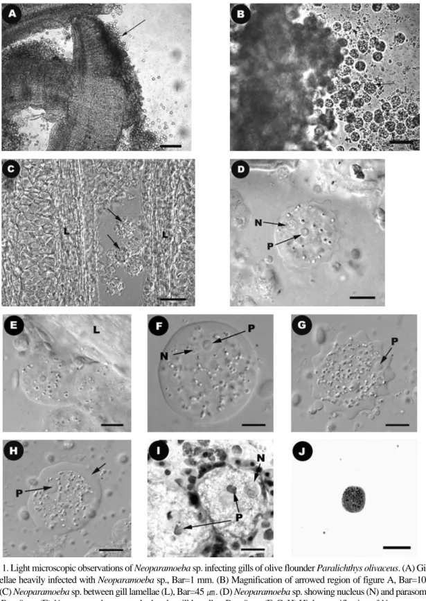
Heavily infected fish with amoebae secreted excess mucus on the gills (Fig. 1A). The gill fila- ments had irregular outlines and abnormal greyish coloration. The trophozoites of amoebae were observed on the secondary lamella of gills by light microscopy (Fig. 1B, C). A nucleus and a parasome
were observed clearly by differential microscopy (Fig. 1D, F). Body shape of trophozoites was spheri- cal with smooth outline (Fig. 1E, F, H) or short blunted projections (lobose pseudopod) from hyalo- plasmic zone (Fig. 1C, D, G). Trophozoite diameter was averaged 38.29 ± 5.03 ㎛. A nucleus (lighter) and a parasome (dark) were also distinguished clear- ly by Diff-Quik stain (Fig. 1I). Cytoplasm of tropho- zoites had numerous vacuoles in the fresh and meth- ylene blue stained amoebae (Fig. 1F, G, H, J).
In transmission electron microscopy, a distinct parasome was presented in the cytoplasm of amoe- bae (Fig. 2A, B, C). Channels and vacuoles present- ed in the center of cytoplasm (Fig. 2D). Also, sever- al mitochondria were found in the cytoplasm of amoebae (Fig. 2E). Surface of amoebae was sur- rounded by glycocalyx (Fig. 2F). Scales or micro- scales were not present on the cell surface.
In histopathological observation, a number of amoebae were found in the gill, which showed a variety of pathological changes. The amoebae were irregular in shapes and had highly-vacuolated cyto- plasm giving them foamy appearance, and a nucle- us with a large, distinct nucleolus (Fig. 3A). Main lesion was extensively fused secondary lamella with hyperplasia of the lamella epithelium. The hyperplastic changes were especially marked over fused tips of secondary lamella where a number of amoebae existed. In heavily infected cases, primary filaments were also fused (Fig. 3B). Fusion of lamellar tips resulted in the formation of a number of discrete interlamellar cavities (Fig. 3A, B).
Desquamated epithelial cells, edematous albu- minous fluid, or various developing stages of amoe- ba were found in the interlamellar cavities. Inflam- mation with the appearance of a few numbers of necrotic cells was occasionally noted in secondary lamella around the parasites (Fig. 3A).
Hyoung Jun Kim, Jae Bum Cho, Mu Kun Lee, Min Do Huh and Ki Hong Kim 126
Fig. 1. Light microscopic observations of Neoparamoeba sp. infecting gills of olive flounder Paralichthys olivaceus. (A) Gill lamellae heavily infected with Neoparamoeba sp., Bar=1 mm. (B) Magnification of arrowed region of figure A, Bar=100
㎛. (C) Neoparamoeba sp. between gill lamellae (L), Bar=45 ㎛. (D) Neoparamoeba sp. showing nucleus (N) and parasome (P), Bar=9 ㎛. (E) Neoparamoeba sp. attached to the gill lamellae, Bar=9 ㎛. (F, G, H) High magnification of Neoparamoe- ba sp., Bar= 9 ㎛. An arow in figure H indicate hyalinic ectoplasm. (I) Neoparamoeba sp. stained with Diff-Quik. N, Nucle- us; P, Parasome, Bar= 9 ㎛. (J) Neoparamoeba sp. stained with methylene blue, Bar= 40 ㎛.
Discussion
The parasitic agent of amoebic gill disease (AGD) of olive flounder in the present study was identified as a species of the genus Neoparamoeba
Page, 1987 based on the morphological and ultra- structural characteristics of trophozoites. The pres- ence of a parasome in the trophozoites of the pre- sent study was visualized by the fresh preparations as well as by the ultrstructural sections. The para-
Hyoung Jun Kim, Jae Bum Cho, Mu Kun Lee, Min Do Huh and Ki Hong Kim 128
Fig. 2. Electron micrographs of Neoparamoeba sp. (A) Cytoplasm containing a large nucleus (N), parasome (P) and small mitochondria (M). (B) Nucleus and parasome with 2 membranes, Bar=1 ㎛. (C) Morphology of parasome, Bar=1 ㎛. (D) Channels and vacuoles (V) in the center of cytoplasm, Bar=1 ㎛. (E) Mitochondria in the cytoplasm, Bar=400 nm. (F) Gly- cocalyx (G) of cell surface, Bar=334 ㎛.
somes in amoebae have been described as endosymbionts of the family Paramoebidae (Dyko- va et al., 2000, 2003, 2005). The presence of para- some separates species of Paramoeba, Neop- aramoeba and Janickina from all other amoebae except P. eilhardi which sometimes lacked para- somes (Dykova et al., 2000), and has been taken as the pivotal diagnostic feature of the genus Paramoe- ba. Among parasome containing amoebae, Janicki- na lacked finger-like lobose pseudopodia (dacty- lopodia) and showed monopodial morphology under light microscope (Hollande, 1980). Amoeba trophozoites in the present study had several dacty- lopodia, similar to the species of the genera Paramoeba and Neoparamoeba.
Cell surface structure of amoebae was extremely imporant for the taxonomy of genus Paramoeba (Page, 1983), because it includes species with sur- face microscale or with surface glycocalyx. Page (1987) transferred P. pemaquidensis and P. aestuar- ina to the newly established genus Neoparamoeba Page, 1987, based on their lacking of microscales
on the surface. In the ultra-thin sections, the surface of Neoparamoeba species is covered with dense glycocalyx consisting of tightly packed tubular ele- ment without microscale.
Based on the presence of parasome, dactylopodia, and surface glycocalyx structure, we assigned the present AGD agent of olive flounder to a species of the genus Neoparamoeba.
The detailed description for the lesions of amoe- bic gill disease (AGD) were previously made by Dykova et al. (1995) and Leiro et al. (1998) in tur- bot, Zilberg and Munday (2000) and Adams et al.
(2001, 2003) in salmonids. AGD is generally char- acterized by the extensive fusion and hyperplasia of primary and secondary lamella. In the present study, the histopathological findings of AGD in farmed flounder were quite similar to those reported previ- ously from other species. Extensive fusion and hyperplasia of gill lamella will lead to increase res- piratory distance, which could be contributable to respiratory distress, especially under unfavorable environments. However, failure of artificial infec- Fig. 3. Hyperplastic and edematous gill tissue associated with Neoparamoeba sp. (A) Neoparamoeba (arrowed) attached to the hyperplastic region of the gill epithelium, Bar=160 ㎛. (B) Extensive gill hyperplasia and lamellar fusion of gill filament with Neoparamoeba trophozoites (arrowed), Bar=1 mm.
tion to olive flounder with the isolated amoebae and lack of data on the epizootiological characteristics of the amoeba make it ambiguous to determine whether the amoeba can act as a primary pathogen of olive flounder. Therefore, further studies are needed to elucidate the pathogenic potential of Neoparamoeba sp. in olive flounder.
Acknowledgements
This work was supported by the Brain Korea 21 Project in 2005, Republic of Korea.
References
Adams, M. and Nowak, B. F.: Distribution and structure of lesions in the gills of Atlantic salmon (Salmo salar L.) affected with amoe- bic gill disease. J. Fish Dis., 24: 535-542, 2001.
Adams, M. and Nowak, B. F.: Amoebic gill disease (AGD): sequential pathology in cultured Atlantic salmon, Salmo salar L. J. Fish Dis., 26: 601-614, 2003.
Dykova, I. and Novoa, B.: Comments on diagnosis of amoebic gill disease(AGD) in turbot, Scophthalmus maximus. Bull. Eur. Assoc.
Fish Pathol., 21: 40-44, 2001.
Dykova, I., Figueras, A. and Novoa, B.: Amoebic gill infection of turbot, Scophthalmus max- imus. Folia Parasitol., 42: 91-96, 1995.
Dykova, I., Figueras, A., Novoa, B. and Fernandez- Casal, J.: Paramoeba sp., an agent of amoe- bic gill disease of turbot Scophthalmus max- imus. Dis. Aquat. Org., 33: 137-141, 1998.
Dykova, I., Figueras, A. and Peric, Z.: Neop- aramoeba Page, 1987: light and electron microscopic observations on six strains of different origin. Dis. Aquat. Org., 43: 217-
223, 2000.
Dykova, I., Fiala, I., Lom, J. and Lukes, J.: Perkin- siella amoebae-like endosymbionts of Neop- aramoeba spp., relatives of the kinetoplastid Ichthyobodo. Europ. J. Protistol., 39: 37-52, 2003.
Dykova, I. Nowak, B. F., Crosbie, P. B. B., Fiala, I., Peckova, H., Adams, M. B., Machackova, B. and Dvorakova, H.: Neoparamoeba branchiphila n. sp., and related species of the genus Neoparamoeba Page, 1987: mor- phological and molecular characterization of selected strains. J. Fish Dis., 28: 49-64, 2005.
Hollande, A.: Identification du parasome (nebenkern) de Janickina pigmentifera a un symbionte (Perkinsella amoebae nov. gen. - nov. sp.) apparente aux flagelles kinetoplas- tidies. Protistologica, 16: 613-625, 1980.
Kent, M. L., Sawyer, T. K. and Hedrick, R. P.:
Paramoeba pemaquidensis (Sarco- mastigophora: Paramoebidae) infestation of the gills of coho salmon Oncorhynchus kisutch reared in seawater. Dis. Aquat. Org., 5: 163-169, 1988.
Leiro, J., Paniaqua, E., Ortega, M., Parama, A., Fer- nandez, J. and Sanmartin, M. L.: An amoeba associated with gill disease in turbot, Scoph- thalmus maximus (L.). J. Fish Dis., 21: 281- 288, 1998.
Munday, B. L., Foster, C. K., Roubal, F. R. and Lester, R. J. G.: Paramoebic gill infection and associated pathology of Atlantic salmon, Salmo salar, and rainbow trout, Salmo gairdneri in Tasmania. (ed. by Perkins F. O.
and Cheng T. C. ), Pathology in Marine Sci- ence. Academic Press, Lodon, UK, pp. 215- 222, 1990.
Munday, B. L., Lange, K., Foster, C., Lester, R. T.
Hyoung Jun Kim, Jae Bum Cho, Mu Kun Lee, Min Do Huh and Ki Hong Kim 130
G. and Handlinger, J.: Amoebic gill disease of sea-caged salmonids in Tasmania waters.
Tasmanian Fish Res., 28: 14-19, 1993.
Nash, G., Nash, M. and Schilotfeld, H. J.: Systemic amoebiasis in cultured European catfish, Sil- urus glanis L. J. Fish Dis., 11: 57-71, 1988.
Noble, A. C., Herman, R. L., Noga, E. J. and Bul- lock, G. L.: Recurrent amoebic gill infesta- tion in rainbow trout cultured in a semi- closed water recirculation system. J. Aquat.
Anim. Health, 9: 64-69, 1997.
Page, F. C.: Marine Gymnamoebae. Institute of Ter- restrial Ecology, Culture Centre of Algae and Protozoa Cambridge, England, 54 pp, 1983.
Page, F. C.: The classification of “naked” amoebae of phylum Rhizopoda. Arch. Protistenkd.,
133: 199-217, 1987.
Roubal, F. R., Lester, R. J. G. and Foster, C. K.:
Studies on cultured and gill-attached Paramoeba sp. (Gymnamoebae: Paramoe- bidae) and the cytopathology of paramoebic gill disease in Atlantic salmon, Salmo salar L., from Tasmania. J. Fish Dis., 12: 481-493, 1989.
Zilberg, D. and Munday, B. L.: Pathology of experi- mental gill disease in Atlantic salmon, Salmo salar L., and the effect of pre-mainte- nance of fish in sea water on the infection. J.
Fish Dis., 23: 401-407, 2000.
Manuscript Received : June 08, 2005 Revision Accepted : August 02, 2005 Responsible Editorial Member : Myung-Joo Oh (Yosu Univ.)