양식 넙치 , Paralichthys olivaceus 에서 분리한 Vibrio scophthalmi 의 감염 특성
김수현⋅우승호*⋅이소정⋅박수일†8)
부경대학교 수산생명의학과, *부경대학교 수산과학연구소
The Infection Characteristics of Vibrio scophthalmi Isolated from Olive Flounder, Paralichthys olivaceus
Su Hyun Kim, Sung Ho Woo*, So Jung Lee and Soo Il Park†
Department of Aquatic Life Medicine, Pukyong National University, Busan 608 737, Korea
*Pukyong National University, Institute of Fisheries Sciences, Busan, 619 911, Korea
Recently high mortality of cultured olive flounder, Paralichthys olivaceus occurred frequently at the fish farms in Ulsan, Korea. The diseased fish showed skinny body and swimming behavior around the water surface with liver atrophy and white enteritis as internal signs. The isolated bacteria were identified to V. scophthalmi by biochemical test, nucleotide analysis of 16S rRNA and dnaJ gene sequencing.
The pathogen of this study showed strong pathogenicity as 75% mortality to olive flounder by intraperitoneal injection of 1 × 10⁶ CFU/fish. The pathological sign was not different between the naturally diseased fish and the artificially infected fish. Histopathological changes were shown to liver atrophy, desquamation of the intestinal mucosa and hyaline droplet like as other previous studies.
Key words : Vibrio scophthalmi, Olive flounder, Enteritis, Bacterial fish pathogen, Pathological sign
2012년 5월부터 울산시 울주군 소재 양식장에서 기르고 있던 넙치가 출혈이나 궤양과 같은 특징적인 육안 증상 없이 대량 폐사가 발생하여 그 원인을 조사 하였다. 감염어에서 여윔과 수면 가까이 유영하는 등의 외부 증상과 간 위축, 장관 백탁 등 내부 병변을 볼 수 있었다. 본 실험에서 감염어의 내부 장기로부터 원인균을 분리한 후, 생화학 성상 시험, 16S rRNA 및 dnaJ gene을 이용한 염기 서열 분석을 통하여 Vibrio scophthalmi로 동정하였다.
†Corresponding author: Soo Il Park
Tel: +82-51-726-1012 Fax: +82-51-726-1009 E-mail: [email protected]
V. scophthalmi는 건강한 turbot 유생의 장관에서 최초로 분리된 이후 (Cerdà-Cuéllar et al, 1997), turbot 장관에서 분리되는 vibrio 균 중에서 가장 분포량이 많은 세균으로 보고되었다 (Blanch et al., 1997;
Cerdà-Cuéllar et al, 2002). 넙치와 관련해서는 해수뿐 만 아니라 건강한 넙치나 병어의 장관에서 분리되고 있다 (강, 2003; 조, 2006; Sugita and Ito, 2006). V.
scophthalmi의 병원성 여부를 알고자 summer flounder 를 대상으로 인위감염 실험을 시행한 결과 시험어의 폐사를 볼 수 없어서 병원성이 없는 상주 세균으로 여겨져 왔다 (Gauger et al., 2006).
우리 나라에서는 2001년 6월부터 2002년 5월까지
Strains Origin Date
Reference strains (n=2)
V. scophthalmi CECT4638
Intestinal track, healthy turbot
(Scophthalmus maximus)
1997, Spain
V. scophthalmi CAIM1797
Common dentex
(Dentex dentex L.) 2005, Spain
Tested isolates (n=7)
A28003 Intestinal track,
Diseased olive flounder (Paralichthys olivaceus) 2012, Ulsan, Korea A28004 Kidney,
Diseased olive flounder 2012, Ulsan, Korea V. scophthalmi
A19006
Kidney,
Diseased olive flounder 2005, Jeju, Korea V. scophthalmi
A19008
Kidney,
Diseased olive flounder 2005, Jeju, Korea V. scophthalmi
A19010
Spleen,
Diseased olive flounder 2005, Jeju, Korea Table 1. Bacterial isolates and reference strains used in this study
질병 동향 조사를 위해 제주도 내 넙치 양식장의 병어 로부터 균을 분리하여 분자 생물학적으로 동정한 결 과, 검출된 전체 Vibrio spp. 중 V. scophthalmi가 26.4%
를 차지하였으며, V. harveyi (32%)에 이어 두 번째로 많이 검출되었다 (강, 2003). 2005년 3월부터 2006년 9월까지 같은 지역의 넙치 병어를 조사한 결과 분리 된 비브리오 균 중 V. scophthalmi가 45.3%로 가장 많이 검출되어 (조, 2006) 해가 갈수록 그 비중이 증가 함에도 불구하고 질병에 의한 피해 발생과 관련된 별다른 언급이 없었다. 그리고 2005년에는 부산, 포 항, 제주도의 넙치 양식장에서 수주일간 매일 40~50 마리씩 지속적인 폐사가 발생하여 병어로부터 분리 한 원인균을 V. scophthalmi로 동정하고 넙치에 인위 감염 시험을 하였을 때 복부팽만, 근육 출혈, 비장, 신장 비대 등의 임상증상을 보였으나 누적 폐사율이 25% 정도로 병원성이 낮게 나타나서 (Qiao, 2012), 넙치의 직접적인 폐사 원인균으로서는 다소 의문이 남았다.
이처럼 V. scophthalmi가 비병원성 혹은 약한 병원
성을 가진 것으로 알려져 왔음에도 불구하고 지속적 으로 병어가 발생하고 있으며, V. scophthalmi의 자연 감염 특성 등에 관한 연구 자료가 부족하여 질병 진단 및 대책 수립에 혼란을 초래하고 있다. 본 연구의 목적은 본 시험에서 분리한 V. scophthalmi의 감염증 특성을 비롯하여 분리 균주에 대한 병원성 및 병리 조직학적 분석을 통하여 질병 진단을 위한 기초 자료 를 확보하는 데 있다.
재료 및 방법
시험균의 분리와 생화학적 성상 검사
실험에 사용한 균주는 2012년 5월 울산광역시 소 재 넙치 양식장과 부경대학교 수산과학연구소의 넙 치에서 분리된 것이다. 넙치사육수온은 16.5~18℃
이었으며, 병어의 전장은 18~25 cm 이었으며 여윔과 입올림, 체색흑화 등의 특징적인 외부 증상을 보였다.
병어의 간, 신장, 비장, 및 장을 도말한 후, 1.5% NaCl 첨가 TSA 배지와 TCBS 배지, SS배지에 도말한 후,
27℃, 24시간 배양하여 병원균을 분리하였다 (Table 1). 그리고 병원체의 형태, 크기, Gram 염색 및 운동성 을 관찰하였으며, 생화학적 성상 검사를 위하여 Macfaddin (2000)의 방법에 따라 배지를 제작하고 시 험균을 접종한 후 24시간, 48시간 배양하여 결과를 확인하였다. 그리고 참조균주와 이전 연구자에 의해 사용된 분리 균주의 생화학적 성상 검사를 실시하여 결과를 비교하였다.
염기서열 분석
분리균의 염기서열 분석을 위하여 분리균을 5 ml 의 1.5% NaCl 첨가 TSB 배지에 접종하여 27℃에서 24시간 진탕 배양한 후 6000rpm에서 5분간 원심 분리 로 상징액을 제거한 다음 Genomic DNA Extraction Kit (Bioneer)를 사용하여 세균의 genomic DNA를 분 리하였다. 16S rRNA gene과 dnaJ gene을 검출하기 위하여 thermal cycler (Perkin-Elmer)로 PCR을 수행하 였다 (Table 2, 3). Agarose gel에서 85V로 30분간 전기 영동한 후 UV transilluminator 상에서 확인한 다음, PCR 산물에 해당하는 부분을 칼로 절취하였다. 그
절취된 gel을 high pure PCR products purification kit (Roche)로 PCR products를 정제하였다. pGEM T-easy vector (Promega)를 사용하고, T4 ligase로 PCR products와 vector를 연결시켜 재조합된 plasmid DNA 를 준비하였다. Escherichia coli DH5α를 이용하여 제 작한 competent cell에 재조합된 plasmid DNA를 Heat shock 방법으로 transformation 시킨 후 white colony를 선별하여 ampicillin이 첨가된 LB broth에 배양하였 다. Plasmid DNA mini-preparation kit (GeneAll)을 사 용하여 plasmid DNA를 추출하였다. Sanger et al.
(1977)의 방법에 따라 model ABI 377 automatic sequencer로 sequencing을 실시하였다. 염기 서열의 분석에는 Bioedit program 을 사용하였으며, NCBI에 서 제공되는 BLAST program 정보를 이용하여 상동 성을 비교하였다.
인위 감염 시험
경상북도 울진군 소재의 종묘장에서 사육 중인 평균 전장 14.5±0.8 cm, 체중 25.4±5.4 g의 건강한 넙치를 분양받아 부경대학교 수산과학연구소의 사
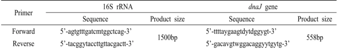
Primer 16S rRNA dnaJ gene
Sequence Product size Sequence Product size Forward 5’-agtgtttgatcmtggctcag-3’
1500bp 5’-ttttaygaagtdytdggygt-3’
558bp Reverse 5’-tacggytaccttgttacgactt-3’ 5’-gacavgtwggacaggyytgytg-3’
Table 2. PCR primers used in this study for 16S rRNA and dnaJ gene
16s rRNA dnaJ gene
Temperature Reaction time Cycle(s) Temperature Reaction time Cycle(s)
Pre-denaturation 94 3min 1 94 3min 1
Denaturation 94 30sec
30
94 30sec
40
Annealing 56 30sec 50 30sec
Extention 72 90sec 72 60sec
Post extention 72 7min 1 72 7min 1
Table 3. Conditions of reaction steps and PCR cycles
Isolates References A28003 A28004 A19006 A19008 A19010 CAIM
1797
CECT 1797
Gram-stain - - - -
Motility + + + + + + +
Catalase + + + + + + +
Oxidase + + + + + + +
TSI A/A A/A A/A A/A A/A A/A A/A
Nitrate + + + + + + +
OF F F F F F F F
Indole - - - -
Methyl red + + + + + + +
Voges-Proskauer - - - -
Citrate - - - -
Arginine + + + + + + +
Lysine - - - -
Ornithine - - - -
Arabinose - - - -
Adonitol - - - -
Cellibiose +w +w - - - +w -
Fructose + + + + + + +
Galactose - + - +w - +w +
Glucose + + + + + + +
Inositol - - - -
Lactose - - - -
Maltose + + + + + + +
Mannitol - - - + - + +
Mannose + + + + + + +
Melibiose - - - -
Raffinose + + - - - - -
Rhamnose - - - -
Salicin +w - - - -
Sorbitol - - - +w -
Sucrose + + + +w + + +
Trehalose + + + + + + +
Xylose - - - -
+; positive rasction, -; negative reaction, W; Weak
Table 4. Biochemical characteristics of the isolates from diseased olive flounder (Paralichthys olivaceus)
육 수조에 옮긴 후 수온 22~23℃에서 반유수식으로 관리하면서 시험 전 1주일간 순치하여 사용하였다.
공격 시험을 위한 균액은 본 연구에서 분리한 균주 A28003, A28004와 이전 분리 균주 A19008 (Qiao et al., 2012)를 1.5% NaCl 첨가 TSA에 27℃, 24시간 배양 한 후 멸균 생리식염수에 현탁하여 일정 농도별로 준비한 것을 사용하였고, 대조 시험구에는 멸균 생리 식염수를 사용하였다. 다양한 농도의 균 현탁액을 어체당 100 ㎕ 씩 복강 주사한 후 14일간 폐사와 변화 정도를 관찰하였다. 시험어는 시험구별로 8마리씩, 50 L 크기의 아크릴 수조에 넣은 다음 서서히 가온하 여 24시간 후부터 25±0.5℃로 사육 수온을 유지하였 으며, 25±0.5℃로 사육 수온을 유지하였으며, 매일 1회 전량 환수시켰다. 시험 기간 중 폐사한 어류는 내부 장기로부터 병원균을 재분리하여 시험균을 확 인하였다.
조직 병리학적 검사
병증을 보이는 자연 감염어 3마리의 전 장기와 인위 감염에 의해서 빈사상태 혹은 죽은 직후의 실험 어 3마리의 간, 신장, 비장, 위 및 장 등을 절취하여 Bouin’s solution에 24시간 1차 고정한 후 다시 동종의 고정액에 48시간동안 2차 고정하였다. 2차 고정 후 상법에 따라 조직 수세, 에탄올 탈수 등을 거쳐 파라 핀으로 포매하였다. 이를 Rotary 형 microtome Reichert-Jung 820 (Leica, Germany)으로 만든 4~5 ㎛ 의 절편을 H&E 염색하여 병리 조직 표본을 제작 후 광학현미경으로 관찰하였다.
결 과
병원균의 분리
자연감염어로부터 분리한 모든 균주는 최초 분리 시 1.5% NaCl 첨가 TSA에서 둥근 집락 형태를 보였으 며, TCBS 배지에서 황색 혹은 녹색을 나타내었다.
또 SS 배지에서도 투명한 콜로니를 형성하며, 이를 순수 분리한 후 1.5% 첨가 BHIA에 배양시켜 얻은 단일 콜로니를 SS 배지에 접종하였을 때에는 자라지 않았다.
생화학적 성상 검사
V. scophthalmi 7개의 분리 균주와 2개의 참조 균주 의 생화학적 성상 시험을 실시한 결과를 Table 4에 나타내었다. 분리 균주들은 Gram 음성의 운동성을 가진 간균으로, indole 생성능, citrate 이용능, Voges-Proskauer test는 negative 반응을 보였고, methyl red 시험, nitrate 환원능은 positive 반응을 보였 으며 모든 균주로부터 동일한 결과를 얻었다. 당의 경우 glucose, maltose, trehalose, sucrose를 분해하여 positive 반응을 보였다. 그러나 cellibiose, mannitol, salicin, sorbitol, sucrose, raffinose, galactose 이용능에 서 균주간의 차이를 보였다.
시험 균주의 염기 서열 분석
분리 균주의 분자생물학적 동정을 위하여 16S rRNA와 dnaJ gene으로 염기 서열을 분석한 다음 Genbank에 등록되어 있는 여러 균주의 sequence를 비교하였다. 현 분리 균주와 이전 분리 균주 모두 V. ichthyoenteri와 가장 유사한 염기서열을 가지고 있으며, 99.2~99.8%의 상동성을 보였다. 다음으로 V.
scophthalmi에 대해 높은 유사성을 보이며, 99.4~99.6%
의 상동성을 나타내었다. 그러나 dnaJ gene으로 분리 균주를 분석하였을 때에는 V. scophthalmi와 98.1~
99.3% 상동성을, V. ichthyoenteri와 91~92%의 상동 성을 보였다.
시험 균주의 병원성 확인
시험균의 넙치에 대한 병원성 시험 결과는 Fig.
1과 같이 본 시험의 분리 균주가 이전 분리 균주보다 병원성이 강한 것으로 나타났다. 즉 1×10⁸ CFU/fish의
Fig. 1. Cumulative mortality of olive flounder, Paralichthys olivaceus, challenged A19008, A28003, and A28004 intraperitoneally with 1×10⁶ and 1×10⁸ CFU/ml respectively.
A
C
Fig. 2. The clinical signs of olive flounder, Paralichthys olivaceus, infected with Vibrio scophthalmi naturally (A and B) and challenge test (C and D) by intraperitoneal injection with A28003 of 1×10⁶ CFU/ml. A, skinny body; B, atrophy of liver (arrow), white enteritis (astrick). C, skinny body; D, white enteritis (astrick).
Fig. 3. Histopathological sections of olive flounder, Paralichthys olivaceus, infected with Vibrio scophthalmi naturally and in the challenge test by intraperitoneal injection with A28003 of 1×10⁶ CFU/ml. A, atrophied liver by natural infection;
B, atrophied liver by artificial infection; C, intestine by natural infection, detachment and destruction of gut epithelium (astricks); D, kidney by artificial infection, hyaline droplet degeneration (astricks); E, intestine by artificial infection, leak of cellular material (astricks); F, spleen by artificial infection, ellipsoid hyperplasia (astricks).
고농도 감염 시험구에서 A28003과 A28004가 100%
의 누적 폐사율을, A19008이 60%의 누적폐사율을
보였다. 1×10⁶ CFU/fish의 저농도 시험구에서는 A28003과 A28004이 75%, A19008은 25%의 누적 폐
사율을 보였다. 멸균 생리식염수로 복강 주사한 대조 구에서는 폐사가 나타나지 않았다.
자연 감염어와 인위 감염어의 증상 비교 자연 감염어의 외부 증상으로는 여윔과 입올림, 체색흑화가 있었고, 내부 증상으로는 간 위축과 장관 백탁이 특징적이었다. 시험 균주 A28003과 A28004 로 인위 감염시킨 시험구의 폐사어에서는 간 위축, 체색 흑화 및 장관 백탁이 관찰되었다. A19008 인위 감염 시험구도 앞의 시험 균주와 같은 증상을 보였으 나, 일부 폐사어에서 복부 팽만의 증세를 보였다. 폐 사어의 장기에서 공격 시험에 사용한 균주가 분리되 었으며, 일부 폐사어에서는 V. harveyi가 동시에 검출 되었다 (Fig. 2).
조직 병리학적 관찰
V. scophthalmi에 감염된 자연 감염어의 조직 병리 학적 병변을 조사한 결과는 Fig. 3에 나타낸 것과 같이 심한 간 위축, 장 상피 박리 및 장내 세포물질 유출 등이 관찰되었다. 인위 감염어에서도 간 위축이 나타났으며, 비장에서 마크로파지 침윤, ellipsoid 비 후 등이 관찰되었다. 그리고 신장에서는 hyaline droplet 변성, 조혈기관 활성이 관찰되었다. 자연 감염 어와 공격 시험에 의한 병어의 병리 조직학적 소견은 유사하게 나타났다.
고 찰
본 연구에서 사용한 자연 감염어는 외부 증상으로 서 수면 가까이 유영, 여윔, 그리고 체색 흑화 등의 일반적인 병변을 보였으나, 내부 증상에서 장관 백탁, 간 위축 등의 특징적인 병변을 나타내었다. 병어로부 터 분리한 시험균으로 인위 감염시킨 어류의 증상이 자연 감염어와 일치하여 본 연구에서 분리한 균이 그 원인이라 판단되며, V. scophthalmi로 동정하였다.
본 시험 균주 A28003과 A28004를 이전 분리 균주 및 참조균주의 생화학적 성상과 비교하였을 때 대체 로 일치하는 결과를 보였으나 cellibiose, galactose, mannitol, raffinose, sorbitol, salicin, sucrose에 대하여 균주에 따라 차이가 있었다. Cerdà-Cuéllar and Blanch (2002)는 터봇 유생기에 분리한 136개의 V. scophthalmi 를 생화학적 성상으로 동정하여 28가지의 생화학적 표현형을 가졌다고 보고하였다. 따라서 당 분해능의 차이를 보이는 것이 V. scophthalmi의 특성으로 볼 때, 본 시험 균주를 V. scophthalmi로 추정 동정할 수 있다.
세균 동정에 널리 사용되는 16S rRNA를 이용한 분리균주의 염기서열을 NCBI에 등록된 sequence와 비교 분석한 결과, V. ichthyoenteri와 99.2~99.8%의 상동성을 보여 매우 유사하였으며, V. scophthalmi에 대해서도 99.4~99.6%의 상동성으로 아주 높은 유사 성을 나타내었다. 그러나 V. ichthyoenteri의 경우 자어 기의 넙치에 대하여 병원성이 강하지만, 위장이 완전 히 발달한 성어에선 병원성이 없다고 알려져 있다 (김, 2002). 또, V. scophthalmi와 V. ichthyoenteri의 16S rRNA 염기 서열이 매우 유사하기 때문에 이를 이용 하여 두 균을 구별하는 것은 매우 어려운 일이다 (Thompson et al., 2005). 따라서 좀더 정확한 동정을 위하여 dnaJ gene을 이용한 동정을 실시하였다. dnaJ gene은 heat shock protein 40을 인코딩하는 house keeping gene으로서, Mycobacterium, Legionella, Streptococcus, Enterobacteriaceae Family 뿐만 아니라 57 종류의 Vibrio 균주를 dnaJ gene을 이용하여 성공 적으로 분류한 바 있다 (Nhung et al., 2007). 본 연구의 분리 균주를 dnaJ gene로 동정하였을 때 V.
scophthalmi에 대하여 98.1%, 99.3%로 가장 높은 상동 성을 나타냈으며, V. ichthyoenteri에 대해서 91~92%
의 상동성을 보여, 16S rRNA에 대한 염기 서열 상동 성과는 달리 V. ichthyoenteri에 대하여 본 균주가 낮은 상동성을 나타내는 것을 알 수 있었다. 위에서 시행한
본 분리 균주의 생화학적 시험과 염기서열을 분석한 분자생물학적 분석 결과를 종합하였을 때, A28003과 A28004는 V. scophthalmi로 동정하는 것이 타당하다 고 본다.
Qiao et al. (2012)는 넙치를 대상으로 인위감염 실험을 시행하여 V. scophthalmi가 넙치에 대하여 25%의 누적폐사율로 비교적 약한 병원성을 가지는 것으로 보고하였다. 본 연구에서는 1 × 10⁶ CFU/fish 로 넙치에 인위감염 시험을 실시하였을 때 최근에 분리한 시험 균주가 누적폐사율 75%로 누적폐사율 25%를 나타낸 이전 분리 균주보다 강한 병원성을 가지는 것으로 밝혀졌고, V. scophthalmi는 균주에 따 라 다양한 병원성을 나타내는 것으로 사료된다.
넙치에게 단독 병원체로서 잘 알려진 V.
anguillarum, V. harveyi, P. damsela damsela 등의 병원 성 세균의 인위 감염 시험 결과, 105~108 CFU/fish의 LD50값을 보였다. 먼저, 넙치에 대한 V. harveyi의 LD50
값은 2.48×10⁵ ~ 8.76×10⁷ CFU/fish (Won and Park, 2008)와 106.1 CFU/fish (Sun et al., 2009)가 보고된 바 있다. 또 권 (2005)의 연구에서 1.2×10⁶ CFU/fish로 균을 복강 주사하였을 때, P. damsela subsp. damsela 는 80%, V. anguillarum은 60%의 누적 폐사율을 나타 냈다. 본 연구의 분리 균주 또한 106 CFU/fish의 공격 실험에서 75%의 폐사율을 보이는 것으로 미루어보 아 비교적 강한 병원성을 가지고 있다고 사료된다.
조직병리학적 병변으로 자연 감염어의 경우, 간 위축과 장 상피 탈락, 장내 세포 물질 유출을 보였고, 인위 감염어는 간 위축, ellipsoid 비후, 마크로파지 침윤, hyaline droplet 변성 등이 발견되었다. 이와 같 은 조직병리학적 병변은 Qiao et al. (2012)의 결과와 일치하며, 본 연구에서도 이전 균주와 현 분리 균주의 조직병리학적 병변의 차이는 없었다. 관찰된 조직병 리학적 변화 중 장 상피 탈락은 본 시험균의 장내 증식으로 병어의 장내 세포 물질이 유출되는 것과 연관성이 있으며, 그들이 응집되어 장관 백탁 증상을
보이는 것으로 사료된다. 따라서 넙치 자어의 장관백 탁증을 일으키는 V. ichthyoenteri와의 유전적 상동성 이 높고 본 연구의 병어에서 나타난 장관 백탁 증상 으로 미루어보아, 이들 세균이 넙치 성장 단계별로 유발하는 장관백탁증의 비교 연구를 통해 병원성 기작이나 생리적 변화를 밝히는 것도 주요할 것으로 사료된다.
본 연구에서 사용한 병어는 특징적인 임상 증상이 없으며, 내부 소견으로서 장관 백탁과 간 위축이 주증 상으로 관찰되었다. 질병의 원인체로 밝혀진 V.
scophthalmi는 균주에 따라 다양한 병원성을 가지지 만, 본 연구의 분리균주는 성장기의 넙치에 장관백탁 증을 유발하며, 강한 병원성을 나타내어 대량 폐사를 일으키는 병원균으로 판단되었다.
요 약
최근 울산광역시 소재의 넙치 양식장에서 체색 흑화, 간 위축, 장관 백탁 등의 증상을 보이며 넙치의 대량 폐사가 빈번히 발생하여, 병원체를 분리하고 감염 특성에 관하여 연구하였다. 2012년 5월 병어로 부터 분리한 원인균은 생화학 시험과 16S rRNA, dnaJ gene을 이용한 염기서열 분석을 통해 V. scophthalmi 로 동정하였다.
병원성 시험 결과, 본 시험 균주가 106 CFU/fish 에서 75%의 누적 폐사율을 보여 강한 병원성이 확인 되었다. V. scophthalmi 감염어는 조직병리학적 병변 으로서 간 위축, 장 상피 탈락, 장내 세포 물질 유출 및 장관백탁증 등이 확인되었다.
감사의 글
이 논문은 부경대학교 자율창의학술연구비(2013 년)에 의하여 연구되었음.
참고문헌
강봉조: 제주지역 양식장의 질병증상 넙치 (Paralichthys olivaceus)로부터 분리되는 세균의 특성에 관 한 연구. 제주대학교, 2003.
권문경: 넙치, Paralichthys olivaceus에서 분리된 Photobacterium damselae subsp. damselae의 어병학적 특성. 부경대학교, 2005.
김도형: 양식 넙치, Paralichthys olivaceus 자어에 대한 Vibrio ichthyoenteri의 병원성. 부경대학교, 2002.
조미란: 제주도 양식넙치에서 분리된 비브리오세균 의 신속동정과 항생제 내성. 제주대학교, 2006.
최혜승, 전려진, 김승민, 정현도, 김이경, 임희영, 여인 규, 정준범: 여윔증상 넙치,
Paralichthys olivaceus로부터 분리된 병원균 의 임상적 고찰. 한국어병학회지, 25, 67-76, 2012.
Blanch, A. R., Alsina, M., Simon, M and Jofre, J.:
Determination of bacteria associated with reared turbot (Scophthalmus maximus) larvae. Appl.
Microbiol., 82, 729-734, 1997.
Cerdà-Cuéllar, M., Rossello-Mora, R. A., Lalucat, J., Jofre, J and Blanch, A.: Vibrio scophthalmi sp. nov., a new species from turbot (Scophthalmus maximus). Syst. Bacteriol., 47, 58-61, 1997.
Cerdà-Cuéllar, M. and Blanch, A. R.: Detection and identification of Vibrio scophthalmi in the intestinal microbiota of fish and evaluation of host specificity. J. Appl. Microbiol., 93, 261-268, 2002.
Gauger, E., Smolowitz, R., Uhlinger, K., Casey, J and Gomez-Chiarri, M.: Vibrio harveyi and other bacterial pathogens in cultured summer flounder, Paralichthys dentatus. Aquaculture, 206, 10-20, 2006.
MacFaddin, J. F.: Biochemical test for identification of medical bacteria (3rd ed. by Williams, L, and Wilkins). USA, 2000.
Nhung, P. H., Shah, M. M., Ohkusu, K., Noda, M., Hata, H., Sun, X. C., Iihara, H., Goto, K., Masaki, T., Miyasaka., J and Ezaki., K.: The dnaj gene as a novel phylogenetic marker for identification of Vibrio species. Syst. Appl. Microbiol., 30, 309-315, 2007.
Qiao, G., Lee, D. C., Woo, S. H., Li, H., Xu, D. and Park, S. I.: Microbiological characteristics of Vibrio scophthalmi isolates from diseased olive flounder, Paralichthys olivaceus. Fish Sci., 78, 853-863, 2012.
Sanger, F., Nicklen, S. and Coulson, A. R.: DNA sequencing with chain-terminating inhibition.
Proc. Natl. Acad. Sci. USA, 74, 5461-5467, 1977.
Sugita, H. and Ito, Y.: Identification of intestinal bacteria from Japanese flounder (Paralichthys olivaceus) and their ability to digest chitin. Lett. Appl.
Microbiol., 43, 336-342, 2006.
Sun, K., Zhang, W., Hou, J. and Sun, L.: Immunoprotective analysis of Vhhp2, a Vibrio harveyi vaccine candidate. Vaccine, 27, 2733-2740, 2009.
Thompson, F. L., Gevers, D., Thompson, C. C., Dawyndt, P., Naser, S., Hoste, B., Munn, C. B. and Swings, J.: Phylogeny and molecular identification of
vibrios on the basis of multilocus sequence analysis. Appl. Environ. Microbiol., 71, 5107-5115, 2005.
Tin, T., Yokoyama H., Ogawa K. and Wakabayashi H.;
Myxosporeans and their hyperparasitic microsporeans in the intestine of emaciated tiger
puffer. Fish Pathol., 35, 145-156, 2000.
Won, K. M. and Park, S. I.: Pathogenicity of Vibrio harveyi to cultured marine fishes in Korea.
Aquaculture, 285, 8-13, 2008.
Manuscript Received : October 11, 2013 Revised : November 18, 2013 Accepted : December 11, 2013