카드뮴이 흰쥐 뇌기저핵의 유전자 발현에 미치는 영향
접수일 : 2009년 11월 23일, 채택일 : 2010년 2월 17일
‡ 교신저자 : 이채관( 부산시 부산진구 개금동 633-165, 인제대학교 부산백병원 산업의학과/환경·산업의학연구소,
Tel: 051-890-6174, H.P: 010-3320-3100, e-mail: [email protected])
Effects of Cadmium on the Gene Expression Profile in the Rat Basal Ganglia
This study was aimed at investigating the gene expression profile in basal ganglia of cadmium exposed rat based on cDNA array analysis. For cDNA array analysis, adult Sprague-Dawley male rats (350 ± 25 g) were intraperitoneally injected with 2.0 mg/kg body weight/day of CdCl2 (0.3 ml) for 5 days. For dose- related gene expression analysis rats were intraperitoneally injected with 0.0, 0.1, 0.3, 1.0 mg/kg body weight/day of CdCl
2for 5 days. Control rats were injected with equal volume of saline. Cadmium concentration of brain was analyzed by atomic absorption spectrophotometer. For cDNA array, RNA samples were extracted from basal ganglia and reverse-transcribed in the presence of [α32P]-dATP. Membrane sets of the Atlas Rat 1.2 array II and Toxicology array 1.2 (Clontech, Palo Alto, CA) were hybridized with cDNA probe sets. RT-PCR was employed to validate the relative gene expression patterns obtained from the cDNA array. Northern blot hybridization methods were employed to assess the dose-related gene expression.
Among the 2352 cDNAs, 671 genes were detected in both array sets and 63 genes of 38 classes showed significant (more than two fold) changes in expression. Thirty five of these genes
were up-regulated and twenty eight were down-regulated in the cadmium exposed group. According to the dose-related gene expression analysis, heat shock 27 kDa protein (HSP27), neurodegeneration-associated protein 1 (Neurodap 1) genes were significantly up-regulated and melatonin receptor 1a (Mel1a), Kinesin family member 3C (KIF3C), novel kinesin- related protein (KIF1D) genes were significantly down- regulated even in the low-dose of cadmium exposed group (0.1 mg/kg body weight/day).
Conclusions Sixty three genes detected in this study can give some more useful informations about the cadmium-induced neurotoxicity in the basal ganglia. As well as, HSP27, Neurodap1, Mel1a, KIF3C and KIF1D genes may be useful for the study of the cadmium-induced neurotoxicity because these genes showed dramatic changes of mRNA levels in response to the low dose of cadmium exposure.
basal ganglia, cadmium, cDNA array, gene expression.
Key Words : Chae-Kwan Lee
Institute of Environmental and Occupational Medicine & Department of Occupational and Environmental Medicine, Busan Paik Hospital, Inje University
이채관
인제대학교 부산백병원 산업의학과/환경·산업의학연구소
Ⅰ. 서 론
카드뮴은 합금, 안료(도료), 전지 등의 생산 공정에서 다양 하게 사용되는 중금속으로서 생체에 흡입되면 체내의 축적 경로를 거쳐 거의 모든 기관에 독성을 나타내는데 특히 감각 중추 신경 장애 및 폐, 간, 신장, 정소, 전립선 등에서 기능 장 애를 유발한다.
카드뮴의 생체독성에 관한 연구는 정소에서의 apoptosis, 간기능 장애 등의 분야에서 먼저 진행되어 왔으며(Messner 등, 2009), 최근에는 카드뮴의 뇌신경계 독성에 관한 연구도 활발히 진행되었다. 흰쥐 등 동물모델을 이용한 연구에서 카 드뮴은 시상하부와 뇌하수체에서 신경내분비계 신호전달 물질과 호르몬 분비를 교란시키며, 특히 프로락틴의 분비를 억제한다고 알려졌다(Esquifino 등, 2001; Lafuente 등, 2000). 태 아기의 카드뮴 노출은 소뇌와 선조체의 기능에 영향을 미친 다(Antonio 등, 2002). 그리고 카드뮴은 해마, 선조체 및 전두 엽 등에서 아미노산 대사와 GABA 및 taurine 활성에 영향을 미치며(Caride 등, 2009; Lafuente 등, 2001) 도파민 등의 신경전 달물질 대사에도 영향을 미침으로써(Lafuente 등, 2005;
Minami 등, 2001) 뇌신경의 신호전달체계를 교란시킨다.
카드뮴을 포함한 중금속의 중추신경계 독성기전은 신경 학적 증상에 근거하므로 지금까지 알려진 연구 결과들로는 카드뮴 중독 진단 또는 증상의 단계를 판단하기에 어려움이
많으며, 따라서 증상을 진단하기 위한 객관적인 자료 확보를 위한 연구가 필요하다. 최근 이를 위해는 다량의 유전자들을 동시에 검색할 수 있는 cDNA array와 같은 연구방법을 통해 중금속의 독성기전에 관련된 다수의 유전자들을 조사하는 연구가 수행되고 있으며, 이를 통해 확인된 관련 유전자 정 보를 활용하여 생물학적지표 개발 등의 연구가 수행되고 있 다. 이 연구에서도 카드뮴에 노출된 흰쥐의 뇌기저핵을 이용 하여 cDNA microarray를 시행하여 카드뮴에 의한 신경내분 비계의 독성기전에 관련된 유전자들의 목록을 확보하여 독 성기전 연구를 위한 유전자 수준의 기초자료를 제공하고자 하였으며, 아울러 발현의 현저한 변화를 나타내는 유전자들 에 대하여 카드뮴 노출농도에 따른 유전자 변화 양상을 조사 하여 생물학적지표 개발에 필요한 기초자료를 제공하고자 하였다.
Ⅱ. 연구방법
1
1.. 실 실험 험동 동물 물
실험동물은 출생 후 15주된 카드뮴 노출력이 없는 sprague-
dawley 수컷 흰쥐(350 ± 25 g) 25마리를 사용하였으며 사료
와 물은 자유롭게 공급되었다. 사육된 온도조건은 24℃~26Parameters Condition
Wave length(nm) Slit width(nm) Lamp current(mA) Measurement mode Inert gas
Sample volume(㎕)
Zeeman background correction
228.8 0.7
6 Peak area
Argon 20 Yes
Table 1. Instrumental parameters of atomic absorption spectrophotometerElement Step Temperature(℃) Time (sec.)
Internal flow
Internal flow Hold
Cd
Drying Drying Ashing Atomization
Cleaning
130 150 550 1200 2400
20 10 30 1 1
40 20 50 5 2
250
250
250
0
250
Table 2. Furnace conditions for measurement of blood and brain cadmium℃, 습도는 55% - 65%, 조명은 명기 14시간, 암기 10시간이었 으며, 실험동물의 관리는 인제대학교 실험동물관리규정에 따랐다.
2
2.. 카 카드 드뮴 뮴 노 노출 출
카드뮴(CdCl2
, Sigma-Aldrich Co, St. Louis MO USA)은 생리
식염수에 녹인 후 복강 주사하였으며 대조군은 동량의 생리 식염수를 주사하였다. 카드뮴 노출조건은 카드뮴에 의한 흰 쥐의 중추신경계 독성연구를 수행한 논문들(Caride 등, 2009;Heilmaier 등, 1987; Yang 등, 2007)과 카드뮴의 no-observed- adverse-effect-level(NOAEL)(Groten 등, 1997)을 참고하여 중추
신경계의 기능을 교란시킬 것으로 예측되는 0.0, 0.1, 0.3, 1.0,2.0 mg/kg body weight/day의 농도로 각 군별 5마리씩 5일간 노
출시켰다. 그리고 cDNA array cDNA array는 2.0 mg/kg bodyweight/day 노출군의 뇌기저핵을 이용하였다.
3
3.. 혈 혈액 액과 과 뇌 뇌 조 조직 직의 의 카 카드 드뮴 뮴 농 농도 도분 분석 석
실험동물은 카드뮴 노출 5일째에 모두 도살하였으며 혈액 은 도살시 K3
EDTA(7.5%) bottle(Beliver Industrial Estate,
Polymouth. UK)
로 채취하여 4℃에 보관하였다. 뇌 조직은 적출 후 생리식염수로 씻고 3M 종이로 수분을 제거한 뒤 정량 하였다. 정량된 조직을 HNO3
/ HCL
4(6:4) 용액과 반응시켜 분
해한 후 백색 침전물을 획득, 적정량의 0.1 N HNO3용액에 녹 인 후 원자흡광광도계(Automic absorption spectrometry, Perkin-Elmer AA 800, USA)로 정량하였다. 원자흡광광도계를 이용
한 카드뮴농도 분석 조건은 Table 1, 2와 같다.4
4.. R RN NA A 추 추출 출
적출된 뇌 조직에 Tri-Reagent (Sigma-Aldrich Co, St. Louis
MO USA, 0.1 g/㎖)를 가한 후 homogenizer (Ingenieurburo Co.
Etzenbach, Germany)
를 사용하여 얼음에 시험관을 담은 상태 로 약 30초간 파쇄하였다. 파쇄액이 담긴 시험관에 chloroform 을 넣고 실온에서 15분간 방치 후, 4℃, 13,500 rpm에서 15분간 원심 분리하였다. 그 후 상층액에 동량의 isopropanol을 넣고 실온에서 방치 후, 다시 4℃, 13,500 rpm에서 10분간 원심 분 리하였다. 원심 분리 후 상층액을 제거하고 남겨진 침전물을75% 에탄올로 2회 세척하였다. 이 침전물을 실온에서 10분
동 안 건 조 한 후 적 당 량 의 formamide나 diethylpyrocarbonate(DEPC)로 처리된 멸균수에 녹였다. mRNA는 mRNA Isolation Kit(Roche Molecular Biochemicals, Mannheim, Germany)
를 사용하였다. 추출된 total RNA와 mRNA는 260 nm와 280 nm에서 흡광도를 측정하여 정량하였고, 280 nm에 대 한 260 nm 흡광도의 비율이 1.6~2.0인 것들을 cDNA array, RT-
PCR 및 northern blot hybridization에 사용하였다.
5
5.. c cD DN NA A m miic crro oa arrrra ayy 분 분석 석
cDNA microarray는 Rat Toxicology array 1.2(Clontech, East Meadow Circle Palo Alto, CA, USA)와 Atlas Rat 1.2 array II(Clontech) Kit 외 일부 보유하고 있던 중추신경계 독성기전
에 관련된다고 알려진 유전자를 사용하였다. 추출한 totalRNA를 DNase Ⅰ(Roche Molecular Biochemicals)를 처리하여 genomic DNA를 제거한 다음 denaturing formaldehyde/
agarose/etidium bromide(EtBr) gel로 전기영동하여 RNA 상태
를 확인하였다. 추출한 total RNA에 대한 genomic DNA의 오 염 여부는 β-actin primer를 이용하여 PCR을 수행한 후agarose/EtBr gel에서 확인하였다. 준비된 total RNA 20 ㎍를 0.5
㎖ micro-centrifuge tube에서 70℃로 2분, 50℃로 2분간
incubation 한 후 α-
32P dATP(Amersharm Pharmacia Biotech AB, Uppsala, Sweden), MMLV reverse transcriptase(Roche Molecular Biochemicals)와 CDS(cDNA synthesis) primer mix를 사용하여 50℃에서 25분간 반응하여 동위원소 표지하였다. 합성된 probe는 Nucleospin Extraction Spin Column(Amersharm Pharmacia Biotech)을 이용하여 정제한 후 Scintillation counter로
cpm값을 측정하였으며 각 실험군 별로 5 × 10
6cpm이상 사
용하였다. Atlas array membrane (Clontech)을 0.5 ㎎ denaturated
salmon sperm DNA(Sigma-Aldrich Co)가 첨가된 ExpressHyb로 68℃에서 30분간 pre-hybridization하였다. probe를 10 × denaturing solution(1 M NaOH, 10 mM EDTA)과 2×neutralizing solution(1 M NaH
2PO
4, pH 7.0)로 68℃에서 각각 20분과 10분씩 incubation 한 뒤 ExpressHyb에 첨가하여 68℃에서 over night hybridization 하였다. Hybridization후 wash solution 1 (2 X SSC, 1% SDS)로 68℃에서 30분간 3회, wash solution 2(0.1 X SSC, 0.5% SDS)로 68℃에서 30분간 1회 washing 한 다음 X-ray film (Kodak X-OMAT AR)으로 -70℃에서 1-3일 감광 시켰다.
Signal을 제공된 program 또는 adobe photoshop image analyser를
이용하여 비교 분석하였다.6
6.. R RT T- -P PC CR R을 을 이 이용 용한 한 c cD DN NA A m miic crro oa arrrra ayy 결 결과 과 검 검증 증
RT-PCR 방법으로 cDNA microarray 분석결과 유의성을 검
증하기 위하여 각각 유전자의 합성에 필요한 primer sequence 를 NCBI GenBank database를 활용하여 MacVector software(Oxford Molecular, Oxford, UK)로 획득, 합성하였다. 합성된 primer들과 추출된 total RNA를 이용하여 RT-PCR을 다음과
··
같이 시행하였다. 추출된 total RNA를 200 unit의 Moloney
Murine leukemia virus (MMLV) reverse transcriptase(Roche Molecular Biochemicals)와 37℃에서 1시간 동안 반응시켜 complementary DNA(cDNA)를 합성하였다. 합성된 cDNA를 10 units의 Taq DNA polymerase Perkin-Elmer Cetus, Wellesley, MA, USA) 합성된 primer 및 dNTP 등과 혼합한 후 30 회 반응 (95℃ 1분, 55℃ 1분, 72℃ 1분)시켰다. 증폭된 cDNA 절편들은 1% agarose gel로 전기 영동한 후 사진 촬영하였다.
7
7.. N No orrtth he errn n b bllo ott h hyyb brriid diizza attiio on n
mRNA(1 - 4 ㎍)를 1% agarose/2.2 M formaldehyde gel에서 50 V로 3시간 동안 전기영동 하였다. 전기영동 후 RNA를 transfer kit(Trans Vac, Hoefer Co. San Francisco CA, USA)
를 이용 하여 nylon membrane(Schleicher & Schuell Inc. Keene, NH,USA)으로 옮긴 후, vacuum oven에서 80℃로 2시간 건조하였
다. RNA가 부착된 nylon membrane을 hybridization buffer로 60℃에서 2시간 prehybridization한 후 합성된 cDNA probe (1 ×
10
9cpm/㎖)를 첨가하여 60℃에서 18시간 hybridization하였다.
Hybridization buffer의 조성은 50% deionized formamide, 5 X SSC (1 X SSC: 0.15 M NaCl and 0.015 M sodium citrate), 5 X Denhardt's solution(1 X Denhardt's solution: 0.01% polyvinyl pyrrolidone, 0.01% Ficoll and 0.01% BSA), 0.1% SDS, 2 ㎎/㎖
salmon sperm DNA 이다. Hybridization 후 비특이적인 결합을
제거하기 위하여 nylon membrane을 0.1 X SSC, 0.1% SDS 용액으로 55℃에서 3회 세척한 뒤 X-ray film(X-OMAT, Eastman
Kodak Co. Rochester, NY, USA)으로 1-4일간 감광시켰다. 사용
된 probe는 RT-PCR로 합성 후 pGEM-T Easy Vector(Promega) 에 클로닝 하여 염기서열을 확인한 각 cDNA 절편들을 제한 효 소 SpeⅠ 으 로 절 단 한 후 추 출 하 여 OligolabellingKit(Amersham Pharmacia Biotech.)와 [α-
32P] dCTP(Amersham Pharmacia Biotech.)를 사용하여 합성하였다. 합성된 cDNA probe를 Nick column(Amersham Pharmacia Biotech.)에 통과시
켜 정제하고, SET buffer (0.1% SDS, 1 mM EDTA, 10 mM Tris,10 mM dithiothreitol)로 추출하였다. cDNA probe의 활성은 1×
109 cpm/
㎍ 이상으로 하였다.8
8.. A Au utto orra ad diio og grra ap ph h 및 및 유 유전 전자 자 발 발현 현비 비율 율 분 분석 석
유전자 발현은 image-analyzer 감광방식과 X선 필름 감광방 식을 병행하여 분석하였다. Image-analyzer 감광방식은
phosphor-스크린을 방사성 동위원소로 표지된 나일론종이에
부착하여 감광시킨 후 Typhoon9400 (Amersharm biosciences,Piscatway, NJ, USA)으로 영상화 하였으며, 사용된 프로그램
은 Image Quant(version 5.0)이었다. X선 필름 감광방식은hybridization과 세척이 끝난 나일론종이를 X-OMAT AR 필름 (Eastman Kodak Co.)으로 -70℃에서 1-3일 감광 시킨 후 X선
필름 현상기(RGII, Fuji Co., Tokyo, Japan)로 현상 후 AdobePhotoshop(version 6.0)을 이용하여 비교 분석하였다.
Parameters Experimental group
Control 0.1 mg 0.3 mg 1.0 mg 2.0 mg
Brain (g) 1.85±0.13 1.90±0.13 1.85±0.12 1.83±0.12 1.82±0.11
Table 3. Changes of brain weight according to the cadmium exposure level
mean ± S.D
These values originated from 5 male rats in each group.
Parameters Experimental group
Control 0.1 mg 0.3 mg 1.0 mg 2.0 mg
Blood (ng/㎖) Basal ganglia (ng/g)
3.56±0.37 1.45±0.15
8.20±0.94
a3.06±0.34
a18.54±1.92
ab5.86±0.62
ab35.66±3.11
abc10.96±1.04
abc50.03±3.85
abcd21.16±2.21
abcd Table 4. Changes of cadmium concentration of blood and brain (basal ganglia) according to the cadmiumexposure level mean ± S.D
These values originated from 5 male rats in each group.
p(<0.05) values were calculated by Mann-Whitney(U) test: a
compared to control group.
bcompared to 0.1 mg exposed group,
ccompared to 0.3 mg
exposed group,
dcompared to 1.0 mg exposed group.
다
다.. 자 자료 료 분 분석 석
자료 분석은 SPSS 프로그램(12K)을 이용하였으며 연속자 료의 두 군간 비교에는 mann-whitney(U) test를, 세 군 이상 간 비교에는 kruskall-wallis test를 시행하였다.
Ⅲ. 연구결과
1
1.. 체 체중 중과 과 뇌 뇌의 의 무 무게 게
각 노출농도군별로 5일간 카드뮴(CdCl2
)에 노출된 흰쥐 뇌
의 무게를 조사한 결과는 Table 3과 같다. 뇌의 무게는 각 실 험군간 유의한 변화는 없었다.2
2.. 혈 혈액 액과 과 뇌 뇌 조 조직 직의 의 카 카드 드뮴 뮴 농 농도 도
혈액과 뇌기저핵의 카드뮴 농도를 분석한 결과는 Table 4 와 같다. 혈액의 카드뮴 농도는 대조군에 비하여 카드뮴 노 출군에서 농도에 따라 통계적으로 유의하게 증가되었다. 뇌 기저핵의 카드뮴 농도도 대조군에 비하여 카드뮴 노출군에 서 농도에 따라 통계적으로 유의하게 증가되었다. 그리고 혈 액에 비하여 뇌기저핵의 카드뮴 농도가 모든 실험군에서 낮 게 조사되었다.
3
3.. c cD DN NA A a arrrra ayy 분 분석 석
흰쥐의 뇌기저핵에서는 분석에 사용된 총 2352개의 유전 자 중 Rat Toxicology array 1.2 kit에서 424개, 그리고 Atlas Rat
1.2 array II kit에서 247개를 합하여 671개 유전자의 발현이 확
인 되었다. 이들 유전자에 대하여 image-analyzer 감광방식과X선 필름 감광방식을 병행하여 대조군과 카드뮴 노출간의
유전자 발현정도를 비교한 결과 대조군에 비해 카드뮴 2.0 ㎎ 노출군에서 2배 이상 증가 또는 감소되어 유의한(p<0.05) 차 이를 나타낸 유전자는 Rat Toxicology array 1.2 kit에서 32개,Atlas Rat 1.2 array II kit에서 31개를 합하여 63개였다. 이들 중
카드뮴 노출군에서 발현이 증가된 유전자는 35개였고 감소 된 유전자는 28개였다(Table 5).4
4.. R Re ella attiivve e R RT T- -P PC CR R 방 방법 법을 을 이 이용 용한 한 c cD DN NA A a arrrra ayy 결 결 과
과검 검정 정
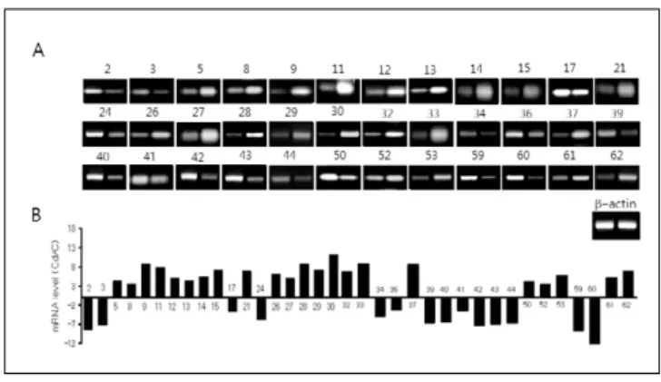
cDNA array결과 (Table 5)에 제시된 63개 유전자 중 36개에
대하여 Relative RT-PCR 방법으로 대조군과 카드뮴 노출군 간의 각 유전자의 발현비율을 검정한 결과 cDNA array와 비교 하여 일부 미약한 차이를 나타내는 유전
5
5.. 카 카드 드뮴 뮴 노 노출 출 농 농도 도에 에 따 따른 른 유 유전 전자 자 발 발현 현 ((D Do os se e- - rre ella atte ed d g ge en ne e e exxp prre es ss siio on n))
Table 5와 Fig. 1에 제시된 카드뮴의 독성기전에 관련된 유
전자들 중 유전자의 발현변화 정도가 크거나, 카드뮴의 뇌지 저핵에 대한 독성기전 연구에서 아직 보고된 적이 없는 heatshock 27-kDa protein(HSP27), neurodegeneration-associated protein 1(Neurodap1), mitochondrial stress-70 protein(MTHSP70),
멜라토닌 수용체 1a (Mel1a), kinesin family member 3C, novelKinesin-related protein(KIF1D)
등 6개 유전자들에 대하여 카드 뮴 노출 농도별 유전자 발현을 조사하였다.Heat shock 27-kDa protein (HSP27, No. 11)은 heat shock protein
군에 속하는 유전자로써 발현은 대조군과 비교하여 카드뮴0.1, 0.3, 1.0 ㎎ 노출군에서 노출농도에 따라 유의하게(p<0.05)
증가되었다. Neurodegeneration-associated protein 1(Neurodap1,No. 21)은 trafficking and targetting protein에 속하는 유전자로써
발현은 대조군과 비교하여 카드뮴 0.1, 0.3, 1.0 ㎎ 노출군에서 노출농도에 따라 유의하게(p<0.05) 증가되었다. Mitochondrialstress-70 protein (MTHSP70, No 30)은 stress response protein에 속
하는 유전자로써 발현은 대조군과 비교하여 카드뮴 0.1 ㎎ 노 출군에서 가장 많이 증가되었으며, 노출량이 증가함에 따라 점차 감소되었으나 1.0 ㎎ 노출군에서도 대조군에 비해 높은 Fig 1. Relative RT-PCR analysis some genes listed in Table 5. (A) Gel scan of the RT-PCR analysis.The β-actin specific cDNA fragment was amplified with the each gene allowing determination of the relative expression (B) The optical densities were analyzed by the NIH Image Software and represented as the mean±S.D. (n=3). The numbers in (A) and (B) refer to the genes listed in Table 5.
Cd: cadmium exposed group; C: control.
GenBank No. Gene/Protein Name Gene/Protein Classification
aFold change D38492
U41662 U41663 U38938 AB012234 AF079314
D83792 X55572 D38380
X14878 M86389 M69246 Z27118 Y00054 X96394 D17521 M16459 M95738 M33003 X05834 D32249
X14209 M17086
U38419 X15958
Y00404 X56600 Y17295
M86870 S78556
M19007 AF003523
1 2 3 4 5 6
7 8 9
10 11 12 13 14 15 16 17 18 19 20 21
22 23
24 25
26 27 28
29 30
31 32
Neural adhesion molecule F3 Neuroligin 2
Neuroligin 3
Element DNA-binding protein 1 (CREBP1) Nuclear factor I-X1 (NF1-X1)
Nuclear factor of kappa light chain enhancer in B- cells p65
Cyclin-dependent kinase inhibitor p27 (p27KIP1) Polipoprotein D (APOD)
Serotransferrin (TF); siderophilin; beta-1-metal- binding
Thioredoxin
Heat shock 27-kDa protein (HSP27) Heat shock 47-kDa protein (HSP47) Heat shock 70-kDa protein (HSP70) Heat shock cognate 71-kDa protein (HSC71) Multidrug resistance protein (MRP) Chloride channel protein 3 (CLCN3; CLC3) Cellular retinol-binding protein 1 (RBP1) Solute carrier family 6 member 11 (SLC6A11);
Solute carrier family 6 member 1 (SLC6A1);
Fibronectin
Neurodegeneration-associated protein 1 (Neurodap 1)
Cytochrome c oxidase subunit IV (COX4) cAMP-dependent protein kinase type I alpha regulatory subunit
Dopa/tyrosine sulfotransferase
Mitochondrial enoyl-CoA hydratase; short chain enoyl-CoA
Soluble superoxide dismutase 1 (SOD1) Mitochondrial superoxide dismutase 2 (SOD2) Thiol-specific antioxidant protein (1-Cys peroxiredoxin)
Endoplasmic reticulum stress protein 72 (ERP72) Mitochondrial stress-70 protein (MTHSP70); 75- kDa
Protein kinase C beta I (PKC-beta 1) BCL2-associated death promoter (BAD)
Cell adhesion proteins
Cell adhesion receptor; cell surface antigens
Basic transcription factor
DNA polymerases, replication factors & topoisomerases Intracellular transducers, effectors & modulators
CDK inhibitors
Complex lipid metabolism
Extracellular transporters & carrier proteins
Immune system proteins Chaperones & heat shock proteins
Drug-resistance proteins Voltage-gated ion channels Facilitated diffusion proteins Symporters & antiporters
Extracellular matrix proteins Trafficking & targetting protein
Energy metabolism Nucleotide metabolism
Metabolism of cofactor Amino acid metabolism
Metabolism enzymes; stress response protein Stress response protein
Bcl family proteins
+ -- -- + ++
+
+ ++
++
- ++
++
++
++
++
+ -- + + - ++
- +
-- +
++
++
++
++
++
- ++
Table 5. List of genes showing changed expression in the cadmium exposed group compared with the control
a The fold change was obtained by comparing signal intensities between control and cadmium exposed in the cDNA expression array. +: increased in cadmium exposed group more than 2 fold; ++: ncreased in cadmium exposed group more than 4 fold; -: decreased in cadmium exposed group more than 1/2; --: decreased in cadmium exposed group more than 1/4.
++
-- - --
++
- -- -- --
-- -- -- -
-
-
- -
-- + ++
++
+
+ - - -
-- -- ++
++
+
GenBank No. Gene/Protein Name Gene/Protein Classification
aFold change
U72350 X82551 D84418 M55291
X59949 M12527 U14409 AF141863
M35077
M36831 J04811 M81766
L08490
L08493
L08494
M18416;
M18330
U34841 M83680 Y14019 AF072935
M12492
D85509 U27201 L31884 X12367
AJ223599 AJ000696 J00750 S65838 M20035
33 34 35 36
37 38 39 40 41
42 43 44 45
46
47
48 49
50 51 52 53 54
55 56 57 58
59 60 61 62 63
BCL2-like protein 1 (BCL2L1); BCLX 60S ribosomal protein L39 (RPL39) High mobility group protein 2 (HMG2) Brain-derived neurotrophic factor/ neurotrophin 3 receptor
Nitric oxide synthase 1
Gonadotropin releasing hormone Melatonin receptor 1a (Mel1a) Melatonin receptor 1b (Mel1b) Dopamine receptor D1A (D1A receptor;
DRD1A)
Dopamine receptor D2 (D2 receptor; DRD2) Growth hormone receptor (GH receptor; GHR) Retinoid X receptor beta (RXR-beta; RXRB) Gamma-aminobutyric-acid receptor alpha 1 subunit (GABA(A) receptor alpha 1; GABRA1) Gamma-aminobutyric-acid receptor alpha 4 subunit (GABA(A) receptor alpha 4; GABRA4) Gamma-aminobutyric-acid receptor alpha 5 subunit (GABA(A) receptor alpha 5; GABRA5) Early growth response protein 1 (EGR1) Protein kinase C delta (PKC-delta; PRKCD;
PKCD)
G protein-coupled receptor kinase 5 (GRK5) Ras-related protein RAB14
Ras-related protein RAB3B Ras-related protein RAB5A
cAMP-dependent protein kinase type II-beta regulatory chain
Matrix metalloproteinase 16 (MMP16);
Tissue inhibitor of metalloproteinase 3 (TIMP3) Tissue inhibitor of metalloproteinase 2 (TIMP2) Cellular glutathione peroxidase I (GSHPX1;
GPX1)
Kinesin family member 3C
A novel kinesin-related protein (KIF1D) Metallothionein 1 (MT1)
Metallothionein 3 (MT3) Prothymosin-alpha (PTMA)
mRNA processing, turnover & transport proteins DNA-binding & chromatin proteins
Intracellular transducers, effectors & modulators
Hormone
Hormone receptor (nuclear receptor)
Neurotransmitter receptor
Extracellular communication proteins
Intracellular transducers, effectors & modulators G-protein
Intracellular kinase network members Metalloproteinases
Enzymes involved in protein turnover Motor protein
Functionally unclassified protein
Table 5. Continueda The fold change was obtained by comparing signal intensities between control and cadmium exposed in the cDNA expression array. +: increased in cadmium exposed group more than 2 fold; ++: ncreased in cadmium exposed group more than 4 fold; -: decreased in cadmium exposed group more than 1/2; --: decreased in cadmium exposed group more than 1/4.
수준을 유지하였다 (Fig. 2).
멜라토닌 수용체 1a (Mel1a, No. 39)은 hormone receptor군에 속하는 유전자로써 발현은 대조군과 비교하여 카드뮴 0.1,
0.3, 1.0 ㎎ 노출군에서 노출농도에 따라 유의하게 감소되었
다. Kinesin family member 3C (KIF3C, No. 59)는 motor protein군 에 속하는 유전자로서 발현은 대조군과 비교하여 카드뮴 노 Fig 2. Dose-related expression of Heat shock 27-kDa protein (HSP27, No. 11), Neurodegeneration- associated protein 1(Neurodap1, No. 21) and Mitochondrial stress-70 protein (MTHSP70, No 30) genes in the rat basal ganglia. (A) Northern blot analysis of mRNA. mRNAs (1 ㎍) were fractionated on a 1% formaldehyde agarose gel, transferred to a nylon membrane, and hybridized with the [α 32P]dCTP-labeled cDNA probe. The β-actin probe was hybridized to confirm equal loading of the mRNA samples. (B) The Northern signals were normalized relative to the β-actin mRNA levels, and expressed as relative units over the C value of 1.0. The experiments were repeated three times, and individual values are expressed as the mean±
S.D. Arabic numbers on the lane indicate the cadmium exposed level/kg body weight/day. C:
control. a, b and c on the bar indicate the significantly difference (p<0.05) compared with control, 0.1 mg and 0.3 mg exposed groups, respectively.
Fig 3. Dose-related expression of Melatonin receptor 1a (Mel1a, No. 39), Kinesin family member 3C (KIF3C, No. 59) and Novel kinesin-related protein (KIF1D, No. 60) genes in the cadmium exposed rat basal ganglia. (A) Northern blot analysis of mRNA. mRNAs (1 ㎍) were fractionated on a 1% formaldehyde agarose gel, transferred to a nylon membrane, and hybridized with the [α 32P]dCTP-labeled cDNA probe. The β-actin probe was hybridized to confirm equal loading of the mRNA samples. (B) The Northern signals were normalized relative to the β-actin mRNA levels, and expressed as relative units over the C value of 1.0. The experiments were repeated three times, and individual values are expressed as the mean±
S.D. Arabic numbers on the lane indicate the cadmium exposed level/kg body weight/day. C:
control. a, b and c on the bar indicate the significantly difference (p<0.05) compared with control, 0.1 mg and 0.3 mg exposed groups, respectively.
출량에 따라 유의하게(p<0.05) 감소되었다. Novel kinesin-
related protein (KIF1D, No. 60)
도 motor protein군에 속하는 유전 자로서 발현은 대조군과 비교하여 카드뮴 노출군에서 노출 량에 따라 감소되었다(Fig. 3).Ⅳ. 고 찰
본 연구에서는 cDNA array방법으로 카드뮴에 노출된 흰쥐 의 뇌기저핵에서 발현이 의미있게 변화된(2배 이상) 63개의 유전지를 확인하였다. 그 중에는 이미 카드뮴의 독성기전과 의 연관성이 알려진 heat shock protein(HSP), superoxide
dismutase(SOD), metallothionein(MT), Bcl-family 등의 유전자도
포함되었으며, gamma-aminobutyric-acid(GABA), 도파민등은 카드뮴의 독성기전과 밀접한 연관이 있는 신경내분비계물 질의 수용체도 확인 되었다.HSP는은 세포환경의 온도변화(열충격)에 대한 적응을 연
구하는 과정에서 확인 되었는데, 온도변화 외에도 독성물질 의 대사에 대한 세포의 적응을 용이하게 하는 기능을 가진 단백질군이다. 본 연구에서는 HSP 27, 47, 70, 71, mitochondrialstress-70 protein(MTHSP70)등 5개 유전자의 발현이 확인되었
으며, 모두 대조군에 비해 카드뮴 노출군에서 발현이 현격 히 증가 되었다. 이러한 결과는 카드뮴에 의한 oxidative stress 에 대한 뇌 세포의 방어기전으로 해석되며 신장의 내피세포 와 배아줄기세포 그리고 뇌종양세포 등을 이용한 선행연구 의 결과들과도 일치 하였다(Bonham 등, 2003; Hung 등, 1998;Wu and Welsh, 1996).
SOD는 세포의 호흡에 의해 생성되는 과산화물대사에 관
여하는 효소로 세포내 과산화물은 미토콘드리아의 호흡에 의해 생성되며 peroxynitrite와 같은 유독성 과산화물을 생성 한다(Keller 등, 1998). 그리고 이러한 과산화물들은 적절히 분 해되지 못하면 세포의 항상성(homeostasis)이 유지되지 못하 며 궁극적으로는 신경손상을 일으킨다(Colombrita 등, 2003).SOD는 과산화물질의 분해 작용을 가진 효소로서 생체에 생
성된 과산화물분자들을 과산화수소(H2O2)로 전환시켜 독성 대사체를 감소시킨다(Kao 등, 2003). 이 연구에서는 SOD 1과2의 발현이 확인되었으며 두 유전자 모두 발현이 증가되었
다. SOD 2의 경우 카드뮴에 의하여 효소활성이 억제 된다고 알려져 있으나, 이 연구에서는 SOD 2등 유전자의 발현이 카 드뮴 노출군에서 증가된 사실로 보아 카드뮴에 의한 SOD유 전자의 전사(transcription), 해독(translation), post-transcriptionalmodification 및 효소활성화 단계에 관한 연구가 필요하리라
생각된다. 그리고 thiol-specific antioxidant protein(1-Cysperoxiredoxin)유전자의 발현도 현격히 증가되었는데 이 또한
카드뮴 노출에 의해 생성된 과산화물 분해과정으로 해석된 다. 그 외 세포내 과산화물 생성억제 효소로 알려진 nitric
oxide synthease(NOS)
유전자가 확인 되었는데 이 효소 또한 카드뮴 노출군에서 유전자 발현이 현격히 증가되었다. 만약SOD와 NOS 등의 활성이 억제되어 세포내 과산화물 분해가
원활치 못하면 세포는 과산화물과 hydroxyl radical 등의 증가 와 이로 인한 지질의 과산화, 세포내 환경의 과산화 및 DNA 와 세포막 파괴 유전자 발현의 교란 등을 일으키게 되어 궁 극적으로는 apoptosis 또는 사멸하게 된다(Chatterjee 등, 2009:Liu 등, 2009). 본 연구에서 확인된 apoptosis 관련 유전자는 BCL-associated death promoter(BAD)와 BCL2-like protein 1(BCLX)이 있는데, BAD는 apoptosis 유도인자로, BCLX는 apoptosis 억제인자로 알려져 있다(Datta 등, 2002; Ma 등, 1995). 그러나 Apoptosis 유도 및 억제 기전은 다양하고 관련
된 유전자 또한 다양하기 때문에 선행연구에서 알려진 유전 자들과 본 연구에서 확인된 유전자들에 대한 추가적인 연구 가 필요하며 이는 카드뮴에 의한 세포사멸(apoptosis) 관련기 전 연구에 많은 도움이 될 것이다.MT는 카드뮴을 비롯한 중금속의 생체내 대사에 관여하는
단백질로서 중금속과 결합하여 간, 신장 및 체내 기관간의 이동에 관여한다(Coyle 등, 2001). 본 연구에서는 MT 1과 3 유 전자의 발현이 확인 되었으며 유전자 발현이 현격히 증가되 었다. 이들은 중금속에 의하여 유전자 발현이 유도되는 것으 로 알려져 있으며(Coyle 등, 2001) 본 연구의 결과와도 유사 하였다.GABA는 glutamate와 함께 중추신경계의 주요한 신경전달
물질로서 GABA는 억제기전에, 그리고 glutamate는 흥분기전 에 관여하며(Calaza Kda 등, 2003), 외인성 물질에 의한 GABA 수용체 유전자의 변화는 중추신경계 신경전달 기전을 교란 시킬 수 있다. GABA수용체들 중 GABAα군은 신경 신호전달 과정에서 postsynaptic clustering과 신경망 형성에도 관여한다.따라서 카드뮴 노출에 의한 GABA 수용체군 유전자의 변화 는 신경망형성 및 신경계 교란의 원인이 될 수 있다
(Schweizer 등, 2003). 본 연구에서 확인된 GABA 수용체 유전
자는 GABAα1, 4, 5등 3개이었으며 cDNA microarray 결과에 의하면 카드뮴노출군의 뇌기저핵에서 GABAα1, 4, 5 수용체 유전자의 발현이 억제되었다. 선행된 연구 자료와 본 연구의 결과로 보아 GABA 수용체 유전자는 카드뮴 노출에 의해 영 향을 받으며 이는 카드뮴에 의한 뇌신경계의 신경망 형성과 신호전달체계를 교란기전의 중요한 자료가 될 것으로 사료 된다.도파민은 카드뮴에 의해 분비교란이 일어나는 대표적인 신경내분비계물질로써 시상하부의 tubero-infundibular neuron 에서 주로 합성되며 프로락틴유전자 발현을 억제한다
(Elsholtz 등, 1991). 본 연구에서는 흰쥐의 뇌기저핵에서 dopa/tyrosine sulfotransferase, 도파민 수용체 1a와 2 유전자의
발현을 확인 하였으며, 카드뮴 노출군에서 유전자발현이 모 두 감소하였다. 이는 카드뮴에 의한 뇌의 도파민과 세로토닌 등 신경전달물질의 분비를 교란으로 해석되었으며(Lafuente 등, 2003; 2005), 본 연구에서 조사된 카드뮴에 의한dopa/tyrosin sulfotransferase 유전자의 발현 감소는 카드뮴에 의
한 도파민 분비 교란기전에 대한 유용한 자료로 해석된다.그 밖에 본 연구에서 확인된 유전자들 중 뇌신경계의 신호 전달기전에 관련된 것으로는 neuroligin 2, 3, kinesine super-
family, 멜라토닌 수용체 등이 있다. Neuroligin은 다수의 subtype을 가진 유전자군으로서 이 연구에서 확인된 subtype 2
와 3은 뇌세포 특이적으로 발현되는 막단백질로(Bolliger 등,2001), neuroligin은 neurexin-beta, postsynaptic scaffolding protein (PSD-95) 등 다양한 종류의 단백질들과 반응한다(Bolliger 등, 2001; Ushkaryov 등, 1992). 이 중 neurexin은 다양한 형태의 뇌
세포 특이적인 막 표면 단백질로서 neuroligin과 함께 뉴런간 의 교량을 형성하며(Missler & Sudhof, 1998), 그 외에도neuroligin의 c-말단 부위는 PSD-95/D1g/ZO-1 (PDZ-95) 단백질
과도 결합하여 뇌세포간의 신호 형성 및 전달에 관여한다(Kornau 등, 1997). 그리고 세포질에서의 PSD-95와 neuroligin
간의 상호작용은 그 자체가 시넵스로 작용한다(Hunt 등,1997). 따라서 neuroligin은 신호전달시 형성되는 단백질 결합
체의 구성을 결정하는 접합인자로 작용한다. 본 연구에서는neuroligin 2, 3와 PSD-95, neurexin 유전자들이 검출되었으며, neuroligin 2, 3유전자의 경우 카드뮴 노출군에서 유전자 발현
이 현격히 감소되었다. 이러한 결과는 카드뮴 노출에 의한neuroligin 유전자의 발현 억제가 카드뮴에 의한 뇌신경계 신
호전달체계 교란의 직접적인 원인중 하나로 작용할 수 있다 고 해석되었다.Kinesin super-family (KIF) 단백질들은 kinesin associated protein3(KAP3)과 작용하여(Yamazaki 등, 1996) 신경세포의 막
구성 물질(plasma membrane, synaptic vesicles, mitochondria의 전구체들) 등의 이동에 관여하는 것으로 알려져 있는데(Brady, 1985; Hirokawa 등, 1991). 본 연구에서는 kinesine family member 3C(KIF3C)와 novel kinesin-related protein 1D(KIF1D)유
전자의 발현이 카드뮴 노출군에서 현격히 감소되어 카드뮴 노출과 KIF 유전자 발현 억제에 관한 해석에는 고려해야 할 간접적 인자들이 많고 추가적인 연구가 필요한 것으로 나타 났다. 다만 kinesin super-family 및 kinesin associated protein 3 유 전자의 발현이 스테로이드 호르몬의 작용과도 관련되어 있 으므로 이 유전자들은 생식기능의 중추인 시상하부에서의 카드뮴의 작용기전에도 관여할 가능성이 높은 것으로 나타 났다.멜라토닌은 송과체에서 주기적으로 분비되는 호르몬으로 써 사람의 경우 일주기(circadian rhythm) 조절에 관여하며, 계 절적 번식(seasonal breeding)을 하는 동물의 경우 번식시기를 조절한다(Arendy, 2000). 멜라토닌의 수용체는 1a, b, c 등이 알 려 져 있 는 데 멜 라 토 닌 수 용 체 1a(mel1a)는 뇌 의
suprechiasmatic nuclei(SCN)과 hypophyseal pars tuberalis(PT)에
서 발현되며 멜라토닌의 일주기조절 및 생식관련 기전에 관 여한다(Roca 등, 1996). 멜라토닌수용체 1b(Mel1b)도 사람의 뇌에서 발현되며 멜라토닌 활성을 매개한다(Reppert 등,1995). 본 연구에서는 멜라토닌 수용체 1a와 1b 유전자의 발
현이 흰쥐의 뇌 기저핵에서 확인되었으며, 두 유전자 모두 카드뮴 노출군에서 현격히 발현이 감소되었다. 이러한 결과 로 보아 카드뮴은 멜라토닌 수용체의 발현을 억제시켜 일주 기 조절기능 및 생식기능에 영향을 미치는 것으로 판단된다.또한 카드뮴 농도별 유전자 발현분석을 통해 6개 유전자에 대하여 카드뮴 노출농도별로 유전자 발현을 조사하였는데
HSP 27, neurodegeneration associated protein 1, 멜라토닌수용체 1a, kinesine family member 3C와 novel kinesin-related protein 1D
유전자는 낮은 농도(0.1 mg/kg body weight)의 카드뮴 노출군 에서 유의한 변화가 관찰 되었다. 유전자 발현이 증가된 것 은 HSP 27과 neurodegeneration associated protein 1인데 HSP 27 은 앞서 언급된 바와 같이 열 충격 또는 독성물질의 대사에 대한 세포의 적응을 용이하게 하는 단백질군의 일종이며,neurodegeneration associated protein 1은 신경세포의 활성 유지
에 필요한 postsynaptic density(PSD) 형성에 관여하는 등 신경 전달 기전에 중요한 역할을 한다(Nakayama 등, 1995). 그리고 유전자 발현이 감소된 것은 멜라토닌 수용체 1a, kinesinefamily member 3C, novel kinesin-related protein 1D등 이었는데
이들은 모두 신경내분비계 신호전달물질 또는 호르몬 수용 체이었다. 따라서 카드뮴 0.1 ㎎ 노출이군 뇌에서 변화가 유 의하게(p<0.05) 나타난 neurodegeneration-associated protein 1, 멜 라토닌 수용체 1a, kinesin family member 3C, novel kinesin-related protein등의 유전자는 직업적 노출 등 사람에서도 노출
이 가능한 저 농도의 카드뮴 노출에 의한 뇌 신경계 독성 기 전에 관한 연구 자료로 활용될 것이다Ⅴ. 결 론
본 연구는 흰쥐의 뇌기저핵에서 카드뮴의 신경독성 기전 과 관련된 유전자들을 조사하여 이 분야의 연구에 필요한 기 초자료를 제공하고자 Rat Toxicology array 1.2 kit 및 Atlas Rat
1.2 array II kit(Clontech)를 이용하여 cDNA microarray 분석을
시행하였으며 다음과 같은 결론을 얻었다.조사된 총 2,352개의 유전자들 중 Rat Toxicology array 1.2 kit 에서 424개, Atlas Rat 1.2 array II kit에서 247개를 합하여 671개 유전자의 발현이 확인 되었으며, 유전자의 발현이 대조군과 비교하여 카드뮴 노출군에서 2배 이상의 유의한 차이를 보 인 유전자는 Rat Toxicology array 1.2 kit에서 32개, Atlas Rat 1.2
array II kit에서 31개를 합하여 63개 유전자였다. 이들 중 카드
뮴 노출군에서 발현이 증가된 유전자는 35개였고 감소된 유 전자는 28개였다. 일부유전자에 대하여 카드뮴 노출농도별 발현을 분석한 결과 heat shock 27 kDa protein(HSP27),neurodegeneration-associated protein 1(Neurodap 1), mitochondrial stress-70 protein (MTHSP70) 유전자는 카드뮴 노출에 따라 유
전자 발현이 통계적으로 유의하게(p<0.05) 증가 되었으며, 멜 라토닌 수용체 1a(Mel1a), kinesin family member 3C(KIF3C),novel kinesin-related protein(KIF1D)
유전자는 통계적으로 유의 하게(p<0.05) 감소되었다. 이러한 결과들로 보아 본 연구에서 조사된 63개 유전자들과 특히 HSP27, Neurodap 1, MTHSP70,Mel1a, KIF3C, KIF1D 유전자는 카드뮴에 의한 뇌기저핵의 신
경독성 기전 연구의 자료로 활용될 것이다.Ⅵ. 감사의 글
본 논문은 2009년도 인제대학교 학술연구조성비 보조에 의한 것임.
REFERENCES
Antonio MT, Lopez N, Leret ML. Pb and Cd poisoning during development alters cerebellar and striatal function in rats.
Toxicology 2002;176:59-66.
Arendy J. Melatonin, circadian rhythm, and sleep. New Eng J Med 2000; 343:1114-1116.
Bolliger MF, Frei K, Winterhalter K H, Gloor SM. Identification of a novel neuroligin in humans which binds to PSD-95 and has a widespread expression. Biochem J 2001;356:581-588.
Bonham RT, Fine MR, Pollock FM, Shelden EA. Hsp27, Hsp70, and metallothionein in MDCK and LLC-PK1 renal epithelial cells: effects of prolonged exposure to cadmium. Toxicol Appl Pharmacol 2003;191:63-73.
Brady ST. A novel brain ATPase with properties expected for the fast axonal transport motor. Nature 1985;317:73-75.
Calaza Kda C, de Mello MC, de Mello FG, Gardino PF. Local differences in GABA release induced by excitatory amino acids
during retina development: selective activation of NMDA receptors by aspartate in the inner retina. Neurochem Res 2003;28:1475-1485.
Caride A, Fernandez-Perez B, Cabaleiroa T, Esquifino AI, Lafuente A.Cadmium exposure disrupts GABA and taurine regulation of prolactin secretion in adult male rats. Toxicol Lett 2009;185:175-179.
Chatterjee S, Kundu S, Sengupta S, Bhattacharyya A. Divergence to apoptosis from ROS induced cell cycle arrest: effect of cadmium. Mutat Res 2009;663:22-31.
Colombrita C, Calabrese V, Stella AM, Mattei F, Alkon DL, Scapagnini G. Regional rat brain distribution of heme oxygenase-1 and manganese superoxide dismutase mRNA:
relevance of redox homeostasis in the aging processes. Exp Biol Med 2003;228:517-524.
Coyle P, Hubert CA, Philcox JC, Rofe AM. Importance of storage conditions for the stability of zinc- and cadmium-induced metallothionein. Biol Trace Elem Res 2001;81:269-278.
Datta SR, Ranger AM, Lin MZ, Sturgill JF, Ma YC, Cowan CW, et al. Survival factor-mediated BAD phosphorylation raises the mitochondrial threshold for apoptosis. Dev Cell 2002;3:631- 643.
Elsholtz HP, Lew AM, Albert PR, Sundmark VC. Inhibitory control of prolactin and Pit-1 gene promoters by dopamine. Dual signaling pathways required for D2 receptor-regulated expression of the prolactin gene. J Biol Chem 1991;266:22919- 22925.
Esquifino AI, Seara R, Fernandez-Rey E, Lafuente A. Alternate cadmium exposure differentially affects the content of gamma- aminobutyric acid (GABA) and taurine within the hypothalamus, median eminence, striatum and prefrontal cortex of male rats. Arch Toxicol 2001;75:127-133.
Groten JP, Schoen ED, van Bladeren PJ, Kuper CF, van Zorge JA, Feron VJ. Subacute toxicity of a mixture of nine chemicals in rats: detecting interactive effects with a fractionated two-level factorial design. Fundam Appl Toxicol 1997;36:15-29.
Heilmaier HE, Drasch GA, Kretschmer E, Summer KH.
Metallothionein, cadmium, copper and zinc levels of human and rat tissues. Toxicol Lett 1987;38:205-211.
Hirokawa N, Sato-Yoshitake R, Kobayashi N. Kinesin associates with anterogradely transported membranous organelles in vivo.
J Cell Biol. 1991;114:295-302.
Hung JJ, Cheng TJ, Chang MD, Chen KD, Huang HL, Lai YK.
Involvement of heat shock elements and basal transcription
´ ´
elements in the differential induction of the 70-kDa heat shock protein and its cognate by cadmium chloride in 9L rat brain tumor cells. J Cell Biochem 1998;71:21-35.
Hunt CA, Schenker LJ, Kennedy MB. PSD-95 is associated with the postsynaptic density and not with the presynaptic membrane 74) Hirokawa N. The mechanisms of fast and slow transport in neurons: identification and characterization of the new kinesin superfamily motors. Curr Opin Neurobiol. 1997;7:605-614.
Kao PF, Lee WS, Liu JC, Chan P, Tsai JC, Hsu YH, et al.
Downregulation of superoxide dismutase activity and gene expression in cultured rat brain astrocytes after incubation with vitamin C. Pharmacology 2003;69:1-6.
Keller JN, Kindy MS, Holtsberg FW, Clair DK, Yen HC, Germeyer A, et al. Mitochondrial manganese superoxide dismutase prevents neural apoptosis and reduces ischemic brain injury:
suppression of peroxynitrite production, lipid peroxidation, and mitochondrial dysfunction. J Neurosci 1998;18:687-697.
Kornau HC, Seeburg PH, Kennedy MB. Interaction of ion channels and receptors with PDZ domain proteins. Curr Opin Neurobiol 1997;7:368-373.
Lafuente A, Fenandez-Rey E, Seara R, Perez-Lorenzo M, Esquifino AI. Alternate cadmium exposure differentially affects amino acid metabolism within the hypothalamus, median eminence, striatum and prefrontal cortex of male rats. Neurochem Int 2001;39:187-192.
Lafuente A, Gonzalez-Carracedo A, Romero A, Cabaleiro T, Esquifino AI.Toxic effects of cadmium on the regulatory mechanism of dopamine and serotonin on prolactin secretion in adult male rats. Toxicol Lett. 2005;155:87-96.
Lafuente A, Gonzalez-Carracedo A, Romero A, Esquifino AI.
Effect of cadmium on 24-h variations in hypothalamic dopamine and serotonin metabolism in adult male rats. Exp Brain Res 2003;149:200-206.
Lafuente A, Marquez N, Pazo D, Esquifino AI. Effects of subchronic alternating cadmium exposure on dopamine turnover and plasma levels of prolactin, GH and ACTH.
Biometals 2000;13:47-55.
Liu J, Qu W, Kadiiska MB. Role of oxidative stress in cadmium toxicity and carcinogenesis. Toxicol Appl Pharmacol 2009;238:209-214.
Ma A, Pena JC, Chang B, Margosian E, Davidson L, Alt FW, et al.
Bclx regulates the survival of double-positive thymocytes. Proc Natl Acad Sci 1995;92:4763-4767.
Messner B, Knoflach M, Seubert A, Ritsch A, Pfaller K, Henderson B et al. Cadmium is a novel and independent risk factor for early atherosclerosis mechanisms and in vivo relevance.
Arterioscler Thromb Vasc Biol 2009;29:1392-1398.
Minami A, Takeda A, Nishibaba D, Takefuta S, Oku N. Cadmium toxicity in synaptic neurotransmission in the brain. Brain Res 2001;894:336-339.
Missler M, Sudhof TC. Neurexins: three genes and 1001 products.
Trends Genet 1998;14:20-26.
Nakayama M, Miyake T, Gahara Y, Ohara O, Kitamura T. A novel RING-H2 motif protein downregulated by axotomy: its characteristic localization at the postsynaptic density of axosomatic synapse. J Neurosci 1995;15:5238-5248.
Reppert SM, Godson C, Mahle CD, Weaver DR, Slaugenhaupt SA, Gusella JF. Molecular characterization of a second melatonin receptor expressed in human retina and brain: the Mel1b melatonin receptor. Proc Natl Acad Sci 1995;92:8734-8738.
Roca AL, Godson C, Weavwe DR, Reppert SM. Structure, characterization and expression of the gene encoding the Mel1a melationi receptor. Endocrinoligy 1996;137:3469-3477.
Schweizer C, Balsiger S, Bluethmann H, Mansuy IM, Fritschy JM, Mohler H, et al. The gamma2 subunit of GABA(A) receptors is required for maintenance of receptors at mature synapses. Mol Cell Neurosci 2003;24:442-450.
Ushkaryov YA, Petrenko AG, Geppert M, Sudhof TC. Neurexins:
synaptic cell surface proteins related to the alpha-latrotoxin receptor and laminin. Science 1992; 257:50-56.
Wu W, Welsh MJ. Expression of the 25-kDa heat-shock protein (HSP27) correlates with resistance to the toxicity of cadmium chloride, mercuric chloride, cis-platinum(II)-diammine dichloride, or sodium arsenite in mouse embryonic stem cells transfected with sense or antisense HSP27 cDNA. Toxicol Appl Pharmacol 1996;141:330-339.
Yamazaki H, Nakata T, Okada Y. Cloning and characterization of KAP3: a novel kinesin superfamily-associated protein of KIF3A/3B. Proc Natl Acad Sci USA 1996;93:8443-8.at forebrain synapses. J Neurosci 1996;16:1380-1288.
Yang Z, Yang S, Qian SY, Hong JS, Kadiiska MB, Tennant RW. et al. Cadmium-induced toxicity in rat primary mid-brain neuroglia cultures: role of oxidative stress from microglia.
Toxicol Sci. 2007;98:488-494.
´