미숙과와 성숙과 복분자의 섭취가 복강 Macrophages의 유전자 발현에 미치는 영향
이정은1․조수묵2․김 진3․김정현1†
1중앙대학교 가정교육과
2농촌진흥청 국립농업과학원 한식세계화연구단 기능성식품과
3숙명여자대학교 식품영양학과
Effects Unripe and Ripe Rubus coreanus Miquel on Peritoneal Macrophage Gene Expression Using cDNA Microarray Analysis
Jung eun Lee1, Soo-Muk Cho2, Jin Kim3, and Jung-Hyun Kim1†
1Dept. of Home Economics Education, ChungAng University, Seoul 156-756, Korea
2Functional Food & Nutrition Division, Rural Development Administration, Gyeonggi 411-853, Korea
3Dept. of Food & Nutrition, Sookmyung Women’s University, Seoul 140-742, Korea
ABSTRACT Rubus coreanus Miquel (RCM) has been used as one of the Korean traditional medicines for prostate health. In addition, recent studies have reported that RCM reduced chronic inflammatory diseases such as cancer, and rheumatoid arthritis. Therefore, in this study, we investigated the effects of unripe and ripe RCM on inflammation- related gene expressions in LPS-stimulated mouse peritoneal macrophages. Mice were fed with 2% unripe RCM (U2), 10% unripe RCM (U10), 2% ripe RCM (R2), and 10% ripe RCM (R10) for 8 weeks. Peritoneal macrophages were isolated and stimulated with LPS then proinflammatory mediators (TNF-α, IL-1β, and IL-6), and prostaglandin E2
(PGE2) productions were assessed. Moreover, gene expression profiles were analyzed by cDNA microarray method.
Unripe and ripe RCM significantly reduced TNF-α production but only unripe RCM decreased IL-1β and IL-6 production. RCM intake significantly reduced inflammatory-related gene expressions such as arachidonate 5-lip- oxygenase, interleukin 11, and nitric oxide synthase 2. Furthermore, unripe and ripe RCM significantly decreased ceruloplasmin, tissue plasminogen activator, thrombospondin 1, and vascular endothelial growth factor A expression which modulates symptoms of chronic inflammatory diseases. RCM intake also significantly increased hypoxia inducible factor 3, alpha which is the negative regulators of hypoxia-inducible gene expression. Furthermore, only unripe RCM reduced chemokine (C-C motif) ligand 8, chemokine (C-X-C motif) ligand 14, and phospholipase A2 expression.
In this study, we showed that RCM had anti-inflammatory effects by suppression of pro-inflammatory mediator ex- pressions and may reduce chronic inflammatory disease progress through regulation of gene expressions. These findings suggest that RCM might be used as a potential functional material to reduce chronic inflammatory responses.
Key words: cDNA microarray, Rubus coreanus Miquel, peritoneal macrophage, inflammation
Received 12 June 2013; Accepted 22 July 2013
†Corresponding author.
E-mail: [email protected], Phone: 82-2-820-5378
서 론
최근 생활양식과 식습관이 서구화됨에 따라 암, 심혈관계 질환, 당뇨병과 같은 만성질환의 발병률이 지속적으로 증가 하고 있는 추세이며(1), 만성질환의 발병 및 증상은 염증반 응과 밀접한 관련이 있는 것으로 보고되고 있다(2). 정상적 인 염증반응은 체내에 bacteria, viruses와 같은 외부물질 이 침입하는 경우 면역세포가 이를 인지하여 다양한 염증매 개물질을 분비함으로써 몸을 보호해주는 기전이다(3). 그러 나 만성질환의 경우, 염증반응으로 인한 염증매개물질이 과
도하게 분비됨으로써 조직이 손상되거나 장기가 정상적으 로 기능하지 못하게 되어 그 증상이 더욱 악화된다고 알려져 있다(4). 특히, 만성질환의 염증반응에 많은 영향을 주는 macrophage는 proinflammatory cytokines, nitric ox- ide(NO), prostaglandin E2(PGE2)와 같은 염증매개물질을 분비하여 염증 반응을 유발하고(3), 염증 부위로 면역 세포 의 이동을 촉진하여 염증 반응을 증폭시켜(5) 암세포의 성장 촉진, 인슐린 저항성 증가, 동맥경화 악화 등 만성염증질환 의 증상을 악화시킨다고 보고되고 있다(6-8).
최근 연구 결과에 의하면, 체내의 산화적 스트레스가 증 가되면 염증반응이 유발되어 만성질환의 유병률을 증가시 킬 뿐 아니라 그 증상을 악화시킨다고 보고되고 있으며(9), 항산화 물질이 다량 함유된 과일과 채소를 많이 섭취할수록
Table 1. Experimental diet
Group Diet
CON U2 U10 R2 R10
AIG-93G
AIG-93G+2% unripe Rubus coreanus Miquel AIG-93G+10% unripe Rubus coreanus Miquel AIG-93G+2% ripe Rubus coreanus Miquel AIG-93G+10% ripe Rubus coreanus Miquel 산화적 스트레스가 감소되어 만성질환의 유병률을 감소시
키고 증상을 완화시킨다고 보고되고 있다(9,10). 특히 항산 화 물질은 체내 항산화력을 향상시켜 만성적인 염증 반응을 감소시킨다고 보고되고 있으며, 여러 항산화 물질 중 poly- phenol이 염증반응을 억제하는데 효과적이라고 보고되고 있다(11-13). 또한 polyphenol이 다량 함유된 딸기, 체리, 포도를 정기적으로 섭취하는 경우 산화적 스트레스 감소와 더불어 염증관련 cytokines의 분비가 억제되고(11-13), PGE2 및 NO의 분비가 감소됨으로써 만성질환의 증상을 완 화시킨다고 보고되었다(14).
복분자(Rubus coreanus Miquel)는 예로부터 한국과 중 국에서 사용되어 온 약용식물로써 미숙과를 물에 달여 발기 부전, 정액루, 유뇨증, 천식, 알레르기 질환 및 당뇨병에 사 용되어 왔다(15-17). 복분자의 효능은 복분자 내 다량 함유 된 polyphenol에 의한 것으로 알려졌으며(16,18-20), 주요 성분으로는 ellagic acid, gallic acid, cinnamic acid, pro- tocatechuic acid, sangiin H-4, sanguiin H-6, 2,3(s)- HHPP-D-glucose, 23-hydroxy tormentic acid 및 niga- ichigoside F1 등이 보고되었다(18,21-23). 특히 성숙과보 다 미숙과 복분자내 polyphenol 함량이 더 높은 것으로 보 고되었으며(23), 복분자의 섭취가 체내 항산화력을 향상시 킬 뿐 아니라 류마티즘, 전립선암 등의 만성질환, 피로, 신장 질환 및 골다공증 등의 증상을 억제하는 것으로 보고되었다 (16,18,19,24-27).
Polyphenol이 다량 함유된 복분자의 섭취가 염증 반응을 억제하는 효과가 있다고 보고되고 있으나(27-29) 아직까지 명확한 분자기전이 규명되지 않고 있으므로, 본 연구에서는 미숙과와 성숙과 복분자 섭취에 의한 복강 대식세포의 염증 반응에 미치는 영향과 함께 관련 유전자 발현의 변화를 mi- croarray 기법을 사용하여 분석함으로써 복분자에 의한 항 염증 효과 및 그 영향을 알아보고자 하였다.
재료 및 방법
실험 동물
성숙도와 섭취량에 따른 복분자의 항염증 효과를 알아보 기 위해 중앙실험동물(Seoul, Korea)에서 5주령 ICR male 을 구입하여 5그룹(대조군(CON), 2% 미숙과 복분자 섭취 군(U2), 10% 미숙과 복분자 섭취군(U10), 2% 성숙과 복분 자 섭취군(R2), 10% 성숙과 복분자 섭취군(R10))으로 나누 고 7일간 적응시킨 후 각 그룹에 해당되는 실험식이를 공급 하였다. 동물은 중앙대학교 내 청정동물연구센터에서 사육 되었으며, 동물실험실은 온도 22±1°C, 습도 50±1%를 유 지하였고 12시간 간격으로 점등과 소등을 반복하였다. 실험 식이에 사용된 미숙과와 성숙과 복분자는 동결건조한 후 분 말화하여 사용하였다. 실험 식이는 AIN-93 purified ro- dent diet base를 기초로 하였으며, 미숙과와 성숙 복분자 를 각각 2%와 10%를 첨가하여 pellet 형태로 제조((주)유니
페이스, 서울)한 후 4°C에서 냉장 보관하였다. 실험식이의 조성은 Table 1과 같다.
Peritoneal macrophage 추출
Peritoneal macrophage는 mouse 복강 내막에 4% thi- oglycollate를 2 mL 투여하고, 3일 후 mouse를 CO2로 희생 시킨 뒤 cold RPMI 1640 media(WelGENE, Seoul, Korea) 를 이용하여 복강으로부터 추출하였다. Macrophage가 포 함된 RPMI를 3,000 rpm에서 10분간 원심분리 한 후, RPMI 로 2번 더 세정하였다. 적정수의 세포를 10% fetal bovine serum(FBS, v/v), 1% penicillin/streptomycin(P/S, Invi- trogen, Carlsbad, CA, USA)이 함유된 RPMI 1640 media 로 희석한 후 37°C, 5% CO2 incubator에서 배양하였다.
Prostaglandin E2(PGE2) 측정
Peritoneal macrophage를 24-well plate에 well당 5×10⁵씩 분주하여 16시간 배양한 후, LPS(1 μg/mL)로 18 시간 염증반응을 유도하였다. 그 후 세포 배양액 상층액을 수거하여 PGE2와 cytokines 측정에 사용하였다. PGE2는 Prostaglandin E2 enzyme immunoassay(EIA) kit(Cay- man Chemical, Ann Arbor, MI, USA)을 사용하여 제시된 실험 방법에 따라 측정하였다. 항체가 코팅된 96-well plate 에 배양액 상층액이나 standard, EIA Buffer, prosta- glandin E2 AChETracer, prostaglandin E2 monoclonal antibody를 넣어 18시간 동안 4°C에서 반응시킨 후 Ellman's reagent를 처리하여 90분 동안 반응시키고 420 nm에서 흡광도를 측정하여 PGE2 분비량을 계산하였다.
Cytokines 측정
TNF-α, IL-1β, IL-6의 발현량은 Enzyme-linked im- munosorbent assay(ELISA) kit(R&D System, Minneap- olis, MN, USA)을 이용하여 측정하였다. 항체가 코팅된 96-well plate에 macrophage의 배양액 상층액이나 stan- dard를 2시간 동안 반응시킨 후 세척하여 conjugate를 넣었 다. 세척 후 substrate solution으로 반응시키고 stop sol- ution을 첨가하여 반응을 정지시켜 450 nm에서 흡광도를 측정함으로써 cytokines의 분비량을 계산하였다.
Peritoneal macrophage RNA 분리
Peritoneal macrophage로 부터 mRNA를 추출하기 위해 60 mm culture dish에 2×10⁶개의 세포를 분주하여 16시간
배양한 후, 1 μg/mL의 LPS로 18시간 활성화 시켰다. 세포 를 cold PBS로 2회 세척하고 Trizol(Invitrogen, Carlsbad, CA, USA)을 이용하여 mRNA를 추출하였다. 세포에 Trizol 을 넣고 실온에 보관한 후 chloroform을 넣고 혼합하여 실 온에 보관하였다. 10,000 rpm에서 원심분리 하여 상층액을 분리한 후 isopropanol과 혼합하여 mRNA를 침전시키기 위 해 10,000 rpm에서 원심분리 하였다. RNA pellet을 75%
ethanol로 세척한 후 원심분리하고, RNAse-free water로 녹여 실험 전까지 -80°C에서 보관하였다. RNA의 농도는 흡광도를 이용하여 측정하였으며, RNA의 quality는 agar- ose gel을 내려 확인하였다.
cDNA Microarray 실험
형광 표지 cDNA 준비: 각 군에서 추출한 RNA(2 μg)에 oligo-dT primer(2 μg/μL)를 첨가하여 65°C에서 10분간 annealing 반응을 시킨 후, pre-reaction mixture(5× first- strand buffer, 0.1 M DTT, unlabeled dNTPs)와 reverse transcriptase enzyme과 함께 넣고 형광표지인 Cy3- dUTP(대조군)와 Cy5-dUTP(실험군)를 넣어 역전사하여 cDNA를 준비하였다.
교잡 반응 및 유전자 발현 측정: 제조된 형광표지 cDNA를 blocking agent, nuclease-free water, fragmentation buffer와 함께 잘 섞은 후 60°C에서 30분간 반응시키고, hybridization buffer HI-RPM을 혼합한다. Agilent Mouse Whole Genome 8×60 Oligo chip(Agilent, Wilmington, DE, USA)을 Agilent prehybridization buffer로 65°C에서 세척한 후, 준비한 cDNA 샘플을 chip에 넣고 65°C에서 17 시간 동안 교잡반응 시켰다. 반응이 끝난 microarray chip 을 GE wash buffer 1으로 세척하고, GE washbuffer 2로 37°C에서 세척한 후, 30°C에서 2분간 말렸다. 유전자 발현 은 laser fluorescence scanner(Agilent Bioanalyzer 2100, Agilent)를 이용하여 cDNA의 형광 정도를 측정하였으며, 총 2번 반복실험 하였다.
통계 분석
복분자 식이에 따른 PGE2의 생성과 cytokines의 발현은 3회 이상 반복실험에 대한 mean±SEM(평균±표준오차)으 로 표시하였으며 그룹간 차이는 Statistical Package for the Social Sciences(SPSS, Chicago, IL, USA) 18.0을 사 용하여 일원배치 분산분석 후 Duncan법으로 사후검정을 실 시하였다. 통계적 유의수준은 P<0.05으로 하였다.
결과 및 고찰
복분자 섭취에 따른 peritoneal macrophage의 proin- flammatory mediator 분비
본 연구에서는 2%와 10%의 미숙과와 성숙과 복분자 섭 취가 염증반응에 미치는 영향을 살펴보았으며, 복분자의 섭
취량은 blueberry가 유선발달 및 유방암 억제 작용을 연구 한 선행연구를 바탕으로 결정하였다(30,31).
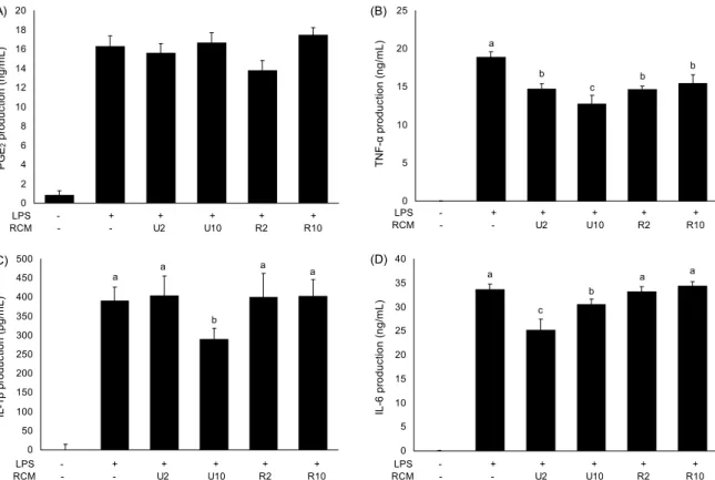
미숙과와 성숙과 복분자를 섭취시킨 쥐에서 peritoneal macrophage를 분리하여 LPS로 염증반응을 유도한 후, 주 요 염증 매개 물질인 PGE2의 분비량과 proinflammatory cytokines인 TNF-α, IL-6와 IL-1β의 분비량을 측정한 결 과는 Fig. 1과 같다. 성숙도와 섭취량에 상관없이 복분자 섭취는 PGE2의 분비에 영향을 주지 않는 것으로 나타났다 (Fig. 1A). 반면 복분자의 섭취는 TNF-α, IL-1β와 IL-6의 분비량을 억제하는 것으로 나타났다(Fig. 1B~D). 우선 TNF-α 분비는 복분자의 성숙도에 관계없이 복분자 섭취에 의해 분비량이 유의적으로 감소하였으며, U2와 U10 섭취 그룹에서는 농도 의존적으로 감소하였다(Fig. 1B). 그러나 IL-1β의 분비는 U10 섭취 그룹에서만 대조군과 비교하여 유의적으로 감소하였으며(Fig. 1C), IL-6의 분비는 미숙과 복분자군(U2와 U10)에서 유의적으로 감소하는 것으로 나 타났다(Fig. 1D). 따라서 성숙도에 따라서는 미숙과 복분자 가 성숙과보다 항염증 반응이 뛰어난 것으로 사료되며, 농도 의존적인 경향을 보이는 것으로 나타났다.
본 실험 결과는 polyphenol을 다량 함유하고 있는 blue- berry의 섭취가 TNF-α와 IL-6의 분비를 감소시켰다는 보 고와 유사한 경향을 보였다(32). 또한 Kim 등(23)과 Park 등(33)은 복분자의 성숙도에 따라 구성 성분과 함유 물질에 차이가 나며, 이는 항산화 효과에 영향을 미친다고 보고하고 있어 성숙도에 따른 차이를 설명하였다(5). 선행연구에 의하 면 복분자의 성숙도에 따라 phenolic compounds의 함유량 이 차이가 있는 것으로 나타났다(23). 미숙과 복분자가 성숙 과 복분자보다 16배의 cinnamic acid, 6배의 ferulic acid와 epicatechine, 4배의 protocatechuic acid, 3배의 gallic acid, 2.5배의 vanillic acid를 함유하는 것으로 나타났다 (23). 본 실험 결과에서도 복분자의 성숙도에 따른 구성 성 분의 차이로 항산화 활성뿐 아니라 항염증 반응에도 영향을 미쳐 미숙과 복분자가 성숙과보다 항염증 효과가 크다는 Yang 등(16)의 연구결과와 본 실험결과와도 같은 경향을 보였다.
따라서 복분자의 섭취는 마우스 대식세포의 TNF-α, IL-1β와 IL-6의 분비를 억제함으로써 항염증 효과를 나타 내며, 성숙도에 따른 생리활성물질의 차이로 인해 미성숙 복분자 섭취의 항염증 효과가 더 뛰어난 것으로 사료된다.
복분자 섭취에 따른 peritoneal macrophage의 유전자 발현 분석
미숙과와 성숙과 복분자 섭취에 의한 복강 대식세포의 유 전자 발현을 분석한 결과, 염증반응 관련 유전자중에서 미숙 과와 성숙과의 섭취에 의해 발현이 감소한 유전자는 Table 2와 같다. 미숙과와 성숙과 복분자는 arachidonate 5-lip- oxygenase(5-LOX)의 발현을 유의적으로 감소시켰으며, 복분자 섭취에 의해 농도 의존적으로 그 발현이 감소하는
0 2 4 6 8 10 12 14 16 18 20
PGE2 production (ng/mL) .
LPS - + + + + + RCM - - U2 U10 R2 R10
(A)
b b c b a
0 5 10 15 20 25
TNF-α production (ng/mL) .
LPS - + + + + + RCM - - U2 U10 R2 R10
(B)
0 50 100 150 200 250 300 350 400 450 500
IL-1β production (pg/mL) .
LPS - + + + + + RCM - - U2 U10 R2 R10
a a a
b
(C) a
0
5 10 15 20 25 30 35 40
IL-6 production (ng/mL) .
a a a
c b
LPS - + + + + + RCM - - U2 U10 R2 R10
(D)
Fig. 1. Effects of unripe and ripe Rubus coreanus Miquel on PGE2, TNF-α, IL-1β, and IL-6 productions in peritoneal macrophage.
Mice were fed with U2, U10, R2, and R10 diet for 8 weeks then peritoneal macrophages were isolated. Isolated peritoneal macrophages were stimulated with 1 μg/mL of LPS for 18 hr then PGE2 (A), TNF-α (B), IL-1β (C), and IL-6 (D) productions were measured from supernatants.
Table 2. Gene down-regulated of inflammatory and immune regulating genes in peritoneal macrophages by unripe and ripe Rubus coreanus Miquel
Genes Functions U2 U10 R2 R10
Arachidonate 5-lipoxygenase
Transforms arachidonic acid into leukotrienes and is a current target for phamaceutical intervention in a number of diseases.
0.87 0.42 0.57 0.47
Ceruloplasmin The major copper-carrying protein in the blood, and in
addition plays a role in iron metabolism. 0.22 0.17 0.26 0.14 Dynamin 1 A member of the dynamin subfamily of GTP-binding
proteins. 0.49 0.65 0.58 0.43
Interleukin 11
A key regulator of multiple events in hematopoiesis, most notably the stimulation of megakaryocyte maturation.
0.20 0.45 0.19 0.29
Nitric oxide synthase 2, inducible
Involved in immune response, binds calmodulin at physiologically relevant concentrations, and produces NO as an immune defense mechanism, as NO is a free radical with an unpaired electron.
0.42 0.78 0.58 0.48
Plasminogen activator, tissue Catalyzes the conversion of plasminogen to plasmin,
the major enzyme responsible for clot breakdown. 0.97 0.40 0.64 0.40
Thrombospondin 1
An adhesive glycoprotein that mediates cell-to-cell and cell-to-matrix interactions. Play roles in platelet aggregation, angiogenesis, and tumorigenesis.
0.43 0.32 0.54 0.49
Vascular endothelial growth factor A
Mediating increased vascular permeability, inducing angiogenesis, vasculogenesis and endothelial cell growth, promoting cell migration, and inhibiting apoptosis.
0.51 0.37 0.48 0.38
Table 3. Gene up-regulated of inflammatory and immune regulating genes in peritoneal macrophages by unripe and ripe Rubus coreanus Miquel
Genes Functions U2 U10 R2 R10
Hypoxia inducible factor 3, alpha subunit
Regulate many adaptive responses to low oxygen tension (hypoxia). Negative regulators of hypoxia-inducible gene
expression. 2.43 2.26 2.05 2.48
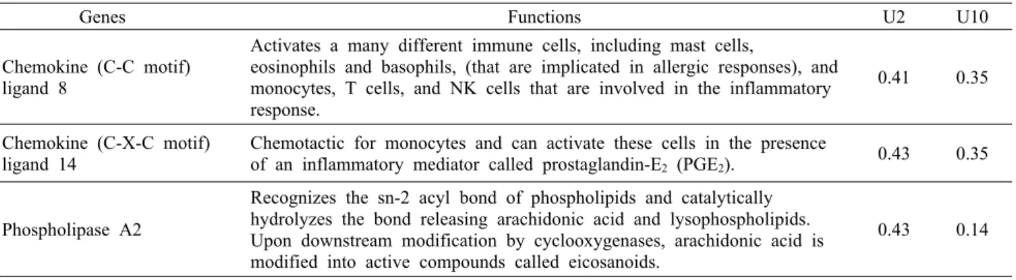
Table 4. Gene down-regulated of inflammatory and immune regulating genes in peritoneal macrophages by unripe Rubus coreanus Miquel
Genes Functions U2 U10
Chemokine (C-C motif) ligand 8
Activates a many different immune cells, including mast cells,
eosinophils and basophils, (that are implicated in allergic responses), and monocytes, T cells, and NK cells that are involved in the inflammatory response.
0.41 0.35
Chemokine (C-X-C motif)
ligand 14 Chemotactic for monocytes and can activate these cells in the presence
of an inflammatory mediator called prostaglandin-E2 (PGE2). 0.43 0.35
Phospholipase A2
Recognizes the sn-2 acyl bond of phospholipids and catalytically hydrolyzes the bond releasing arachidonic acid and lysophospholipids.
Upon downstream modification by cyclooxygenases, arachidonic acid is modified into active compounds called eicosanoids.
0.43 0.14 것으로 나타났다. 5-LOX는 arachidonic acid 대사체를 생
성하는 효소로 leukotrienes를 합성하여 염증반응을 유발 할 뿐 아니라, 만성질환자의 경우 5-LOX의 발현과 활성이 높아 동맥경화증이나 암세포의 성장을 촉진하는 것으로 알 려져 있다(34-38). 또한 복분자의 섭취는 interleukin 11 (IL-11)과 nitric oxide synthase 2, inducible(iNOS)의 발 현도 80%와 50% 정도 감소시키는 것으로 나타났다. IL-11 은 염증반응과 함께 암세포의 침윤을 조절하는 cytokine으 로 알려져 있으며, iNOS 또한 nitric oxide(NO)를 생성하여 NO-sensitive enzyme을 활성화시켜 생리적인 반응을 유 발하거나 만성적인 염증질환에서 과량 분비되어 림프구의 증식을 억제하고, 정상세포와 조직에 손상을 주는 것으로 보고되고 있다(39-41).
복분자의 섭취는 염증 관련 물질 이외에도 만성질환을 조 절하는 인자인 ceruloplasmin, vascular endothelial growth factor A(VEGF A), tissue plasminogen activator(tPA), thrombospondin 1의 발현을 감소시키는 것으로 나타났다.
우선, 혈장에서 구리를 운반하는 단백질로 알려진 cer- uloplasmin의 발현을 80~90%까지 감소시켰는데, 최근 보 고에 의하면 ceruloplasmin은 염증 반응뿐 아니라 심혈관계 질환과 같은 만성질환과도 관련이 있는 것으로 보고되었으 며(42-45), 신생혈관생성을 조절하는 중요한 성장인자인 VEGF A도 암세포의 성장과 전이에 영향을 주는 것으로 보 고되었다(46-48). 또한 tPA는 혈액 응고와 관련 있는 성분 으로 plasminogen을 plasmin으로 전환하여 혈액 응고를 유 도할 뿐 아니라 세포의 이동과 세포 remodeling을 유도하 고, 혈액응고로 인한 뇌졸중, myocardial infarction, acute ischemic strock와도 관련이 있는 것으로 보고되고 있으며, tPA의 발현을 억제함으로써 심혈관계 질환을 억제하려는
연구가 수행되고 있다(49-52). 마지막으로 복분자 섭취는 adhesion molecules인 thrombospondin 1의 발현을 감소 시키는 것으로 나타났는데, thrombospondin 1은 대식세포 가 extracellular matrix에 부착되기 위해 필요한 mem- brane receptor로 세포의 모양이나 움직임, 세포주기를 조 절하는 것으로 알려져 있으며, 혈액 응고와도 관련이 있는 것으로 보고되고 있다(53-57). 따라서 미숙과와 성숙과 복 분자의 섭취가 염증반응 관련 물질의 발현을 억제할 뿐 아니 라 만성질환, 특히 심혈관계 관련 물질인 ceruloplasmin, VEGF A, tPA, thrombospondin 1의 발현을 억제하는 것으 로 보아 혈전생성이나 심혈관계질환 예방에 효과가 있을 것 으로 사료된다.
마지막으로 복분자의 섭취가 유의적이지는 않았으나 TNF-α, IL-17, IFNγ 등의 pro-inflammatory cytokines 의 발현을 억제되었으며, phopholipase C(PLC) beta와 mitogen-activated protein kinase kinase 6(MAPKK)의 발현도 복분자 섭취에 의해 감소하는 것으로 나타나(data not shown), PLC와 MAPKK signaling에 의해 염증반응이 억제되었을 것으로 사료된다.
미숙과와 성숙과 복분자 섭취에 의해 hypoxia inducible factor 3, alpha(HIF3A)가 2배 이상 발현이 증가된 것으로 나타났다(Table 3). 이 유전자는 산소의 농도가 낮은 경우 발현되어 HIF의 기능을 억제하는 전사조절인자로 암의 성 장과 전이를 억제하는 것으로 알려져 있다(58-60).
미숙과 복분자의 섭취에 의해서만 발현이 조절되는 유전 자로는 chemokine(C-C motif) ligand 8(CCL8), chemo- kine(C-X-C motif) ligand 14(CXCL14), phospholiase A2(PLA2) 등이 있는 것으로 나타났다(Table 4). CCL8은 monocyte chemoattractant protein(MCP) 중 하나로 피부
에서 eosinophilic inflammation을 유도하고, T(H)2 세포 의 recruitment 등 염증반응을 조절하는 것으로 알려져 있 으며(61), CXCL14은 염증 반응뿐 아니라 체중 조절과 중추 신경계에서 식행동을 조절하는 chemokine으로 CXCL14의 분비가 적을수록 비만과 식욕이 억제되는 것으로 보고되었 다(62). 또한 PLA2는 인지질의 sn-2에 위치한 arach- idonic acid(AA)를 분리시키는 효소로 분리된 AA는 cy- clooxygenase나 lipoxygenase에 의해 PG나 leukotrienes 를 생성하여 염증반응을 유도한다고 알려져 있으며, PLA2 의 활성이 높을수록 류마티스나 심장질환, 암과 같은 만성질 환의 발생이나 증상이 악화되는 것으로 보고되고 있다(63).
현재 성숙과는 발효청이나 주스, 추출주로 활용되고 있으 며, 미숙과의 경우 예전에는 차로 섭취하였으나, 현재 간 건 강에 도움을 주는 기능성 원료로 개별인정을 받아 발효유에 첨가되어 기능성 유제품으로 판매되고 있다. 본 연구 결과 복분자 특히 미숙과 복분자 섭취는 염증관련 유전자의 발현 뿐 아니라 동맥경화나 심혈관계 관련 물질의 발현을 억제하 는 것으로 나타나 차후 항염증성 약물이나 만성염증성 질환 을 위한 기능성 식품을 개발하는데 활용될 수 있을 것으로 사료된다.
요 약
본 연구에서는 미숙과와 성숙과의 복분자 섭취에 의한 쥐 복강 대식세포의 염증반응을 조사하였다. 8주간 농도별 미 숙과와 성숙과 복분자 식이를 섭취시킨 후 복강대식세포를 분리한 다음, LPS로 염증반응을 유도하여 염증매개 cyto- kines인 TNF-α, IL-1β, IL-6의 분비와 PGE2의 분비량을 측정하였으며, cDNA microarray 방법으로 유전자 발현을 측정하였다. 미숙과와 성숙과 복분자 섭취는 TNF-α의 생성 을 유의적으로 억제하였으나, IL-1β, IL-6는 미숙과 복분자 섭취에 의해서만 감소하였으며 PGE2의 분비에는 영향을 주 지 않았다. 본 연구결과, 미숙과와 성숙과 복분자 섭취에 의 해 8개의 유전자 발현이 감소된 것으로 확인되었는데, 이 중 세포의 면역반응과 관련된 5-LOX, iNOS, IL-11의 발현 이 유의적으로 감소되었으며, 만성질환 특히 심혈관계 질환 을 유발하는 인자인 tPA, thrombospondin 1, cerulo- plasmin과 암의 성장 및 전이와 관련된 VEGF A의 발현을 유의적으로 억제하였다. 한편 혐기성 관련 유전자의 발현을 억제하는 HIF3A의 발현을 유의적으로 증가시켰다. 또한 미 숙과 복분자의 섭취만이 CCL8, CXCL14, PLA2의 발현을 감소시키는 것으로 나타났다. 따라서 복분자의 섭취, 특히 미숙과 복분자의 섭취는 항염증 효과를 보일 뿐 아니라 만성 염증성 질환 관련 인자의 발현을 유의적으로 감소시키므로 이와 관련된 기능성 식품 개발에 활용될 수 있을 것으로 사 료되며, 추후 복분자내 항염증 효능을 갖는 생리활성 성분에 대한 연구가 더 진행되어야 할 것으로 판단된다.
감사의 글
본 연구는 농촌진흥청 아젠다과제(과제번호: PJ006387) 예 산으로 추진된 연구의 일부로서 연구비를 지원해 주신 농촌 진흥청에 감사를 드립니다.
REFERENCES
1. Statistics Korea. 2011. Available from: https://www.index.
go.kr/egams/stts/jsp/potal/stts/PO_STTS_IdxMain.jsp?idx_
cd=1438&bbs=INDX_001.
2. Reuter S, Gupta SC, Chaturvedi MM, Aggarwal BB. 2010.
Oxidative stress, inflammation, and cancer: how are they linked? Free Radic Biol Med 49: 1603-1616.
3. Laroux FS. 2004. Mechanisms of inflammation: the good, the bad and the ugly. Front Biosci 9: 3156-3162.
4. Blackwell TS, Christman JW. 1997. The role of nuclear fac- tor-κB in cytokine gene regulation. Am J Respir Cell Mol Biol 17: 3-9.
5. Barnes PJ, Karin M. 1997. Nuclear factor-κB: a pivotal tran- scription factor in chronic inflammatory diseases. N Engl J Med 336: 1066-1071.
6. Ben-Neriah Y, Karin M. 2011. Inflammation meets cancer, with NF-κB as the matchmaker. Nat Immunol 12: 715-723.
7. Schmidt MI, Duncan BB, Sharrett AR, Lindberg G, Savage PJ, Offenbacher S, Azambuja MI, Tracy RP, Heiss G. 1999.
Markers of inflammation and prediction of diabetes mellitus in adults (Atherosclerosis Risk in Communities study): a cohort study. Lancet 353: 1649-1652.
8. Pearson TA, Mensah GA, Alexander RW, Anderson JL, Cannon RO 3rd, Criqui M, Fadl YY, Fortmann SP, Hong Y, Myers GL, Rifai N, Smith SC Jr, Taubert K, Tracy RP, Vinicor F. 2003. Markers of inflammation and cardiovascular disease: application to clinical and public health practice:
A statement for healthcare professionals from the Centers for Disease Control and Prevention and the American Heart Association. Circulation 107: 499-511.
9. Knekt P, Kumpulainen J, Järvinen R, Rissanen H, Heliövaara M, Reunanen A, Hakulinen T, Aromaa A. 2002. Flavonoid intake and risk of chronic diseases. Am J Clin Nutr 76: 560- 568.
10. Arts ICW, Hollman PCH. 2005. Polyphenols and disease risk in epidemiologic studies. Am J Clin Nutr 81: 317S- 325S.
11. Ellis CL, Edirisinghe I, Kappagoda T, Burton-Freeman B.
2011. Attenuation of meal-induced inflammatory and throm- botic responses in overweight men and women after 6-week daily strawberry (Fragaria) intake. A randomized place- bo-controlled trial. J Atheroscler Thromb 18: 318-327.
12. Terra X, Montagut G, Bustos M, Llopiz N, Ardèvol A, Bladé C, Fernández-Larrea J, Pujadas G, Salvadó J, Arola L, Blay M. 2009. Grape-seed procyanidins prevent low-grade in- flammation by modulating cytokine expression in rats fed a high-fat diet. J Nutr Biochem 20: 210-218.
13. Seymour EM, Lewis SK, Urcuyo-Llanes DE, Tanone II, Kirakosyan A, Kaufman PB, Bolling SF. 2009. Regular tart cherry intake alters abdominal adiposity, adipose gene tran- scription, and inflammation in obesity-prone rats fed a high fat diet. J Med Food 12: 935-942.
14. Santangelo C, Varì R, Scazzocchio B, Di Benedetto R, Filesi C, Masella R. 2007. Polyphenols, intracellular signalling
and inflammation. Ann 1st Super Sanita 43: 394-405.
15. Ra J, Lee HY, Choi MK, Park HG, Kang KS. 2004. Effect of decreasing body weight with plant extracts containing Rubi fructus. J Toxicol Pub Health 20: 167-172.
16. Yang HM, Oh SM, Lim SS, Shin HK, Oh YS, Kim JK. 2008.
Antiinflammatory activities of Rubus coreanus depend on the degree of fruit ripening. Phytother Res 22: 102-107.
17. Yang HM, Lim SS, Lee YS, Shin HK, Oh YS, Kim JK.
2007. Comparison of the anti-inflammatory effects of the extracts from Rubus coreanus and Rubus occidentalis.
Korean J Food Sci Tech 39: 342-347.
18. Kim YH, Choi JH, Rim HK, Kang HJ, Chang SG, Park JH, Park HJ, Choi JW, Kim SD, Lee KT. 2011. 23-Hydroxytor- mentic acid and niga-ichgoside f1 isolated from Rubus cor- eanus attenuate cisplatin-induced cytotoxicity by reducing oxidative stress in renal epithelial LLC-PK1 cells. Biol Pharm Bull 34: 906-911.
19. Sohn SI, Rim HK, Kim YH, Choi JH, Park JH, Park HJ, Choi JW, Kim SD, Jeong SY, Lee KT. 2011. The amelio- rative effect of 23-hydroxytormentic acid isolated from Rubus coreanus on cisplatin-induced nephrotoxicity in rats.
Biol Pharm Bull 34: 1508-1513.
20. Choi J, Lee KT, Ha J, Yun SY, Ko CD, Jung HJ, Park HJ.
2003. Antinociceptive and antiinflammatory effects of niga- ichigoside F1 and 23-hydroxytormentic acid obtained from Rubus coreanus. Biol Pharm Bull 26: 1436-1441.
21. Lee J, Dossett M, Finn CE. 2012. Rubus fruit phenolic re- search: The good, the bad, and the confusing. Food Chem 130: 785-796.
22. Pang KC, Kim MS, Lee MW. 1996. Hydrolyzable tannins from the fruits of Rubus coreanum. Kor J Pharmacogn 27:
366-370.
23. Kim HS, Park SJ, Hyun SH, Yang SO, Lee J, Auh JH, Kim JH, Cho SM, Marriott PJ, Choi HK. 2011. Biochemical mon- itoring of black raspberry (Rubus coreanus Miquel) fruits according to maturation stage by 1H-NMR using multiple solvent systems. Food Res Int 44: 1977-1987.
24. Nam JH, Jung HJ, Choi J, Lee KT, Park HJ. 2006. The anti-gastropathic and anti-rheumatic effect of niga-ichigo- side F1 and 23-hydroxytormentic acid isolated from the un- ripe fruits of Rubus coreanus in a rat model. Biol Pharm Bull 29: 967-970.
25. Jung KA, Han D, Kwon EK, Lee CH, Kim YE. 2007. Anti- fatigue effect of Rubus coreanus Miquel extract in mice.
J Med Food 10: 689-693.
26. Do SH, Lee JW, Jeong WI, Chung JY, Park SJ, Hong IH, Jeon SK, Lee IS, Jeong KS. 2008. Bone-protecting effect of Rubus coreanus by dual regulation of osteoblasts and osteoclasts. Menopause 15: 676-683.
27. Park JH, Oh SM, Lim SS, Lee YS, Shin HK, Oh YS, Choe NH, Park JH, Kim JK. 2006. Induction of heme oxygen- ase-1 mediates the anti-inflammatory effects of the ethanol extract of Rubus coreanus in murine macrophages. Biochem Biophys Res Commun 351: 146-152.
28. Biesalski HK. 2007. Polyphenols and inflammation: basic interactions. Curr Opin Clin Nutr Metab Care 10: 724-728.
29. Rahman I, Biswas SK, Kirkham PA. 2006. Regulation of inflammation and redox signaling by dietary polyphenols.
Biochem Pharmacol 72: 1439-1452.
30. Wu X, Rahal O, Kang J, Till SR, Prior RL, Simmen RC.
2009. In utero and lactational exposure to blueberry via ma- ternal diet promotes mammary epithelial differentiation in prepubescent female rats. Nutr Res 29: 802-811.
31. Adams LS, Kanaya N, Phung S, Liu Z, Chen S. 2011. Whole
blueberry powder modulates the growth and metastasis of MDA-MB-231 triple negative breast tumors in nude mice.
J Nutr 141: 1805-1812.
32. Xie C, Kang J, Ferguson ME, Nagarajan S, Badger TM, Wu X. 2011. Blueberries reduce pro-inflammatory cytokine TNF-α and IL-6 production in mouse macrophages by in- hibiting NF-κB activation and the MAPK pathway. Mol Nutr Food Res 55: 1587-1591.
33. Park Y, Kim SH, Choi SH, Han J, Chung HG. 2008. Changes of antioxidant capacity, total phenolics, and vitamin C con- tents during Rubus coreanus fruit ripening. Food Sci Bio- technol 17: 251-256.
34. de Gaetano G, Donati MB, Cerletti C. 2003. Prevention of thrombosis and vascular inflammation: benefits and limi- tations of selective or combined COX-1, COX-2 and 5-LOX inhibitors. Trends Pharmacol Sci 24: 245-252.
35. Hoque A, Lippman SM, Wu TT, Xu Y, Liang ZD, Swisher S, Zhang H, Cao L, Ajani JA, Xu XC. 2005. Increased 5-lip- oxygenase expression and induction of apoptosis by its in- hibitors in esophageal cancer: a potential target for pre- vention. Carcinogenesis 26: 785-791.
36. Melstrom LG, Bentrem DJ, Salabat MR, Kennedy TJ, Ding XZ, Strouch M, Rao SM, Witt RC, Ternent CA, Talamonti MS, Bell RH, Adrian TA. 2008. Overexpression of 5-lip- oxygenase in colon polyps and cancer and the effect of 5-LOX inhibitors in vitro and in a murine model. Clin Cancer Res 14: 6525-6530.
37. Ding XZ, Iversen P, Cluck MW, Knezetic JA, Adrian TE.
1999. Lipoxygenase inhibitors abolish proliferation of hu- man pancreatic cancer cells. Biochem Biophys Res Commun 261: 218-223.
38. Manev H, Manev R. 2007. 5-lipoxygenase as a possible bio- logical link between depressive symptoms and athero- sclerosis. Arch Gen Psychiatry 64: 1333.
39. Korhonen R, Lahti A, Kankaanranta H, Moilanen E. 2005.
Nitric oxide production and signaling in inflammation. Curr Drug Targets 4: 471-479.
40. Nussler AK, Billiar TR. 1993. Inflammation, immunor- egulation, and inducible nitric oxide synthase. J Leukoc Biol 54: 171-178.
41. MacMicking J, Xie QW, Nathan C. 1997. Nitric oxide and macrophage function. Annu Rev Immunol 15: 323-350.
42. Giurgea N, Constantinescu MI, Stanciu R, Suciu S, Muresan A. 2005. Ceruloplasmin-acute-phase reactant or endogenous antioxidant? The case of cardiovascular disease. Med Sci Monit 11: RA48-51.
43. Denko CW. 1979. Protective role of ceruloplasmin in in- flammation. Agents Actions 9: 333-336.
44. Fox PL, Mazumder B, Ehrenwald E, Mukhopadhyay CK.
2000. Ceruloplasmin and cardiovascular disease. Free Radic Biol Med 28: 1735-1744.
45. Goldstein I, Kaplan HB, Edelson HS, Weissmann G. 1979.
A new function for ceruloplasmin as an acute-phase reactant in inflammation: a scavenger of superoxide anion radicals.
Trans Assoc Am Physicians 92: 360-369.
46. Hirakawa S, Kodama S, Kunstfeld R, Kajiya K, Brown LF, Detmar M. 2005. VEGF-A induces tumor and sentinel lymph node lymphangiogenesis and promotes lymphatic metastasis. J Exp Med 201: 1089-1099.
47. George ML, Tutton MG, Janssen F, Arnaoutz A, Abulafi AM, Eccles SA, Swift RI. 2001. VEGF-A, VEGF-C, and VEGF-D in colorectal cancer progression. Neoplasia 3: 420- 427.
48. Carmeliet P. 2005. VEGF as a key mediator of angiogenesis
in cancer. Oncology 69: 4-10.
49. Mirshahi M, Soria J, Soria C, Faivre R, Lu H, Courtney M, Roitsch C, Tripier D, Caen JP. 1989. Evaluation of the in- hibition by heparin and hirudin of coagulation activation during r-tPA-induced thrombolysis. Blood 74: 1025-1030.
50. Demchuk AM, Tanne D, Hill MD, Kasner SE, Hanson S, Grond M, Levine SR. 2001. Predictors of good outcome after intravenous tPA for acute ischemic stroke. Neurology 57: 474-480.
51. Saver JL, Yafeh B. 2007. Confirmation of tPA treatment effect by baseline severity-adjusted end point reanalysis of the NINDS-tPA stroke trials. Stroke 38: 414-416.
52. Tanne D, Gorman MJ, Bates VE, Kasner SE, Scott P, Verro P, Binder JR, Dayno JM, Schultz LR, Levine SR. 2000.
Intravenous tissue plasminogen activator for acute ischemic stroke in patients aged 80 years and older: the tPA stroke survey experience. Stroke 31: 370-375.
53. DiPietro LA, Polverini PJ. 1993. Angiogenic macrophages produce the angiogenic inhibitor thrombospondin 1. Am J Pathol 143: 678-684.
54. Lawler J. 2000. The functions of thrombospondin-1 and -2.
Curr Opin Cell Biol 12: 634-640.
55. Salvesen HB, Akslen LA. 1999. Significance of tumour- associated macrophages, vascular endothelial growth factor and thrombospondin-1 expression for tumour angiogenesis and prognosis in endometrial carcinomas. Int J Cancer 84:
538-543.
56. Majack RA, Goodman LV, Dixit VM. 1998. Cell surface thrombospondin is functionally essential for vascular smooth muscle cell proliferation. J Cell Biol 106: 415-422.
57. Yamauchi M, Imajoh-Ohmi S, Shibuya M. 2007. Novel an- tiangiogenic pathway of thrombospondin-1 mediated by suppression of the cell cycle. Cancer Sci 98: 1491-1497.
58. Dang CV, Kim JW, Gao P, Yustein J. 2008. The interplay between MYC and HIF in cancer. Nat Rev Cancer 8: 51-56.
59. Zhong H, De Marzo AM, Laughner E, Lim M, Hilton DA, Zagzag D, Buechler P, Isaacs WB, Semenza GL, Simons JW. 1999. Overexpression of hypoxia-inducible factor 1α in common human cancers and their metastases. Cancer Res 59: 5830-5835.
60. Yeo EJ, Chun YS, Park JW. 2004. New anticancer strategies targeting HIF-1. Biochem Pharmacol 68: 1061-1069.
61. Islam SA, Chang DS, Colvin RA, Byrne MH, McCully ML, Moser B, Lira SA, Charo IF, Luster AD. 2011. Mouse CCL8, a CCR8 agonist, promotes atopic dermatitis by re- cruiting IL-5+ T(H)2 cells. Nat Immunol 12: 167-177.
62. Tanegashima K, Okamoto S, Nakayama Y, Taya C, Shitara H, Ishii R, Yonekawa H, Minokoshi Y, Hara T. 2010.
CXCL14 deficiency in mice attenuates obesity and inhibits feeding behavior in a novel environment. PLoS One 5:
e10321.
63. Magrioti V, Kokotos G. 2013. Phospholipase A2 inhibitors for the treatment of inflammatory diseases: a patent review (2010-present). Expert Opin Ther Pat 23: 333-344.