- 53 - J. Exp. Biomed. Sci. 12 (2006) 53–56
Effect of mPER1 on the Expression of HSP105 Gene in the Mouse SCN
Han-Gyu Kim and Kiho Bae†
Department of Life Science, Yonsei University, Wonju 220-710, Korea
The suprachiasmatic nucleus (SCN) of the anterior hypothalamus is the circadian pacemaker entrained to the 24-hr day by environmental time cues. Major circadian genes such as mPeriod (mPer1~3) and mCryptochrome (mCry1~2) are actively transcribed by the action of CLOCK/BMAL heterodimers, and in turn, these are being suppressed by the mPER/mCRY complex. In the study, the locomotor activity rhythms of mPer1 Knockout (KO) mice are measured, and the expression profiles of Heat Shock Protein 105 kDa (HSP105) genes in the SCN were measured by in situ hybridization.
In agreement with previous reports, the locomotor activity rhythm of mPer1 KO mice was much shorter than that of wildtype. In addition, the total bout of activity of mPer1 KO was less in comparison to control mice. The expression of HSP105 in the SCN of mPer1 KO mice was ranged from CT6 to CT22, with a peak level at CT14, implying that the gene are under the control of circadian clock. However, the expression of HSP105 in the SCN of wildtype could not be detected in our study. Further analysis will reveal the direct or indirect regulation by mPer1 on the expression in the SCN and the role of the gene in the circadian clock.
Key Words: Circadian clock, Activity rhythm, mPer1, HSP105, in situ hybridization
서 론
사람을 비롯한 모든 생물은 빛, 온도 등과 같은 환경 변화 (environmental time cues)에 자기만의 주기적인 생활 (~24시 간)을 나타내고 있다. 이러한 환경의 주기적인 변화는 오랜 진화과정을 통하여 생명체 내의 유전자로 각인되었으며, 이 것을 조절하는 생체시계 유전자들은 생물의 발생과정을 포 함한 성장과정에서 서로 다른 리듬으로 발현한다. 이렇게 생 체시계를 조절하는 뇌 속의 박동조절자로 시교차상핵 (supra- chiasmatic nucleus, SCN)이 밝혀졌다 (Weaver et al., 1998). 생 체시계 유전자는 1970년 초 미국의 Ron Konopka에 의해 초파리에서 period (per)를 확인함으로써 최초로 알려졌으며 (Konopka and Benzer, 1971), 그 후 timeless (tim), Clock (Clk), cycle (cyc), cryptochrome (cry) 등 초파리의 생체시계를 조절 하는 많은 유전자가 확인되었다 (Sehgal, 2004; Bae et al., 1998; Edery et al., 1999).
포유류에서는 처음 생쥐에서 Clock 유전자 돌연변이 생쥐 를 얻었으며, 이후 여러 gene knockout (KO)생쥐가 연구되
었다. 포유류 생체시계를 조절하는 중요한 단백질 CLOCK 과 BMAL1은 상호결합하여 E box (CANNTG)라고 불리는 promoter 부위에 결합하여 mPER, mCRY 등을 만드는 basic helix-loop-helix (bHLH)형의 전사 인자이다. 생체 내에 mPER, mCRY가 많이 축척되어 있을 경우 이들은 핵 안으로 이동하 여 CLOCK과 BMAL1의 작용을 억제한다 (positive and nega- tive feedback loop) (Gekakis et al., 1998; King et al., 1997;
Shearman et al., 2000). 2000년대에 들어서는 DNA chip을 통 하여 초파리의 head와 body, 포유류에서는 SCN과 fibroblast, liver 등에서 유전자의 발현양상을 비교함으로써 생체시계에 관여하거나 이의 조절을 받는 유전자들의 각 기관에 따른 리 듬을 비교하는 단계까지 이르렀다 (Panda et al., 2002; Storch et al., 2002; Rudic et al., 2005). 아직까지 뇌와 말단 조직들 간의 시계의 연락망은 밝혀지지 않았지만, 생체시계의 흐름 으로 보아 심장의 박동원처럼 시계의 조절중추에 의해서 뇌 가 말단시계를 조절할 것이라는 것을 가설로 연구가 진행되 고 있다 (Balsalobre et al., 2000).
생체시계 유전자 mPer1은 SCN에서 나타나는데 특히, 빛 에 의해서 mPer1의 경우는 빠르게 유도가 된다. 본 연구그 룹의 선행연구에 의하여 wildtype (WT)과 생체시계가 완전히 소실된 mPer1, mPer2 double knockout (DKO)생쥐의 말초조 직인 간에서 단백질의 2차원 전기영동 분석을 통하여 단백 질 발현양상의 차이가 뚜렷한 후보 단백질들을 찾았다 (Lee et al., 2005). 아직은 일주기리듬과 관련하여 이들이 직접 혹
*논 문 접 수: 2006년 2월 20일 수정재접수: 2006년 3월 3일
†교신저자: 배기호, (우) 220-710 강원도 흥업면 매지리 234, 연세대학교 생명과학과
Tel: 82-33-760-2280, Fax: 82-33-760-2183 e-mail: [email protected]
Brief Communication
- 54 - 은 간접적으로 관련되는지 알려지지 않았다. 이러한 결과를 토대로, 생체시계를 조절하는 중심 유전자인 mPer1 KO 생 쥐를 이용하여, 본 실험에서는 우선 WT과 mPer1 KO 생쥐 의 뇌 속 SCN 내에서 Heat Shock Protein 105 kDa (HSP105) mRNA (Yasuda et al., 1999)를 in situ hybridization (ISH)방법으 로 발현양상을 비교하였다. 본 연구 결과 생체시계의 중심 조절중추인 SCN에서 말초조직인 간으로 이어지는 연결고 리를 밝히는 실마리를 제공하리라 기대한다.
본 연구에 사용된 재료 및 방법은 다음과 같다. 실험동물 은 생쥐 129/sv 품종을 사용하였다 (Bae et al., 2001). 동물의 생체리듬을 확인하기 위하여 12-h light : 12-h dark (LD) 반복
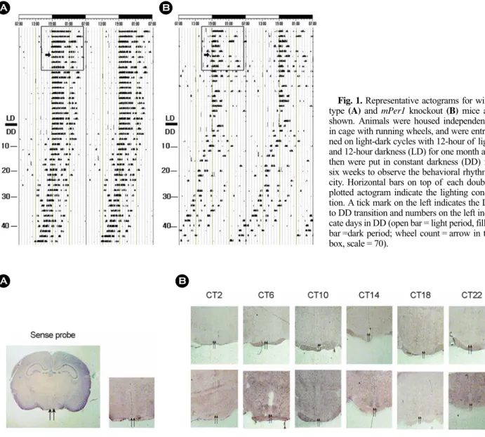
조건에서 Exercise wheel을 장착한 cage 내에 4주 이상 적응 시켰다. 이후 constant darkness (DD)에서 6주 동안 wheel의 회전정도를 관찰하고 이를 actogram으로 double plot하여 (ActiView v1.3, Mini Mitter Co, USA) 각 개체의 행동리듬을 분석하였다. WT 생쥐의 경우 LD에서 DD로 바뀐 후, 행동 리듬이 24시간보다 약 30분 정도 앞당겨져 유지됨을 볼 수 있다 (Fig. 1A). mPer1 KO의 경우는 동일한 조건에서 행동 리듬이 더욱 빨라졌으며 또한, WT과 비교하여 리듬이 불안 정하였다 (Fig. 1B). 생쥐들의 운동양은 wheel의 회전수로 알 수 있는데, 회전 양을 보면 WT 보다는 mPer1 KO의 경우 운동능력이 떨어진다는 것을 또한 확인할 수 있었다 (Fig. 1
Fig. 1. Representative actograms for wild- type (A) and mPer1 knockout (B) mice are shown. Animals were housed independently in cage with running wheels, and were entrai- ned on light-dark cycles with 12-hour of light and 12-hour darkness (LD) for one month and then were put in constant darkness (DD) for six weeks to observe the behavioral rhythmi- city. Horizontal bars on top of each double- plotted actogram indicate the lighting condi- tion. A tick mark on the left indicates the LD to DD transition and numbers on the left indi- cate days in DD (open bar = light period, filled bar =dark period; wheel count = arrow in the box, scale = 70).
A B
Fig. 2. Expression of Hsp105 mRNA in a circadian cycle in the mouse SCN. A whole brain image that is probed with sense riboprobe as a control is shown in A. Enlargement of the SCN area is also shown at right. The SCN area in the brain are marked in two arrows. B.
Enlargement of the SCN area from either wildtype (upper row) or mPer1 knockout (bottom row) mice are shown. Brains (n=5) were collected at indicated circadian time points (CT), sectioned, and probed with antisense riboprobe against HSP105. A hot-wash at 45℃ was carried out for in situ hybridization at 55℃ and the detection time was extended to 36 hours.
B A
- 55 - box안 화살표 부분에서 측정). 그러므로 mPer1 유전자의 소실이 근육의 운동능력에도 다소 영향을 미칠 것으로 사료 된다.
생쥐 HSP105 유전자의 전체 염기서열을 Genbank에서 얻 었으며 (Genbank no. BC018378), 이에 근거하여 Upstream 5'-aataagcttgccgttttgttgttcagaatg-3' (Tm = 58.7℃)과 Downstream 5'-agcggaattcctctgtctcaatatacatg-3' (Tm = 58.1℃) oligo를 제작하 였다 (Bioneer). WT 생쥐 간의 mRNA를 정제하여 이를 주형 으로 두 oligo로 RT-PCR을 한 후, PCRII-Topo Vector (In- vitrogen)에 Ligation하였다. 이때 클로닝한 유전자는 양 말단 에 NotI, BamHI 인식부위를 넣어 ISH probe의 방향성 (sense 또는 antisense)을 주었다. 형질 전환한 세균을 선택배지에 배양하고 Miniprep하여 plasmid DNA를 얻었다. 이후 Dig RNA labeling Kit (Roche, #11 175 025 910)를 사용하여 ISH probe를 제작하고, Anti Dig-AP (Roche, #11175041910)와 BCIP/
NBT (Roche, #1201093)를 사용하여 발현을 측정하였다. 한 편, 실험동물 WT과 mPer1 KO 생쥐를 각각 5마리씩 시간대 별로 잡고 (CT2, CT6, CT10, CT14, CT18, CT22 [CT=circadian time, CT2 means 2-hr after lights-off in subjective daytime in DD]) 뇌가 손상되지 않게 영하 30도로 유지한 2-Methylbutane (JUNSEI, #4A2156)에 적출하였고, Cryostat을 사용하여 관상 면의 뇌절편을 준비하였다. ISH은 Roche에서 제공한 실험방 법을 일부 변형하여 50% deionized formamide (Sigma, #F9037), 10% dextran sulfate (Amersham, #99250), 2x Denhardt's solution (Sigma, D2532), 4x SSC, 10 mM DTT (DL-Dithiothreitol, #43815), 50 µg/ml yeast total RNA, 50 µg/ml salmon sperm DNA (Pro- mega, #D181B), 0.1% SDS (DUCHEFA, #003468.03)를 첨가한 hybridization buffer에 10~20 ng/µl 농도의 ISH probe를 희석 하고, HSP105 detection에 맞는 온도값을 계산하여 55℃에서 수행하였다. Anti-Digoxigenin-Alkaline phosphatase (AP; 750 U/
ml)와 NBT/BCIP를 사용한 발색반응으로 유전자의 발현을 확인하였다.
본 실험의 목적은 시계조절중추인 SCN 내에서 HSP105의 발현양상을 보고자 함이다. HSP105의 경우 아직 뇌에서의 발현양상에 대한 연구가 많이 알려져 있지 않다. 예비실험을 통해 살펴본 HSP105의 발현양상은 대뇌피질, 시상하부, 해 마 부분에서 보였으며, 본 실험에서 사용한 sense probe에서 도 일부 발현을 관찰하였다 (Fig. 2A). 선행연구에 의하면 생 체 내에서 스트레스나 열을 받을 경우 주요한 대응 단백질이 HSP70이고, 이 HSP70의 발현을 줄이는 단백질이 HSP105/
HSP110로 알려져 있다 (Hylander et al., 2000; Yamagishi et al., 2004). 또한, HSP70는 Morphine을 처리하였을 경우 대뇌피 질과 시상하부에서 발현이 크게 증가한다는 보고도 있다 (Ammon et al., 2002). HSP105의 경우 HSP70의 억제 역할을 하려면 이들이 뇌의 같은 위치에서 발현되는 것이 타당하나
분포가 다양함으로 보아, HSP110과 같은 다른 단백질과의 상호작용이 있어야 할 것으로 생각한다. 한편, HSP110은 대 뇌피질에서도 발현양상을 보였지만, 특히 운동의 조절에 주 된 역할을 담당하는 소뇌에서 HSP110을 갖는 다수의 세포를 발견할 수 있었다 (Xue et al., 1998). 아울러 본 연구의 결과 로, WT 생쥐의 SCN 내에서 HSP105의 발현이 관찰되지 않 았고, mPer1 KO 생쥐의 SCN에서는 CT6~CT22 사이에 발 현을 볼 수 있으며, CT14에서 최대로 발현됨을 알았다 (Fig.
2B 화살표 부분). 흥미롭게도, HSP105 유전자의 promoter 에는 E box 서열이 두 곳 있다 (Oishi et al., 2003). 이는 포 유류 생체시계 유전자를 조절하는 전사단백질 CLOCK과 BMAL1 복합체가 이들의 전사에도 직접 관여할 수 있음을 시사한다. 이러한 사실들을 종합해보면 HSP105 유전자는 시 계 유전자 mPer1에 의해서 직접 혹은 간접적으로 영향을 받 을 것으로 생각된다. HSP105 유전자의 주기적인 조절을 통 해 우리 몸의 생체시계가 반복적인 스트레스에 효과적으로 대응할 가능성을 제시한다. 후속연구를 통해 이들 유전자 및 그 단백질들 사이의 상호작용을 밝힐 수 있다면 앞으로 생체 시계 유전자에 의해서 조절되는 다른 유전자들의 연구에도 많은 도움이 될 것이다.
감사의 글
본 연구는 2002학년도 연세대학교 학술 연구비 지원에 의 해 수행되었습니다. 연구에 도움을 준 김한석, 서영욱, 엄기 혁에게 감사를 전합니다.
REFERENCES
Ammon S, Mayer P, Riechert U, Tischmeyer H, Hollt V. Micro- array analysis of genes expressed in the frontal cortex of rats chronically treated with morphine and after naloxone preci- pitated withdrawal. Brain Res Mol Brain Res. 2003. 112: 113 -125.
Bae K, Lee C, Sidote D, Chuang KY, Edery I. Circadian regula- tion of a Drosophila homolog of the mammalian Clock gene:
PER and TIM function as positive regulators. Mol Cell Biol.
1998. 10: 6142-6151.
Bae K, Jin X, Maywood ES, Hastings MH, Reppert SM, Weaver DR. Differential functions of mPer1, mPer2 and mPer3 in the SCN circadian clock. Neuron 2001. 30: 525-536.
Balsalobre A, Marcacci L, Schibler U. Multiple signaling pathways elicit circadian gene expression in cultured rat-1 fibroblasts.
Curr Biol. 2000. 10: 1291-1294.
Hylander BL, Chen X, Graf PC, Subjeck JR. The distribution and localization of hsp110 in brain. Brain Res. 2000. 869: 49-55.
- 56 - Edery I. Role of posttranscriptional regulation in circadian clocks:
lessons from Drosophila. Chronobiol Intl. 1999. 16: 377-414.
Gekakis N, Staknis D, Nguyen HB, Davis FC, Wilsbacher LD, King DP, Takahashi JS, Weitz CJ. Role of the CLOCK pro- tein in the mammalian circadian mechanism. Science 1998.
280: 1564-1569.
King DP, Zhao Y, Sangoram AM, Wilsbacher LD, Tanaka M, Antoch MP, Steeves TD, Vitaterna MH, Kornhauser JM, Lowrey PL, Turek FW, Takahashi JS. Positional cloning of the mouse circadian Clock gene. Cell 1997. 89: 641-653.
Konopka RJ, Benzer S. Clock mutants of Drosophila melanogaster.
Proc Natl Acad Sci USA 1971. 68: 2112-2116.
Lee J-W, Kim H-K, Bae K. Proteomic analysis of circadian clock mutant mice. J Exp Biomed Sci. 2005. 11: 493-501.
Oishi K, Miyazaki K, Kadota K, Kikunno R, Nagase T, Atsumi GI, et al. Genome-wide expression analysis of mouse liver reveals CLOCK-regulated circadian output genes. J Biol Chem. 2003. 278: 41519-41527.
Reppert SM, Weaver DR. Molecular analysis of mammalian circa- dian rhythms. Ann Rev Physiol. 2001. 63: 647-676.
Rudic RD, McNamara P, Reilly D, Grosser T, Curtis A-M, Price TS, Panda S, Hogenesch JB, FitzGerald GA. Bioinformatic analysis of circadian gene oscillation in mouse aorta. Circula- tion 2005. 112: 2716-2724.
Sehgal A. Molecular biology of circadian rhythms. 2004. pp 33 -74. John Wiley & Sons, Inc. Hoboken, NJ, USA.
Shearman LP, Sriram S, Weaver DR, Maywood ES, Chaves I, Zheng B, Kume K, Lee CC, van der Horst GTJ, Hastings MH, Reppert SM. Interacting molecular loops in the mamma- lian circadian clock. Science 2000. 288: 1013-1019.
Storch KF, Lipan O, Laykin I, Viswanathan N, Davis FC, Wong WH, Weitz CJ. Extensive and divergent circadian gene expression in liver and heart. Nature 2002. 417: 78-83.
Weaver DR. The suprachiasmatic nucleus: a 25-year retrospective.
J Biol Rhythms. 1998. 13: 100-112.
Xue JH, Fukuyama H, Nonoguchi K, Kaneko Y, Jido T, Fuku- moto M. Induction of Apg-1, a member of the heat shock protein 110 family, following transient forebrain ischemia in the rat brain. Biochem Biophys Res Comm. 1998. 247: 796 -801.
Yamagishi N, Ishiara K, Nakashima K, Hatayama T. Hsp105α suppresses Hsc70 chaperone activity by inhibiting Hsc70 ATPase activity. J Biol Chem. 2004. 279: 41727-41733.
Yasuda K, Ishiara K, Nakashima K, Hatayama T. Genomic cloning and promotor analysis of the mouse 105-kDa heat shock protein (HSP105) gene. Biochem Biophys Res Comm. 1999.
256: 75-80.