공기노출 및 절식시기 동안 참담치,
Mytilus coruscus
에서 Hsp70 및 GST 유전자 발현에 대한 연구김철원, 강한승
1한국농수산대학교, 1엠에스바이오랩
The Expression of Hsp70 and GST Genes in Mytilus coruscus during Air Exposure and Starvation
Chul Won Kim and Han Seung Kang
1Department of Aquaculture, Korea National College of Agriculture and Fisheries, Kongjwipatjwi-ro 1515, Wansan-gu, Jeonju, Jeollabuk-do 54874, Korea
1MS BioLab, 49 Dongseo-daero 1730 beon-gil, Dong-gu, Daejeon 34574, Korea
ABSTRACT
Heat shock proteins (HSPs), one of the most highly conserved groups of proteins characterized to date, play crucial roles in protecting cells against environmental stresses, such as heat shock, salinity and oxidative stress.
The glutathione S-transferases (GST) have important role in detoxification of oxidative stress, environmental chemicals and environmental stress. GST mRNA expression have been used as biomarkers on environmental stress. The purpose of this study was to investigate the death rate and the gene expression of Hsp70 and GST during air exposure and starvation. Results showed that, the expression of Hsp70 mRNA was significantly changed in the experiment groups, such as air exposure and starvation. GST mRNA expression was significantly increased in the experimental group of starvation. These results suggest that Hsp70 and GST were played roles in biomarker gene on the air exposure and starvation.
Keywords: Mytilus coruscus, Hsp70, GST, air exposure, tarvation
서 론
우리나라, 일본, 북태평양 및 중국 황해 연안에 서식하는 참 담치 (Mytilus coruscus, Gould) 는 이매패강 (Bivalvia), 홍 합목 (Mytiloida), 홍합과 (Mytildae) 에 속하며 생식은 자웅 이체로서 체외수정을 한다 (Yoo, 1988). 접착성이 강한 단백 질성 섬유 다발인 표류용 족사 (drifting thread) 및 부착용 족사 (attachment thread) 를 가지고 있어서 암초에 군집으 로 부착해서 서식하는 특성이 있으며, 모양은 긴 계란형으로서
껍질이 두껍고 칠흑색의 형태적 특징을 가지고 있다 (Lane et al., 1985).
백합을 비롯한 다양한 패류는 갯벌에서 서식하는데, 갯벌에 서식하는 생물의 환경에서 가장 중요한 환경요인은 온도이다. 갯벌에서의 온도의 변화는 조석주기에 따라 기온, 수온, 지온 등으로 서식 패류의 대사활동, 번식생리 및 생체 방어기능 등 에 영향을 준다 (Song et al., 2008).
참담치는 갯벌에서 서식하지 않으나 암초에 군집으로 부착 서식하는 특성으로 인하여 조석주기에 의해 일정한 주기로 공 기노출에 따른 온도, 염분 및 먹이공급 등의 환경요인의 변화 가 많은 조건에 노출되어 스트레스를 받고 있다. 이러한 환경 요인의 변화에 의한 패류의 생체에 미치는 영향에 관한 연구로 는, 온도에 의한 바지락의 생체방어기작 (Paillard et al., 2004), 온도와 염분이 참굴의 혈구 활성에 미치는 영향 (Gagnaire et al., 2006), 온도와 먹이공급에 따른 버지니아굴 혈구의 종류별 조성과 혈구 활성에 대한 영향 (Ashton-Alcox and Ford, 1998) 및 온도와 염분의 변화에 의한 생화학 및 생 리적 변화의 유도에 따른 대사유지를 위한 에너지 불균형에 의 Received: June 1, 2016; Revised: June 20, 2016; Accepted:
June 29, 2016
Corresponding author : Han Seung Kan
Tel: +82 (42) 632-9753, e-mail: [email protected] 1225-3480/24613
This is an Open Access article distributed under the terms of the Creative Commons Attribution Non-Commercial License with permits unrestricted non-commercial use, distribution, and reproducibility in any medium, provided the original work is properly cited.
한 생산력 감소 (Shin and Wi, 2004) 등이 있다.
이들 환경요인 중 온도는 직접적인 요인으로 대사 및 에너지 균형에 영향을 미치며 (Newell and Kofoed, 1977; Loomis et al., 1995; Chapple et al., 1998) 염분의 변화는 세포 내 수분과 염분의 불균형을 일으켜 참담치의 대사율을 감소시키 는 원인이 된다고 알려져 있다 (Bailey et al., 1996). 또 다른 환경요인인 공기노출 시, 패류는 일반적으로 패각을 닫는 형태 와 여는 형태로 나타나며 공기노출 후 다시 물에 잠기면 빠르 게 대사를 회복시킨다 (Widdows et al., 1979; Widdows and Shick, 1985). 따라서 수온, 염분 및 공기노출 등의 환경 요인은 참담치의 생산에 있어서 매우 중요한 요인으로, 이러한 환경요인이 생물에 과도하게 스트레스로 작용하면 항상성에 문제를 일으켜 궁극에는 죽음을 초래하게 된다.
환경요인의 변화에 따른 생체 내 스트레스 반응의 정도를 세 포수준의 분자생물학적인 연구방법을 통해 연구를 진행하는 경향이 최근 두드러지고 있다. 수온의 변화에 따른 스트레스 연구는 열충격단백질 (Heat shock protein, Hsp) 의 분석을 통해 많이 이루어지고 있다. Hsp는 온도, 염분, 중금속 등의 환경요인의 영향에 따라 발현에 차이를 보이는 대표적인 유전 자이며, 면역상태의 지표를 나타내는 유전자이다 (Parsell and Lindquist, 1993; Feder and Hofmann, 1999;
Kregel, 2002; Fangue et al., 2006; Colinet et al., 2010;
Xu and Qin, 2012; You et al., 2013). Hsp는 분자량을 기 준으로 Hsp110, Hsp100, Hsp90, Hsp70, Hsp60 및 저분자 량 Hsp 등이 있다 (Georgopoulos and Welch, 1993;
Parsell and Lindquist, 1993; Feder and Hofmann, 1999). 이중에서 Hsp90은 세포분화 및 발생에 관여한다고 알 려져 있으며, 다양한 생물에서 스트레스 반응 시 단백질조절, 세포사 (apoptosis) 조절 및 신호전달에 관여한다고 알려져 있 다 (Jakob et al., 1995; Galea-Lauri et al., 1996; Imai and Yahara, 2000; Richter and Buchner, 2001;
Soetaert et al., 2006; Wu and Chu, 2008; Li et al., 2009). Hsp 70은 분자량이 70 kDa 으로 외부환경에 대한 해 양생물 반응의 생체지표유전자로 많이 활용되고 있으며, 온도 및 염분의 영향에 따라 발현에 차이를 보이는 대표적인 유전자 이다 (Kim et al., 2006; Nam et al., 2013). 또한, 모든 스트 레스 단백질 중 가장 진화적으로 잘 보존되어 있으며 (Bukau and Horwich, 1998), 진핵생물의 주요 세포 내 기관에 존재 하여 스트레스에 의한 손상으로부터 세포를 보호하는 기능을 수행한다 (Chirico et al., 1988). Hsp70과 환경요인 스트레 스에 의한 생리적 변화에 따른 유전자의 발현에 있어서 수온상 승은 해삼에서 Hsp70 유전자 발현을 증가시키고 (Dong et al., 2008: Kim et al., 2015), 또한 빈산소에 노출된 성게에 서 Hsp70 유전자의 발현이 증가한다고 보고되었다 (Lee et
al., 2012).
어류 및 패류에서 약물대사효소계 Phase Ⅱ로 알려진 glutathione-S-transferase (GST) 는 다양한 기능을 가진 단백 질로서 비생체물질 (xenobiotic), 중금속 (heavy metal), 병원 체 (pathogen), 온도 (temperature), 염분 (salinity) 및 산화 스트레스 (oxidative stress) 등 외부에서 유입된 독성물질이나 대사과정에서 생성된 독소를 체외로 쉽게 배출되게 하거나 또는 반응성이 적은 물질로의 분해를 통해 제거하는 해독작용에 관여 하여 세포를 보호하는 기능을 가진다 (Clark, 1989: Hayes et al., 2005: Hayes and Pulford, 1995). 패류 및 어류에서는 독 성시험의 지표인자로 이용되고 있으며 polychlorinated biphenyls (PCBs), dichlorodiphenyltrichloroethane (DDT), polycyclic aromatic hydrocarbons (PAHs) 와 같은 내분비계 장애물질에 노출 시 GST의 발현이 증가한다는 연구결과가 있다 (Looise et al., 1996: Hansson et al., 2006; Hamed et al., 2003).
본 연구에서는 공기노출 및 절식 등의 요인에 따른 참담치의 생존 및 스트레스 정도를 관찰하는 연구의 일환으로 공기노출 및 절식에 따른 참담치 치패의 폐사율 및 스트레스 관련 유전 자인 Hsp70과 GST 단백질의 mRNA 유전자 발현을 조사하 고자 한다. 본 연구를 통한 결과는 환경요인인 수온과 공기노 출 및 절식 과의 상관성을 밝히고 또한 참담치 스트레스 정도 의 파악에 Hsp70 및 GST 유전자가 생체지표유전자 (Biomarker gene) 의 활용 가능성에 대해 알아보고자 한다.
재료 및 방법
1. 실험동물
본 연구에 사용된 참담치 (Mytilus coruscus, Gould) 는 초기부착치패 (1.0 ± 0.2 mm 내외) 로 국립한국농수산대학교 생물사육장에서 사육한 생물이며 사육을 위한 해수는 멸균해 수를 사용하였다. 사육용기로는 2 L 유리소재 비이커를 사용 하였으며, 실험생물인 참담치는 각각의 실험구에 100마리씩 수용하여 다연실배양기 (multi room incubator) 에서 사육하 였다.
2. 실험동물의 공기노출 및 절식 실험
공기노출 실험의 조건은 17℃, 20℃ 및 23℃에서 3일 (72 시간) 동안 노출시켰으며, 절식실험은 15℃, 20℃ 및 25℃에 서 5일 동안 먹이를 공급하지 않으면서 사육하였다. 폐사개체 확인은 패각이 벌어진 상태인 것을 죽은 개체로 판단하였다.
모든 실험구는 3반복 수행하였다.
3. Hsp70 및 GST 유전자 발현을 위한 실험동물의 준비
유전자 발현 분석을 위한 참담치의 수집은 공기노출실험의 경우 23℃ 온도 실험구에서 대조군, 1시간, 3시간, 6시간, 12 시간 및 24시간 경과의 실험동물을 각각 10마리씩 수집하여 실험에 이용하였다. 대조군은 23℃에서 시간이 경과하지 않은 0시간 실험구에서 수집하여 실험에 사용하였다. 공기노출 실험 동물을 대상으로 한 유전자 발현 분석에 있어서 23℃ 온도의 실험구를 선택한 이유는 다른 실험구 온도인 17℃ 및 20℃에 비하여 시간의 경과에 따른 스트레스에 대한 실험동물의 폐사 율이 급격히 변하는 양상을 보여 23℃ 온도 실험구를 선택하 였다. 절식실험을 위한 참담치의 수집은 25℃ 온도 실험구에서 대조군, 1일, 2일, 3일, 4일 및 5일 경과의 실험동물을 각각 10마리씩 수집하여 실험에 이용하였다. 대조군은 25℃에서 시 간이 경과하지 않은 0시간 실험구에서 수집하여 실험에 사용 하였다. 절식상태의 실험동물을 대상으로 한 유전자 발현 분석 에 있어서 25℃ 온도의 실험구를 선택한 이유는 공기노출 상 태의 실험구와 마찬가지로 다른 실험구 온도인 15℃ 및 20℃
에 비하여 시간의 경과에 따른 실험동물의 폐사율이 급격히 변 하는 양상을 보여 25℃ 온도 실험구를 선택하였다. 모든 실험 구는 3반복 수행하였다.
4. Total RNA 추출
Total RNA 추출을 위한 시료의 준비는 실험구당 3개의 사 육용기별로 각각 10개체씩 실험동물의 조직을 준비하였다. 각 사육용기에 따른 10 개체의 조직은 함께 혼합하여 total RNA 의 추출을 위해 사용하였다. Total RNA의 추출을 위한 조직 은 패각을 제외한 육질 부위 및 부속 조직 전체를 수집하여 사 용하였다. 수집한 조직은 식염수로 깨끗하게 세척 및 식염수를 제거한 다음, 액체질소에 침지시켰다. 침지시킨 조직은 마이크 로 튜브에 넣은 후에 실험에 사용하기 전까지 - 80℃ 초저온냉 동고에 보관하였다. Total RNA의 추출은 RNAiso Plus (TaKaRa Co. Shiga, Japan) 시약 용액을 이용하여 제조사 가 준비하고 제시한 방법에 따라 시행하였다. 추출한 total RNA는 분광광도계 (NanoVue, GE Healthcare) 를 이용하 여 정량 하였고, RNA quality는 260/280 ratio 1.8 이상을 확인하였으며, 역전사반응 (Reverse Transcription: RT) 전 까지 - 80℃ 초저온냉동고에 보관하였다.
5. 역전사-중합효소연쇄반응 (RT-PCR)
추출한 total RNA에서 1 μg 을 취하여 oligo(dT)18(0.5 μg) primer와 AccuPower RT premix (Bioneer Co.
Daejeon, Korea) 를 이용하여 최종 반응용액 20 μl 로 42℃
에서 1시간 시행하여 cDNA를 합성하였다. 중합효소연쇄반응 (Polymerase Chain Reaction: PCR) 은 RT를 통해 얻은 20 μl 의 cDNA 중에서 cDNA 1 μl 와 각각의 유전자
primer인 Hsp70 (Accession No: KF322135) F (5’-TGGAATACCTCCAGCACCAA-3’), R (5’-AGCATCATT GACCATGCGTT-3’), GST (Accession No: KC525103) F (5’-GAAATTGTCTTAGTAGGTGGATC-3’), R (5’-CAGG CTGGTTGTCGGAGTAAGT-3’), 내재표준유전자로 사용한 β-actin (Liu et al. 2014) primer F (5'-ATGAAACCA CCTACAACAGT-3'), R (5'-TAGACCCACCAATCCAGA CG-3’) 및 AccuPower HotStart PCR premix (Bioneer Co. Daejeon, Korea) 를 이용하여 최종 반응용액 20 μl 로 시행하였다. 각 유전자의 RT-PCR 조건은 Hsp70 및 GST 의 경우 pre-denaturation 95℃, 5분; denaturation, annealing, extension 각각 95℃ 30초, 50℃ 30초, 72℃ 30 초, 28회; extension 72℃, 10분 수행하였다. 내재표준유전자 β-actin의 PCR 조건은 pre-denaturation 95℃, 5분;
denaturation, annealing, extension 각각 95℃ 30초, 55℃
30초, 72℃ 30초, 24회; extension 72℃, 10분 수행하였다.
PCR 수행을 위한 annealing 온도의 설정은 gradient PCR 을 수행하여 최적의 온도를 선택하였으며, PCR 수행의 반복 횟수는 각각 18회부터 32회 수행한 PCR 산물을 전기영동하 여 PCR 산물의 포화상태가 이루어지기 전의 횟수를 선택하여 결정하였다. RT-PCR은 실험구당 3개의 total RNA를 이용하 여 각각 3회 반복 수행하였다. 증폭된 PCR산물은 ethidiumbromide (100 ng/ml) 가 포함된 2% 아가로즈겔 전 기영동을 통해 확인하였으며 band intensity는 Gel DocTM XR+ System (Bio-Rad, California, USA) 을 이용하여 분 석하였다.
6. 통계학적 분석
공기노출 및 절식에 따른 폐사율 개체의 통계처리는 Statistical Analysis System (SASInc., 1999) program을 이용한 ANOVA 및 Duncan's Multiple Range Test 방법으 로 유의성 (P < 0.05) 을 분석하였다. 유전자 발현에 대한 대조 군과 실험구와의 유의성 검정을 위해 Student's t-test를 수행 하였으며, p값이 0.05이하인 경우에 유의한 것으로 판정하였다.
결 과
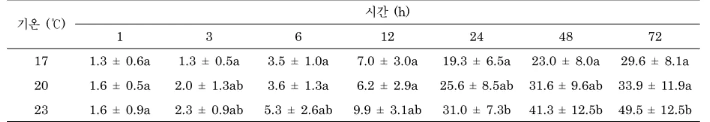
1. 참담치 초기부착치패의 공기노출에 따른 폐사율 참담치 초기부착치패의 노출시간에 따른 폐사율을 관찰한 결과 실험 종료 시에 기온 23℃에서 49.5%로 가장 높은 폐사 율을 보였으며 기온 17℃에서 29.6%로 가장 낮게 나타났다.
그리고 20℃에서는 33.9%로 나타났다. 폐사 양상은 노출 24 시간에 모든 기온 실험구에서 폐사율이 급격히 높아진 것으로 나타났고 24시간부터 72시간까지 17℃와 20℃ 실험구에서 폐
사율이 각각 10.3%와 8.3% 상승한 것으로 나타났으나 23℃
실험구에서는 18.4% 상승한 것으로 나타났다. 참담치 초기부 착치패의 노출시간에 따른 폐사율은 노출시간 대비 기온이 높 을수록 폐사율이 높아지는 것으로 나타났으며 참담치 치패의 안정적인 생존이 유지되는 기온범위에서는 24시간 이후 비교적 안정화 되는 것으로 나타났으나 23℃처럼 높은 기온 대에서는 지속적으로 폐사율이 높아지는 것으로 나타났다 (Table 1).
2. 참담치 초기부착치패의 절식기간에 따른 폐사율 참담치 초기부착치패의 절식기간에 따른 폐사율을 관찰한 결과 실험 종료시 수온 25℃에서 54.0%로 가장 높은 폐사율 을 보였으며 수온 15℃에서 39.3%로 가장 낮게 나타났다. 그 리고 20℃에서는 41.9%로 나타났다. 기간별 폐사 양상은 수온 25℃에서는 절식 3일부터 폐사개체가 많이 관찰되는 것으로 나타났으며 절식 3일부터 절식 5일까지의 폐사율은 38.9%로 매우 높게 나타났다. 그러나 수온 18℃와 20℃에서는 절식 4 일부터 폐사개체가 많아지기 시작하였으며 절식 5일에 각각 22.2%와 22.5%로 높아지는 것으로 나타났다 (Table 2).
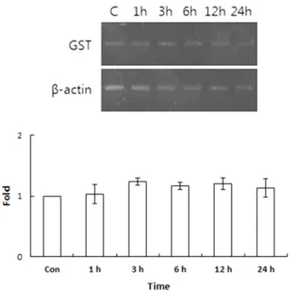
3. 23℃ 기온에서 공기노출 시킨 참담치 초기부착치패 개 체에서의 Hsp70 및 GST 유전자 발현
공기노출에 따른 개체의 생체 내 스트레스 수준을 알아보기 위하여 23℃ 기온에 24시간 공기노출 시킨 참담치 초기부착치 패에서 Hsp70 및 GST 유전자의 발현을 살펴보았다. Hsp70 유전자의 발현은 대조군과 비교하여 1시간 및 3시간 경과 실 험구에서는 유의적인 차이가 나타나지 않았다. 그러나 6시간 경과의 실험구에서부터 유의적인 발현의 증가가 나타나기 시 작하여 24시간 경과 실험구에서 가장 높은 발현 양상이 나타
났다 (Fig. 1). 대조군과 비교하여 실험구에서의 Hsp70 발현 은 1시간, 3시간, 6시간, 12시간 및 24시간에서 각각 1.10, 1.30, 3.83, 5.4 및 6.17배로 나타났다.
GST 유전자의 발현은 대조군과 비교하여 시간의 경과에 따 른 실험구에서 유의적인 차이가 나타나지 않았다 (Fig. 2). 대 조군과 비교하여 실험구에서의 GST 발현은 1시간, 3시간, 6 시간, 12시간 및 24시간에서 각각 1.03, 1.23, 1.17, 1.2 및 1.13배로 나타났다.
기온 (℃) 시간 (h)
1 3 6 12 24 48 72
17 1.3 ± 0.6a 1.3 ± 0.5a 3.5 ± 1.0a 7.0 ± 3.0a 19.3 ± 6.5a 23.0 ± 8.0a 29.6 ± 8.1a 20 1.6 ± 0.5a 2.0 ± 1.3ab 3.6 ± 1.3a 6.2 ± 2.9a 25.6 ± 8.5ab 31.6 ± 9.6ab 33.9 ± 11.9a 23 1.6 ± 0.9a 2.3 ± 0.9ab 5.3 ± 2.6ab 9.9 ± 3.1ab 31.0 ± 7.3b 41.3 ± 12.5b 49.5 ± 12.5b Table 1. Death rate (%) of Mytilus coruscus spat during air exposure
수온(℃) 일(day)
1 2 3 4 5
15 0.3 ± 0.1a 2.0 ± 0.6a 5.5 ± 1.5a 17.1 ± 8.0a 39.3 ± 7.6a
20 0.6 ± 0.2a 1.3 ± 0.3a 9.6 ± 3.5ab 19.4 ± 7.7a 41.9 ± 11.1a
25 2.1 ± 0.3a 3.3 ± 1.16a 15.1 ± 5.3b 25.5 ± 11.3b 54.0 ± 10.5b
Table 2. Death rate (%) of Mytilus coruscus spat during starvation
Fig. 1. Expression of Hsp70 mRNA in cultured at 23℃ during 1 day exposure to air. Con: control, 1h: 1 hour, 3h: 3 hours, 6h: 6 hours, 12h: 12 hours, 24h: 24 hours (** P <
0.01).
4. 25℃ 수온에서 절식 시킨 참담치 초기부착치패 개체에 서의 Hsp70 및 GST 유전자 발현
절식기간에 따른 개체의 생체 내 스트레스 수준을 알아보기 위하여 25℃ 수온에 5일간 절식 시킨 참담치 초기부착치패에 서 Hsp70 및 GST 유전자의 발현을 살펴보았다. Hsp70 유전
자의 발현은 대조군과 비교하여 1일 및 2일 경과 실험구에서 는 유의적인 차이가 나타나지 않았다. 그러나 3일 경과의 실험 구에서부터 유의적인 발현의 증가가 나타나기 시작하여 5일 경과 실험구에서 가장 높은 발현 양상이 나타났다 (Fig. 3). 대 조군과 비교하여 실험구에서의 Hsp70 발현은 1일, 2일, 3일, 4일 및 5일에서 각각 1.20, 1.16, 3.07, 3.23 및 3.97배로 나 타났다.
GST 유전자의 발현은 대조군과 비교하여 1일에서 4일 경 과까지의 실험구에서 유의적인 차이가 나타나지 않았다 (Fig.
4). 대조군과 비교하여 실험구에서의 GST 발현은 1일, 2일, 3 일, 4일 및 5일에서 각각 1.07, 1.07, 1.10, 1.17 및 2.93배로 나타났다.
고 찰
참담치와 같은 이매패류의 분포와 성장, 생존에 영향을 미치 는 환경요인은 수온 (Wilson and Elkaim, 1991; Shin et al., 2000), 염분 (Deaton et al., 1989; Maslin, 1989, Navarro and Gonzalez, 1998), 용존산소 (Shin et al., 2002), 공기노출 (Davenport and Wong, 1986) 등이 있다.
이러한 환경요인이 그 생물이 수용할 수 없을 정도로 항상성이 초과했을 때 스트레스를 받게 되고 이것으로 인하여 대사율이 나 내성이 감소된다. 특히 공기 중에 노출되었을 경우나 먹이 공급이 원활히 공급되지 않은 경우 저산소와 영양결핍 등으로 Fig. 3. Expression of Hsp70 mRNA in cultured at 25℃ during
5 days in starvation. Con: control, 1d: 1 day, 2d: 2 days, 3d: 3 days, 4d: 4 days, 5d: 5 days (** P < 0.01).
Fig. 4. Expression of GST mRNA in cultured at 25℃ during 5 days in starvation. Con: control, 1d: 1 day, 2d: 2 days, 3d: 3 days, 4d: 4 days, 5d: 5 days (** P < 0.01).
Fig. 2. Expression of GST mRNA in cultured at 23℃ during 1 day exposure to air. Con: control, 1h: 1 hour, 3h: 3 hours, 6h: 6 hours, 12h: 12 hours, 24h: 24 hours.
인하여 생리활성이 크게 감소되기 때문에 공기에 노출되는 환 경에서 패류는 노출기간 동안 패각을 닫아서 수분발산을 최대 한 억제하면서 패류 체내의 산소로 호흡하게 되고, 먹이공급이 되지 않는 경우에는 체내 축적된 영양분을 활용하여 대사활동 을 최소화하면서 생존한다. 그러나 이러한 환경스트레스가 장 시간 지속되었을 경우 패류 개체는 성장부진 등의 현상이 나타 나며 궁극에는 폐사 등이 발생하게 된다.
본 연구에서는 참담치 초기부착치패 (1.0 ± 0.2 mm 내외) 를 실험생물로 이용하여 공기노출 및절식에 따른 생존율을 조 사한 결과 참담치 초기부착치패의 공기노출시간에 따른 폐사 율은 노출시간 대비 기온이 높을수록 폐사율이 높아졌으며 노 출 24시간부터 폐사개체가 급격히 많아지는 것으로 나타났다 (Table 1). 그리고 17℃와 20℃ 실험구는 24시간 이후 비교적 폐사율이 서서히 증가하는 양상을 보여 안정화 되는 것으로 나 타났으나, 23℃ 실험구에서는 폐사율이 지속적으로 높아지는 것으로 나타났다 (Table 1). 이러한 결과는 해양생물의 대사율 이 수온에 직접적인 영향을 받으며 개체크기에 따라서는 반비 례한다는 보고와 일치한다 (Fry, 1971; Dabrowski, 1986).
그러나 Shin and Wi, (2004) 는 참담치 치패의 상한 반수치 사수온이 27℃라고 보고한 결과와 차이를 보이고 있다. 이러한 차이는 치패의 크기가 다를 뿐 아니라 수온 이외에 공기노출이 라는 두 개의 스트레스 요인이 작용되었기 때문으로 추정된다.
한편 이매패류의 인공종묘생산 산업이 활성화되면서 치패의 성장과 생존에 식물먹이생물 (Hur et al., 2008, Hur et al., 2010) 의 중요성이 강조되고 있다. 그러나 실내에서 대량으로 치패를 사육하는 과정에서는 충분한 먹이생물공급이 원활하지 못한 경우가 빈번하게 발생되고 있다. 본 연구에서는 절식기간 에 따른 참담치 초기부착치패의 폐사율을 조사한 결과 절식기 간 대비 수온이 높을수록 폐사가 일찍 일어나며, 폐사율도 높 아지는 것으로 나타났다 (Table 2). 그러나 절식이 4일∼5일 동안 지속되면 수온과 관계없이 폐사율이 급격히 높아지는 것 으로 나타났다 (Table 2). 이러한 결과는 이매패류 인공종묘생 산 과정 중에 먹이생물공급이 원활하지 않을 경우 에너지 부족 과 영양적인 스트레스로 대사활력이 감소되어 폐사로 결부된 다는 보고와 일치한다 (Hur et al., 2010).
따라서 참담치 초기부착치패의 경우 24시간 이상 공기노출 과 4일 이상 절식은 생존에 치명적인 영향을 미치기 때문에 안 정적인 사육을 위해서는 이러한 일들이 발생되지 않도록 유의 하여야 할 것이다.
참담치 등의 이동성이 제한적인 패류 개체에는 수온, 염분, 공기노출 및 먹이 등의 환경요인은 스트레스로 작용하여 활성 산소 생성 등의 산화적 스트레스를 유발한다. 스트레스 관련 유전자인 Hsp70 및 GST 유전자는 환경요인에 따른 생체 내 스트레스의 상태를 알아볼 수 있는 생체지표유전자로 활용이
가능하다. 공기노출에 대한 Hsp70 및 GST 유전자의 발현양 상을 살펴본 결과 Hsp70은 23℃ 기온에서 6시간 경과부터 유 의적인 발현양상이 나타났으며, GST 유전자의 경우는 24시간 경과 시까지 유의적인 발현의 차이가 나타나지 않았다 (Fig.
1, 2). 패류의 생존에 있어서 상한치사수온을 살펴보면 전복 pinto abalone (Haliotis kamtschatkana) 은 26.5℃ 및 홍 합 Mytilus edulis는 28.5℃로 알려져 있다 (Chapple et al., 1998). 홍합 (M. edulis) 이 28℃ 이상의 고온에 노출되면 Hsp70의 경우 6시간 경과부터 유의적인 발현의 증가가 나타 나기 시작하여 48시간 가장 높은 수준의 발현을 보이며 서서 히 발현이 감소하기 시작한다는 보고가 있다 (Chapple et al., 1998). 종합해보면 Hsp70은 고온에 노출되어 스트레스를 받 게 되면 세포의 보호를 위해 발현을 하는 것을 알 수 있다. 본 연구에서는 23℃ 온도 설정 상태에서 시간의 경과에 따른 Hsp70의 발현을 살펴본 결과 시간의 경과에 따라 발현이 증 가되어 나타나는 결과는 패류가 공기에 노출되면서 산소의 부 족을 느껴 체내 스트레스를 받게 되므로 나타나는 현상으로 빈 산소에 노출된 해양생물 성게에서 Hsp70 유전자의 발현이 증 가한다는 보고와 경향성이 같음을 알 수 있다 (Lee et al., 2012). 또한 지중해담치 Mytilus galloprovincialis의 공기노 출에 따른 빈산소의 영향에 대한 연구결과 18℃ 48시간까지는 개체의 폐사는 관찰되지 않았으나, 32℃에서는 20시간부터 폐 사가 증가하기 시작하였고, 분자반응에서는 Hsp70 및 Hsp90 이 공기노출에 따라 발현이 증가한다는 보고와 경향이 같음을 알 수 있다 (Anestis et al., 2010). Teixeira 등 (2013) 의 보 고에 의하면 해양생물 산호가 공기에 노출되면 heat shock cognate 70 (Hsc70), Hsp70 및 GST의 발현이 유도된다고 보고하였다. 본 연구에서 24시간 경과까지 GST 발현은 유의 적인 증가가 없었다. 이러한 결과는 참담치와 산호의 종의 다 름도 원인이 되며, 공기노출에 대응한 GST 발현은 Hsp70보 다 늦게 반응이 일어날 수 있음을 제시할 수 있다. 향후 공기노 출 기간의 재설정을 통한 연구의 필요성이 있다.
절식에 대한 Hsp70 및 GST 유전자의 발현양상을 살펴본 결과 Hsp70은 25℃ 수온에서 3일 경과부터 유의적인 발현양 상이 나타났으며, GST 유전자의 경우는 5일 경과 시부터 유 의적인 발현이 나타났다 (Fig. 3, 4). 해양생물에 있어서 먹이 의 부족과 관련해서 생체 내 분자반응은 Hsp70 및 Hsp90 등 과 산화적 스트레스 관련 단백유전자들이 관여한다고 알려져 있다 (Antonopoulou et al., 2013). 일반적으로 영양상태는 세포내의 항산화적 방어기전에 중요한 역할을 한다. 절식에 의 한 영양결핍에 따른 심한 단백질결핍상태에서는 산화적 스트 레스와 lipid peroxidation의 증가에 따른 간 손상 및 단백질 대사교란에 의해 유발되는 산화적 스트레스를 증가시킨다. 또 한 화합물 대사에 관여하는 대사효소계의 변화를 유도하여 약
물대사효소 발현의 변화에 따라 유독화합물에 대한 보호기전 을 작동할 수 없게 된다. 패류 및 해양생물의 절식에 따른 생체 내 기전에 대한 연구는 매우 미비한 상태이다. 본 연구에서는 절식관련 Hsp70 및 GST의 발현 변화에 있어서 Hsp70이 GST 보다 빠르게 반응하는 것을 볼 수 있으며, Hsp70 및 GST는 절식에 따른 지표로 나타냄을 알 수 있었다.
본 연구에서는 공기노출 및 절식과 관련하여 참담치의 폐사 와 Hsp70 및 GST 유전자의 스트레스 관련 지표유전자로서의 가능성을 살펴보았다. 공기노출과 절식의 환경요인이 참담치를 비롯한 해양생물에 미치는 생체 내 기전 연구결과는 현재 미비 한 상태이다. 향후, 많은 연구의 필요성을 느낀다.
사 사
본 논문은 한국해양과학기술진흥원 수산실용화기술개발사업 (과제번호: 312017-4) 의 일환으로 수행되었으며 연구비 지원 에 대하여 깊은 감사를 드립니다.
REFERENCES
Anestis, A., Portner, H.O. and Michaelidis, B. (2010) Anaerobic metabolic patterns related to stress responses in hypoxia exposed mussels Mytilus galloprovincialis. Journal of experimental marine biology and ecology, 394: 123-133.
Antonopoulou, E., Kentepozidou, E., Feidantsis, K., Roufidou, C., Despoti, S. and Chatzifotis, S. (2013) Starvation and re-feeding affect Hsp expression, MAPK activation and antioxidant enzymes activity of European Sea Bass (Dicentrarchus labrax).
Comparative biochemistry and physiology. A, 165:
79-88.
Ashton-Alcox, K.A. and Ford, S.E. (1998) Variability in molluscan hemocytes: a flow cytometric study. Tissue and Cell, 30: 195-204.
Bailey, J., Parsons, J. and Couturier, C.A. (1996) Salinity tolerance in the blue mussel, Mytilus edulis.
Bull. Aquacult. Assoc. Can., 96: 74-76.
Bukau, B. and Horwich, A.L. (1998) The Hsp70 and Hsp60 chaperone machines. Cell, 92: 351-366.
Chapple, J.P., Smerdon, G.R., Berry, R.J. and Hawkins, A.J.S. (1998) Seasonal changes in stress-70 protein levels reflect thermal tolerance in the marine bivalve Mytilus edulis L. J. Exp. Mar. Biol. Ecol., 229:
53-68.
Chirico, W.J., Waters, M.G. and Blobel, G. (1988) 70K heat shock related proteins stimulate protein translocation into microsomes. Nature, 332: 805-810.
Clark, A.G. (1989) The comparative enzymology of the glutathione S-transferases from non-vertebrate organisms. Comp. Biochem. Physiol., 92: 419-446.
Colinet, H., Lee, S.F. and Hoffmann, A. (2010) Temporal
expression of heat shock genes during cold stress and recovery from chill coma in adult Drosophila melanogaster. FEBS J., 277: 174-185.
Dabrowski, K.R. (1986) Active metabolism in larval and juvenile fish: ontogenetic changes, effect of water temperature and fasting. Fish Physiology and Biochemistry, 1: 125-144.
Davenport, J. and Wong, T.M. (1986) Responses of the blood cockle Anadara granosa (L) (Bivalvia: Arcidae) to salinity, hypoxia and aerial exposure. Aquaculture, 56: 151-162.
Deaton, L.E., Derby, J.G.B., Subhedar, N. and Greenberg, M. (1989) Os-moregulation and salinity tolerance in two species of bivalve mollusc:
Limnoperna fortunei and Mytilopsis leucophaeta. J.
Exp. Mar. Ecol., 133: 67-79.
Dong, Y.W., Dong, S.L. and Ji, T.T. (2008) Effects of different thermal regimes on growth and physiological performance of the sea cucumber Apostichopus japonicus Selenka. Aquaculture, 275:
329-334.
Fangue, N.A., Hofmeister, M. and Schulte, P.M. (2006) Intraspecific variation in thermal tolerance and heat shock protein gene expression in common killifish, Fundulus heteroclitus. J. Exp. Biol., 209: 2859-2872.
Feder, M.E. and Hofmann, G.E. (1999) Heat-shock proteins, molecular chaperones, and the stress response: evolutionary and ecological physiology.
Annu. Rev. Physiol., 61: 243-282.
Fry, F.E.J. (1971) The effects of the environmental factors on the physiology of fish. In: Fish Physiology, Vol. 6. (ed. by Hoar WS and Randall DJ), pp. 1-98.
Academic Press, New york.
Gagnaire, B., Heloise, F., Kebin, M., Helene, T.G. and Tristan, R. (2006) Effects of temperature and salinity on hemocyte activities of the Pacific oyster, Crassostrea gigas (Thunberg). Fish & Shellfish Immunology, 20: 536-547.
Galea-Lauri, J., Richardson, A.J., Latchman, D.S. and Katz, D.R. (1996) Increased heat shock protein 90 (hsp90) expression leads to increased apoptosis in the monoblastoid cell line U937 following induction with TNF-alpha and cycloheximide: a possible role in immunopathology. J. Immunol., 157: 4109-4118.
Georgopoulos, C. and Welch, W. (1993) Role of the major heat shock proteins as molecular chaperones. Annu.
Rev. Cell Biol., 9: 601-634.
Hamed, R.R., Farid, N.M., Elowa, S.E. and Abdalla, A.M. (2003) Glutathione related enzyme levels of freshwater fish as bioindicators of pollution. The Environmentalist, 23: 313-322.
Hansson, T., Schiedek, D., Lehtonen, K.K., Vuorinen, P., Liewenborg, J., Noaksson, E., Tjarnlund, U., Hansson, M. and Balk, L. (2006) Biochemical biomarkers in adult female perch (Perca fluviatilis) in a chronically polluted gradient in the Stockholm recipient (Sweden). Marine Pollution Bulletin, 53:
451-468.
Hayes, J.D. and Pulford, D.J. (1995) The glutathione S-transferase supergene family: regulation of GST and the contribution of the isoenzymes to cancer chemprotection and drug resistance. Critical Reviews in Biochemistry and Molecular Biology, 30: 445-600.
Hayes, J.D., Flanagan, J.U. and Jowsey, I.R. (2005) Glutathione transferases. Annual Review of Pharmacology and Toxicology, 45: 51-88.
Hur, Y.B., Min, K.S., Kim, T.E., LEE, S.J. and Hur, S.B. (2008) Larvae growth and biochemical composition change of the Pacific Oyster Crassostrea gigas, Larvae during artificial seed production. J.
Aquaculture, 21: 203-212.
Hur, Y.B., Kim, T.E., Lee, S.J. and Hur, S.B. (2010) Variations in Reserved Nutrient Consumption and Growth of Pacific Oyster (Crassostra gigas) Larvae during Starvation. Korean J. Fish. Aquat. Sci., 43(5):
489-494.
Imai, J. and Yahara, I. (2000) Role of HSP90 in salt stress tolerance via stabilization and regulation of calcineurin. Mol. Cell Biol., 20: 9262-9270.
Jakob, U., Lilie, H., Meyer, I. and Buchner, J. (1995) Transient interaction of Hsp90 with early unfolding intermediates of citrate synthase. J. Biol. Chem., 270: 7288-7294.
Kim, C.W., Jin, Y.G., Kim, T.I., Jeong, D.S. and Kang, H.S. (2015) The expression of Hsp90 and ferritin genes under thermal stress in the sea cucumber (Apostichopus japonicas). Korean J. Environ. Biol., 33: 433-440.
Kim, T.H., Kim, K.J., Choi, M.K. and Yeo, I.K. (2006) Physiological changes of juvenile abalone, Haliotis sieboldii exposed to acute water-temperature stress.
J. of Aquaculture, 77-83.
Kregel, K.C. (2002) Heat shock proteins: modifying factors in physiological stress responses and acquired thermotolerance. J. Appl. Physiol., 92 :2177-2186.
Lee, G., Hwang, J., Chung, Y., Kim, D., Moh, S.H., Chang, M. and Lee, T.K. (2012) Effects of hypoxia on the fertilization and early development of sea urchin, Strongylocentrous nudus. Journal of the Korea Academia-Industrial cooperation Society, 13:
3785-3791.
Li, P., Zha, J. and Zhou, K.Y. (2009) Molecular cloning, mRNA expression, and characterization of HSP90 gene from Chinese mitten crab Eriocheir japonica sinensis. Comp. Biochem. Physiol. B, 153: 229-235.
Liu, H., He, J., Chi, C. and Shao, J. (2014) Differential HSP70 expression in Mytilus coruscus under various stressors. Gene, 543: 166-173.
Looise Bas, A.S., Holwerda, D.A. and Foekema, E.M.
(1996) Induction of glutathione S-transferase in the freshwater bivalve Sphaerium corneum as a biomarker for short-term toxicity tests? Comp.
Biochem. Physiol. C, 113: 103-107.
Loomis, S.H., Ansell, A.D., Gibson, R.N. and Barnes, M.
(1995) Freezing tolerance of marine invertebrates.
Oceanogr. Mar. Biol. Annu. Rev., 33: 337-350.
Maslin, J.L. (1989) The salinity tolerance of Corbula trigona (Bivalvia: Corbulidae) from a West-African lagoon and its variations. Arch. hydrobiol., 117:
205-223.
Nam, B.H., Park, E.M., Kim, Y.O., Kim, D.G., Jee, Y.J., Lee, S.J. and An, C.M. (2013) Analysis of heat, cold or salinity stress-inducible genes in the Pacific abalone, Haliotis discus hannai, by suppression subtractive hybridization. Korean J. Malacol., 29:
181-187.
Navarro, J.M. and Gonzalez, C.M. (1998) Physiological responses of the chilean scallop Argopecten purpuratus to decreasing salinities. Aquaculture, 167:
315-327.
Newell, R.C. and Kofoed, L.H. (1977) Adjustment of the components of energy balance in the gastropod Crepidula fornicate in response to thermal acclimation. Mar. Boil., 44: 275-286.
Paillard, C., Allam, B. and Oubella, R. (2004) Effect of temperature on defense parameters in Manila clam, Ruditapes philippinarum challenged with Vibrio tapes. Dis. Aquat. Org., 59: 249-262.
Parsell, D. and Lindquist, S. (1993) The function of heat-shock proteins in stress tolerance: degradation and reactivation of damaged proteins. Annu. Rev.
Genet., 27: 437-496.
Richter, K. and Buchner, J. (2001) Hsp90: chaperoning signal transduction. J. Cell Physiol., 188: 281-290.
Shin, Y.K., Kim, Y., Chung, E.Y. and Hur, S.B. (2000) Temperature and salinity tolerance of the Manila clam, Ruditapes philippinarum. Korean J. Fish. Soc., 34: 190-193.
Shin, Y.K., Moon, T.S. and Wi, C.H. (2002) Effects of the dis-solved oxygen concentration on the physiology of the blood cockle, Tegilarca granosa. Korean J.
Fish. Soc., 35: 485-489.
Shin, Y.K. and Wi, C.H. (2004) Effect of temperature and salinity on survival and metabolism of the hard shelled mussel Mytilus coruscus, Bivalve: Mytilidae.
J. of Aquaculture, 17: 103-108.
Soetaert, A., Moens, L.N., Van der Ven, K., Van Leemput K., Naudts, B., Blust, R. and De Coen, W.M. (2006) Molecular impact of propiconazole on Daphnia magna using a reproduction-related cDNA array. Comp. Biochem. Physiol. C, 142: 66-76.
Song, J.H., Kim, C.H., Park, S.W., Yu, J.H. and Jo, Y.J.
(2008) Seasonality of the biological activity factors of the hard clam Meretrix lusoria in the western coast of Korea. J. Aquaculture, 21: 111-122.
Teixeira, T., Diniz, M., Calado, R. and Rosa, R. (2013) Coral physiological adaptations to air exposure: Heat shock and oxidative stress responses in Veretillum cynomorium. Journal of experimental marine biology and ecology, 439: 35-41.
Widdows, J. and Shick, J.M. (1985) Physiological responses of Mytilus edulis and Cardium edule to aerial exposure. Marine Biology, 85: 217-232.
Widdows, J., Bayne, B.L., Livingstone, D.R., Newell,
R.I.E. and Domkin, P. (1979) Physiological and biochemical responses of bivalve molluscs to exposure to air. Comp. Biochem. Physiol., 62: 301-308.
Wilson, J.G. and Elkaim, B. (1991) Tolerances to high temperature of infaunal bivalves and the effect of geographical distribution, position on the shore and season. J. Mar. Biol. Ass. U.K., 71: 169-177.
Wu, L.T. and Chu, K.H. (2008) Characterization of heat shock protein 90 in the shrimp Metapenaeus ensis:
evidence for its role in the regulation of vitellogenin synthesis. Mol. Reprod. Dev., 75: 952-959.
Xu, Q. and Qin, Y. (2012) Molecular cloning of heat
shock protein 60 (PtHSP60) from Portunus trituberculatus and its expression response to salinity stress. Cell Stress Chaperones, 17: 589-601.
Yoo, S.K., Kang, K.H. and Lee, D.Y. (1988) Occurrence and survival rate of sea mussel, Mytilus edulis. Bull.
Korean Fish. Soc., 21: 35-41.
You, L., Ning, X., Liu, F., Zhao, J., Wang, Q. and Wu, H. (2013) The response profiles of HSPA12A and TCTP from Mytilus galloprovincialis to pathogen and cadmium challenge. Fish Shellfish Immunol., 35:
343-350.