1Division of Hepatobiliary and Pancreatic Surgery, Department of Surgery, Asklepios Hospital Barmbek,
2Semmelweis University of Medicine, Asklepios Campus Hamburg,
3MVZ Hanse Histologikum GmbH, Hamburg, Germany
Haemangiomas of the liver are benign tumours, which are often diagnosed randomly. With an increase in size hae- mangiomas could become symptomatic. In this case therapeutic options, surgical or interventional, have to be weighted to a conservative approach. We present a case of a 36-year old woman with a symptomatic giant haemangioma of the right liver lobe. Because of the size of the tumor and the small future liver remnant we decided to perform a major liver resection after hypertrophy induction with a preoperative portal vein embolization; an option mainly used for major hepatectomies in malignant tumors of the liver. But however, this case shows, that using a hypertrophy con- cept also for benign liver tumours is the safer approach, if an extended resection is necessary and the future liver remnant is critical. (Ann Hepatobiliary Pancreat Surg 2020;24:357-361)
Key Words: Haemangioma; Liver resection; Portal venous embolization
Received: February 8, 2020; Revised: April 19, 2020; Accepted: April 29, 2020 Corresponding author: Tim Reese
Division of Hepatobiliary and Pancreatic Surgery, Department of Surgery, Asklepios Hospital Barmbek, Rübenkamp 220, Hamburg 22307, Germany
Tel: +49-40-1818-82-8114, Fax: +49-40-1818-82-2819, E-mail: [email protected]
Copyright Ⓒ 2020 by The Korean Association of Hepato-Biliary-Pancreatic Surgery
This is an Open Access article distributed under the terms of the Creative Commons Attribution Non-Commercial License (http://creativecommons.org/
licenses/by-nc/4.0) which permits unrestricted non-commercial use, distribution, and reproduction in any medium, provided the original work is properly cited.
Annals of Hepato-Biliary-Pancreatic Surgery ∙ pISSN: 2508-5778ㆍeISSN: 2508-5859
INTRODUCTION
As the most common type of benign mesenchymal liver tumors, haemangiomas are often asymptomatic and diag- nosed incidentally.1 In general, a conservative therapy ap- proach with imaging follow-up, using ultrasound or MRI scan at regular intervals can be recommended. Due to its benign course, imaging follow-up is not even required for typical haemangiomas. The necessity for treatment of he- mangiomas can be discussed by the presence of symptoms.2 When treatment is indicated, surgery is ther- apy of choice and enucleation of the haemangioma should be performed. As a non-surgical approach a transarterial chemoembolization (TACE) can be performed to reduce the tumor growth.3 In our case the haemangioma was giant and symptomatic, therefore surgical therapy was in- dicated, which was, however, challenging due to size and location.
CASE
A 36-year old female was diagnosed with a size pro- gressive giant haemangioma of the right liver lobe.
Initially it was diagnosed in 2008 during a routine ultra- sound. It measured 6.5 cm and was asymptomatic. In January 2019 the patient started to develop abdominal pain of the right upper quadrant, bloating, nausea and vomiting, which led to a weight loss of 10 kg within 6 months. On physical examination the liver was greatly en- larged with a palpable mass into the pelvic region (Fig.
1). No ascites, signs of jaundice or lower extremity edema where observed. She had no history of viral hepatitis, had taken oral contraceptives in early years and carried a preg- nancy 11 years ago. An abdominal computed tomography (CT) scan revealed a 16×22×23 cm haemangioma filling the right liver lobe and segment 1 almost completely (Fig.
2). Therefore the only treatment option was the resection.
A safe enucleation of the haemangioma with preservation
Fig. 1. Clinical findings. (A) Photograph from the right pa- tient side. Distended abdomen due to enlarged liver. (B) Marking of the palpabel liver.
Fig. 2. Portal Vein Embolisat- ion of all right hepatic portal veins. (A) Angiography of the portal vein with a five French- Pigtail-Catheter. The catheter is placed in the main portal vein.
(B) Portal vein embolization with Glubran of the right ante- rior and posterior branch.
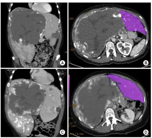
Fig. 3. CT-Scans. (A) Giant haemangioma in the right lobe of the liver and smaller hae- mangioma in segment III. (B) Volumetric CT scan of the left liver lobe. (C) CT Scan 2 weeks after PVE showing an enlarged left liver lobe. (D) Volumetric CT scan of the left liver lobe after PVE.
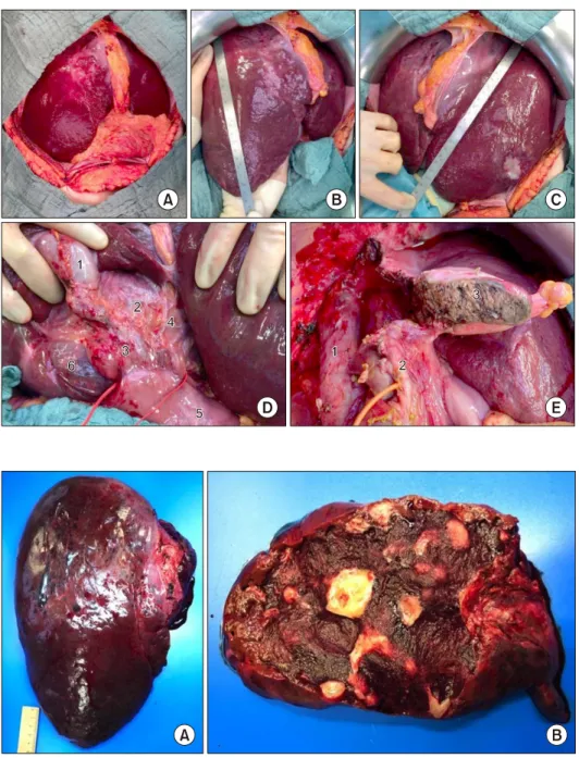
Fig. 5. Pathological findings.
(A) Specimen of the extended right resection (liver segment IV-VIII and I); (B) Cross sec- tion with characteristic structure of haemangioma with small streak of regular liver paren- chyma on the left margin.
Fig. 4. Intraoperative situs. (A) Open abdomen; (B) Left liver lobe with haemangioma, meas- uring 29.5 cm; (C) Hypertro- phied left liver lobe, measuring 23 cm; (D) Ligamentum hep- atoduodenale: Gallbladder (1);
Ductus hepaticus communis (2); Ductus choledochus (3);
Left hepatic arteries (4); Duo- denum (5); Segment I hae- mangioma (6); (E) Situs after extended right resection: Infer- ior vena cava (1); Left bile duct with inserted T-drain (2); Re- section area (3).
operative liver failure. To reduce that risk we chose a hy- pertrophy concept and the patient received a portal venous embolisation (PVE) of all right portal vein branches to in- duce the growth of the future liver remnant (FLR) (Fig.
2). After PVE the volume of the FLR increased from 873 ml to 1.030 ml (sFLR of 69.9%; BSA (Mosteller): 1.79 m2; TELV (Vauthey): 1473 cm3) within 2 weeks (Fig. 3B,
lobe. After dissecting the right hepatic artery, the right portal vein branches, the right bile duct and the right and middle hepatic veins liver segments IV-VIII and segment I were removed en bloc. A T-drainage was inserted into the common hepatic bile duct. The inferior vena cava was clamped for 45 min, because of bleeding complications from the hepatic veins. Pringle maneuver was applied for
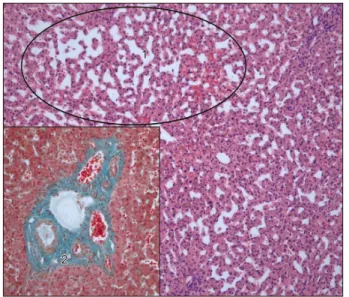
Fig. 6. (A) HE-stained sections showed the classic morphol- ogy of a cavernous haemangioma with widely dilated vas- cular channels lined by flattened inconspicuous endothelial cells and fibrous walls, (B) focal organized thrombi (1) and
partial sclerotic areas (2) could be recognised. Fig. 7. Adjacent liver parenchyma with peliosis hepatis like sinusoidal dilatation (1) and mild fibrosis of the portal tracts in Elastica van Gieson stain (2).
10 min. The intraoperative blood loss was 1100 ml. The ICU-stay was 3 days. The postoperative course was uneventful. There was no sign of a bile-leakage, so the T-drain was removed early. The patient was discharged from the hospital after 12 days without any further com- plications or complaints.
The gross processing showed a liver specimen of the 2,100 g weight with a solitary red-brown and well-demar- cated 17 cm tumour with spongy cut surface and some fibrotic areas (Fig. 5). Histological examination showed a haemangioma with a predominantly cavernous archi- tecture (most common subtype) with focal thrombi and re- gressive fibrosis (Fig. 6). The adjacent liver tissue pre- sented an overall preserved architecture with peliosis hep- atis like change and low to moderate portal field fibrosis, lymphocytic portal field infiltrates and minimal choles- tasis (Fig. 7).
DISCUSSION
In general haemangiomas of the liver are small (<1 cm), asymptomatic and do not require any treatment.
Most commonly monitoring via imaging techniques is recommended. But however, so called giant haemangio- mas are different. They are defined as greater than 5 cm, hypergiant haemangiomas as >10 cm.4 Due to their size they can cause various complaints and often need some form of treatment. Transarterial chemoembolization (TACE),
radiofrequency (RFA) or microwave ablations (MWA) are possible non-surgical treatment strategies for haemangio- mas, which can be applied to decrease the growth.
Nevertheless, these treatments are reserved for minor haemangiomas.5-7 Although non-operative management might be the right approach for the majority of patients, patients with abdominal compression symptoms are more likely to benefit from surgery, as it is the most effective therapy.8 In our case the patient was not likely to benefit from any non-surgical treatment. The complaints led to an immense loss of quality of life, as food-intake was almost not possible. Therefore, different surgical strategies need- ed to be discussed. A conventional right hemihepatectomy (segment V-VIII) would not have been sufficient, because the haemangioma advanced into liver segments IVa and b. An enucleation of the tumour would have been due to the high risk of major complication, severe blood loss for example, and only marginal healthy liver parenchyma not favourable either. Hence, we decided to perform a right trisectorectomy, using a preoperative PVE to induce hy- pertrophy of the remaining segments II and III. PVE is a valuable tool to successfully induce hypertrophy of the future liver remnant prior to surgical resection. In hepatic surgery hypertrophy concepts before resection are almost exclusively applied to malignant tumours of the liver.
PVE was initially used in order to increase the resect- ability rates in patients with cholangiocarcinoma (CCC)
atectomy liver failure. To our knowledge, this case de- scribes the first major hepatectomy following PVE in a patient with a hypergiant haemangioma in the literature.
Previously described surgical treatments were TACE be- fore surgical resection, enucleation or conventional right or left liver resection.4,10
ORCID
Alina Strohmaier: https://orcid.org/0000-0001-5260-6037 Kim C. Wagner: https://orcid.org/0000-0001-7109-1833 Tim Reese: https://orcid.org/0000-0002-9163-5962 Mohammad Fard-Aghaie:
https://orcid.org/0000-0002-5798-6590
Georgios Makridis: https://orcid.org/0000-0001-6187-9336 York von Rittberg: https://orcid.org/0000-0001-6804-274X Katja Horling: https://orcid.org/0000-0003-4939-7312 Karl J. Oldhafer: https://orcid.org/0000-0002-4878-265X
3. Furumaya A, van Rosmalen BV, Takkenberg RB, van Delden OM, Dejong CHC, Verheij J, et al. Transarterial (chemo-)embo- lization and lipiodolization for hepatic haemangioma. Cardiovasc Intervent Radiol 2019;42:800-811.
4. Adhikari DR, Thakur V, Telavane PP, Kulkarni R, Singh R, Joshi RM. Hypergiant hepatic hemangiomas: case series. Indian J Surg 2015;77(Suppl 1):40-42.
5. Firouznia K, Ghanaati H, Alavian SM, Nassiri Toosi M, Ebrahimi Daryani N, Jalali AH, et al. Management of liver he- mangioma using trans-catheter arterial embolization. Hepat Mon 2014;14:e25788.
6. Gao J, Ke S, Ding XM, Zhou YM, Qian XJ, Sun WB.
Radiofrequency ablation for large hepatic hemangiomas: initial experience and lessons. Surgery 2013;153:78-85.
7. Ziemlewicz TJ, Wells SA, Lubner MA, Musat AI, Hinshaw JL, Cohn AR, et al. Microwave ablation of giant hepatic cavernous hemangiomas. Cardiovasc Intervent Radiol 2014;37:1299-1305.
8. Duxbury MS, Garden OJ. Giant haemangioma of the liver: ob- servation or resection? Dig Surg 2010;27:7-11.
9. Glantzounis GK, Tokidis E, Basourakos SP, Ntzani EE, Lianos GD, Pentheroudakis G. The role of portal vein embolization in the surgical management of primary hepatobiliary cancers. A systematic review. Eur J Surg Oncol 2017;43:32-41.
10. Ketchum WA, Lin-Hurtubise KM, Ochmanek E, Ishihara K, Rice RD. Management of symptomatic hepatic "mega" heman- gioma. Hawaii J Med Public Health 2019;78:128-131.