大 韓 氣 管 食 道 科 學 會 誌
□종 설□
Vol.14. No. 2, Dec. 2008
Endobronchial Ultrasound Guided Transbronchial Needle Aspiration의 임상적 유용성
국립암센터 폐암센터 호흡기내과
황 보 빈=Abstract=
Clinical Application of Endobronchial Ultrasound Guided Transbronchial Needle Aspiration
Bin Hwangbo, M.D.
Department of Pulmonology, Center for Lung Cancer, National Cancer Center, Goyang, Korea
Endobronchial ultrasound guided transbronchial needle aspiration is a recently introduceddiag- nostic method which enables real time aspiration of peritracheal and peribronchial lesions. The high sensitivity and the high diagnostic accuracy of EBUS-TBNA in the mediastinal staging of lung cancer have been reported. EBUS-TBNA also showed the high diagnostic yield in the diagnosis of lung parenchymal masses adjacent to the trachea or the large airways.
EBUS-TBNA is a good diagnostic method for mediastinal diseases, such as sarcoidosis. Until now, no major complications of EBUS-TBNA have been reported. EBUS-TBNA should be considered for the mediastinal staging of lung cancer and the diagnosis of mediastinal lymphadenopathies.
교신저자 : 황보빈, 경기도 고양시 일산구 마두1동 809 국립암센터, 폐암센터
Tel : 031-920-1618 Fax : 031-920-1298 E-mail : [email protected]
Ⅰ. 서 론
Endobronchial ultrasound guided transbronchial nee- dle aspiration (EBUS-TBNA)은 기관(trachea)과 기관 지 주변의 초음파 영상을 직접 보면서 세침 검사를 하는 진단 방법이다. 2003년 문헌 상으로 처음 소 개된 최근에 개발된 검사 방법이나 그 임상적 유용 성이 보고됨에 따라 세계적으로 점차 사용이 늘어 나고 있다. 이 글에서는 먼저 EBUS-TBNA의 방법 을 소개하고, EBUS-TBNA의 폐암의 종격동 병기 결정, 폐암의 진단 그리고 기타 종격동 질환 진단 에서 EBUS-TBNA의 임상적 유용성에 대해 알아보
고자 한다.
Ⅱ. EBUS-TBNA 검사 방법
1. EBUS-TBNA 기관지 내시경
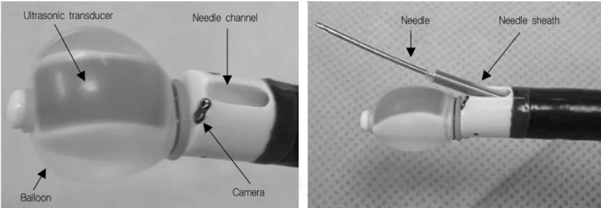
Prototype EBUS-TBNA 기관지 내시경(BF-UC260F- OL8; Olympus)은 기관지 내시경은 말단부에7.5 MHz의 선형(linear) 초음파 transducer가 부착되어 있어 convex probe (CP)- EBUS 기관지 내시경으로 불린다. 이 기관지 내시경의 외경은 6.7 mm이며 working channel의 내경은 2mm이다. 말단부에는 balloon을 장착할 수 있어 transducer의 밀착이 어려 운 부위에서 좋은 초음파 영상을 얻는데 도움이 된 다(Fig. 1).
EBUS-TBNA 기관지 내시경으로 백색광 영상과
Fig. 1. EBUS-TBNA 기관지 내시경(BF-UC260F-OL8; Olympus)
Fig. 2. Fig. 2. EBUS-TBNA 영상 예
초음파 영상을 동시에 얻을 수 있다. 백색광 영상 은 내시경 진행 방향의 30도 사선 방향의 영상을 보여 준다. 따라서 기존의 기관지 내시경과는 조작 방법에 약간의 차이가 있다. 초음파로 50도 각도의 영상을 얻을 수 있으며, 관찰 가능한 깊이는 약 5cm이다. 초음파 영상에는 도플러 기능이 있어 주 변 혈관을 쉽게 감별할 수 있다(Fig. 2). EBUS- TBNA에 사용하는 needle은 외경이 22G 굵기이며, 내경은 21G이다. 음압을 발생시킬 수 있는 syringe 를needle set의 근위부에 부착할 수 있도록 되어 있 다.
2. EBUS-TBNA 검사 방법
EBUS-TBNA는 주로 국소 마취 및 미다졸람을 이용한 진정 상태에서 시행하며 검사는 외래 환자 에 대해서도 가능하다. 일부 센터에서는 후두 마스
크 등을 이용하여 전신 마취 상태에서 시행하기도 한다. EBUS-TBNA 기관지 내시경의 굵기 때문에 코를 통해 삽입하지 않고 입을 통해 내시경을 삽입 한다.
기관으로 내시경을 삽입한 후 balloon을 생리 식 염수로 적당 크기로 부풀린 후 검사를 원하는 부위 에 transducer를 대고 초음파 영상을 관찰한다. 검사 부위가 확인되면 needle set을 기관지 내시경에 장 착하여 고정한다. Needle을 병변에 삽입한 후 음압 syringe를 needle set 근위부에 부착한다. 병변 안에 서 needle을 앞뒤로 움직이면서 검체를 얻는 후 음 압 syringe를 제거하고 needle set을 제거한다.
폐암의 병기 결정을 위해 EBUS-TBNA를 시행할 때, 보통 N3 병변에 대해 먼저 시행한 후 N2 병변 에 대해 검사를 시행한다. 한 병변에서 보통 2-3회 의 흡인을 시행한다.1)
Ultrasonic transducer Needle channel
Camera Balloon
Needle Needle sheath
참고
문헌 폐암 환자수, 환자군 임파절 크기
평균(범위)# 유병율 민감도 특이도 양성
예측도
음성
예측도 정확도
Yasufuku
20052 108, pts with positive
node(s) on CT 19mm
(10-38) 68.5% 94.1% 100% 100% 90.9% 96.3%
Yasufuku
20063 102, candidates for
surgery 8.7mm
(5-25) 25.5% 92.3% 100% 100% 97.4% 98.0%
Herth
20064 100, pts with normal
mediastinum on CT 8.1mm
(4-10) 17.0% 94.1% 100% 100% 98.8% 99.0%
Herth
20085 97, normal CT & PET
on mediastinum 7.9mm
(5-10) 6.2% 100% 100% 100% 100% 100%
Bauwens
20086 106, pts with PET(+)
mediastinal nodes - 58% 95% 100% 100% 91% 97%
Lee
20081 102, potentially
operable pts 8.6mm
(5.1-19.3) 29.4% 93.8% 100% 100% 96.9% 97.9%
* N2/N3 진단율을 논문에 주어진 자료로 계산함. 환자 중심으로 계산된 진단율이며, 임파절 중심으로 계산 하지 않음. N1은 제외함. # EBUS-TBNA를 시행한 임파절의 CT 상 단경
Table. 1. 폐암의 종격동 임파선 전이 확인에서 EBUS-TBNA의 진단율*
3. EBUS-TBNA 검체 처리 방법
Needle set 근위부에서 stylet을 넣어 needle 입구로 부터 검체가 나오게 한다. 유리 슬라이드에 도말하.
여 95% 알코올에 고정한 후 세포 검사를 의뢰한다.
검체의 70-80%에서 tissue core 검체를 얻을 수 있으 며, 이는 포르말린에 고정하여 조직 병리 검사를 의 뢰한다. On-site cytopathologic examination이 가능한 경우에는 즉시 염색하여 결과를 확인할 수 있다.
4. EBUS-TBNA 검사 가능 부위
EBUS-TBNA 기관지 내시경이 진입할 수 있는 기관지 주변의 병변에 대해서 검사가 가능하다. 종 격동 임파절(N2 또는 N3) 중에는 1, 2, 3P, 4, 7번 임파절이 접근 가능하며, 일부8번 임파절도 접근이 가능하다. 3A, 5, 6, 9번 임파절은 EBUS-TBNA로 접근이 불가능하며, 일부 8번 임파절도 접근이 불 가능하다. N1 위치의 임파절 중에는 10번, 11번과 일부 12번 임파절이 접근이 가능하다(Fig. 3).
5. 폐암에서의 종격동 병기 결정에서 EBUS-TBNA 의 유용성
종격동 병기 결정에서 EBUS-TBNA를 이용한 대 표적 전향적 연구는 표 1에 요약되어 있다. 연구에 포함된 환자군에는 차이가 있으나, 이들 연구들은
폐암 환자에서 종격동 임파절 전이를 진단하는데 있어 EBUS-TBNA의 정확도를 95%이상으로 높게 보고하고 있다.2-6) EBUS-TBNA를 비침습적 검사방 법인 CT와 PET과 비교한 연구에서도 EBUS-TBNA 는 CT 또는 PET보다 더 높은 진단율을 보여, EBUS-TBNA는 이들 비침습적 검사 후에 시행할 수 좋은 침습적 검사 방법임이 확인되었다.3) EBUS- TBNA의 높은 진단율을 고려할 때, EBUS-TBNA는 폐암의 종격동 병기 결정에 적극적으로 고려되어 야 할 검사 방법이다.
EBUS-TBNA가 종격동경을 대체할 수 있을 지 여부는 큰 관심사다. 현재까지 EBUS-TBNA와 종 격동과의 비교 연구는 발표되지 않았다. 종격동경 은 전신 마취 하에서 시행하는 침습적 검사로 2-3%
에서 합병증이 보고되어 있다. 또한 모든 종격동 임파절을 종격동경 검사로 진단할 수 있는 것은 아 니며 종격동경으로 접근 가능한 부위는 EBUS- TBNA로도 진단이 가능하다(Fig. 3). 그러나 종격동 경은 작은 크기의 임파절 전이를 진단하는데 있어 EBUS-TBNA에 비해 장점이 있을 것으로 생각된다.
현재까지 EBUS-TBNA가 종격동경 검사를 완전히 대체할 수 있다는 근거는 없으나 EBUS-TBNA의 높은 진단율을 고려할 때, 종격동경 검사의 대부분 을 대체할 것으로 보인다.
EBUS-TBNA: 1, 2, 3P, 4, 7, (8), 10, 11, (12)
EUS-FNA: (1), (2), 3P, (4R), 4L, (5), 7, 8, 9, (10L)
Cervical mediastinoscopy: 1, 2, 4, (7)
괄호 없는 임파절은 대부분 검사 가능함
괄호 안의 임파절은 일부에서만 검사 가능함.
Fig. 3. 검사에 따른 종격동 임파절의 접근 가능 위치
EBUS-TBNA와 상호 보완적 관계에 있는 검사 방법이 EUS-FNA이다. EUS-FNA는 기관 앞쪽 (pr- etracheal)의 임파절에 대해 접근이 불가능하다는 단점이 있지만 EBUS-TBNA로 검사가 불가능한 5, 9번 및 일부 8번에 대해 검사가 가능하다는 것이 장점이다(Fig. 3). 따라서 EBUS-TBNA와 EUS-FNA 를 같이 시행하는 것이 일부 환자에서는 도움이 될 것이라 생각된다.7)
종격동 재 병기결정에서 EBUS-TBNA의 역할을 본 한 개의 연구가 있다. Herth 등은 항암 치료 후 EBUS-TBNA를 시행한 89명의 N2 환자를 대상으로 후향적 연구를 시행하였다. 이 연구에서 EBUS- TBNA의 종격동 재병기 결정에서 민감도, 음성 예 측도, 정확도는 각각 76%, 20%, 77%로 나타났다.8) 종격동 재병기 결정에서 EBUS-TBNA의 역할에 대 해서는 더 연구가 필요하다.
6. 폐암의 진단에서의 EBUS-TBNA의 유용성 EBUS-TBNA는 폐암의 종격동 병기 결정 분 아 니라 폐암의 진단에도 이용할 수 있다. Annema 등 는 60명의 폐 중심부 종괴를 가진 환자에서 EBUS-TBNA의 진단율을 77%로 보고하였다.9) Nak- ajima 등은 기관지 주변 폐종괴의 진단에 있어 EBUS-TBNA의 민감도를 94.1%로 보고하였다.10)
폐암 자체 뿐 아니라, 전이된 임파절에서도 EBUS- TBNA로 진단이 가능하므로, 폐암의 진단 방법으 로도 적극적으로 고려되어야 한다.
7. 기타 종격동 질환에서 EBUS-TBNA의 유용성 EBUS-TBNA는 유육종증, 임파종, 임파선 결핵 등 기타 종격동 병변의 진단에도 유용하다. 유육종 증의 진단에 있어 EBUS-TBNA의 민감도는 85-93%
로 높게 보고되어 있다.11, 12) 종격동 임파종 진단에 있어서도 민감도가 91%로 보고된 바 있다.13)
8. EBUS-TBNA의 합병증
현재까지 문헌 상 EBUS-TBNA에 의한 중대한 합병증은 거의 보고되지 않았다. 문헌상 언급된 합 병증으로는 시술 중 저산소증, 저절로 소실된 심방 세동, 기도 부종/발적, stridor 등이 있다.1, 9, 11) 한 예 에서 시술 후 기흉이 보고된 바 있다.6) 그 외 가능 한 합병증으로는 pneumo-mediastinum, 혈종, 종격 동염 등이 있다.
Ⅲ. 맺음말
EBUS-TBNA는 폐암의 병기 결정 및 진단 및 기 타 종격동 질환의 진단에 있어 유용한 검사 방법이
다. 합병증이 드물게 보고되어 있는 안전한 검사 방법이다. 따라서 EBUS-TBNA 는 폐암의 병기 결 정, 폐암 진단, 기타 종격동 병변 질환의 진단에 적 극적으로 활용되어야 하겠다.
References
1. Lee HS, Lee GK, Lee HS, et al. Real-time en- dobronchial ultrasound-guided transbronchial needle aspiration in mediastinal staging of non-small cell lung cancer: how many aspirations per target lymph node station? Chest. 2008;134:368-74.
2. Yasufuku K, Chiyo M, Koh E, et al. Endobronchial ultrasound guided transbronchial needle aspiration for staging of lung cancer. Lung Cancer. 2005;50:
347-54.
3. Yasufuku K, Nakajima T, Motoori K, et al. Com- parison of endobronchial ultrasound, positron emission tomography, and CT for lymph node stag- ing of lung cancer. Chest. 2006;130:710-8.
4. Herth FJ, Ernst A, Eberhardt R, et al. Endo- bronchial ultrasound-guided transbronchial needle aspiration of lymph nodes in the radiologically no- rmal mediastinum. Eur Respir J. 2006;28:910-4.
5. Herth FJ, Eberhardt R, Krasnik M, et al. Endobron- chial ultrasound-guided transbronc hial needle aspiration of lymph nodes in the radiologically and positron emission tomography-normal mediastinum in patients with lung cancer. Chest. 2008;133:887-91.
6. Bauwens O, Dusart M, Pierard P, et al. Endo-
bronchial ultrasound and value of PET for pr- ediction of pathological results of mediastinal hot spots in lung cancer patients. Lung Cancer. 2008;61:
356-61.
7. Wallace MB, Pascual JM, Raimondo M, et al. Min- imally invasive endoscopic staging of suspected lung cancer. JAMA. 2008;299:540-6
8. Herth FJ, Annema JT, Eberhardt R, et al. Endo- bronchial ultrasound with transbron hial needle aspiration for restaging the mediastinum in lung cancer. J Clin Oncol. 2008;26:3346-50.
9. Tournoy KG, Rintoul RC, van Meerbeeck JP, et al.
EBUS-TBNA for the diagnosis of central paren- chymal lung lesions not visible at routine bron- choscopy. Lung Cancer. 2008 in press.
10. Nakajima T, Yasufuku K, Fujiwara T, et al.
Endobronchial ultrasound-guided transbronchial needle aspiration for the diagnosis of intrapul- monary lesions. J Thorac Oncol. 2008;3:985-8 11. Garwood S, Judson MA, Silvestri G, et al.
Endobronchial ultrasound for the diagnosis of pulmonary sarcoidosis. Chest. 2007;132:1298-304.
12. Wong M, Yasufuku K, Nakajima T, et al. Endo- bronchial ultrasound: new insight for the diagnosis of sarcoidosis.Eur Respir J. 2007;29:1182-6.
13. Kennedy MP, Jimenez CA, Bruzzi JF, et al.
Endobronchial ultrasound-guided transbronchial needle aspiration in the diagnosis of lymphoma.
Thorax. 2008;63(4):360-5.