International Journal of Gastrointestinal Intervention
journal homepage: www.ijgii.org
Review Article
Recent developments in endoscopic ultrasound-guided
radiofrequency ablation for pancreatic lesions
Jae Hee Cho*, Sung Ill Jang, and Dong Ki Lee
A B S T R A C TRadiofrequency ablation (RFA) has been regarded as an established technique to treat various diseases such as hepatocellular carcinoma, renal cell carcinoma and Barret’s esophagus. Although the application of RFA in the pancreas has been limited due to increased risk of adverse events, endo-scopic ultrasound-guided RFA (EUS-RFA) has generated interest as a novel minimally invasive treatment modality which combines real-time visu-alization with a precise locvisu-alization of the treatment procedure. For over a decade, the optimization of RFA devices have made EUS-RFA relatively safe, and several studies have supported its feasibility. However, there is insufficient evidence to suggest the appropriate indications and to describe long-term outcomes of EUS-RFA for various pancreatic neoplasms such as pancreatic neuroendocrine tumor, ductal adenocarcinoma, and cystic le-sions. Therefore, this review focuses on the technical aspects and clinical applications of EUS-RFA for each pancreatic disease.
Copyright © 2020, Society of Gastrointestinal Intervention. Keywords: Endoscopic ultrasound; Pancreatic cystic lesions; Pancreatic neopla; Pancreatic neuroendocrine tumor; Radiofrequency ablation
Department of Internal Medicine, Gangnam Severance Hospital, Yonsei University College of Medicine, Seoul, Korea Received July 27, 2020; Revised August 31, 2020; Accepted August 31, 2020
* Corresponding author. Department of Internal Medicine, Gangnam Severance Hospital, Yonsei University College of Medicine, 211 Eonju-ro, Gangnam-gu, Seoul 06273, Korea.
E-mail address: [email protected] (J.H. Cho).
pISSN 2636-0004 eISSN 2636-0012 https://doi.org/10.18528/ijgii200030
This is an open-access article distributed under the terms of the Creative Commons Attribution Non-Commercial License (http://creativecommons.org/licenses/by-nc/4.0) which permits unrestricted noncommercial use, distribution, and reproduction in any medium, provided the original work is properly cited.
Introduction
Surgery is the only potentially successful treatment option for various pancreatic neoplasms; however, a minority of these have a surgical indication. In pancreatic ductal adenocarcinoma (PDAC), only 20% of patients have the opportunity to undergo a surgical resection because most patients are diagnosed with unresectable disease at presentation. More so, surgery-related morbidity and mortality are not negligible. In pancreatic neuroendocrine neo-plasm (PNEN) and pancreatic cystic lesion (PCL), patients with a low malignant potential require life-long surveillance rather than extensive surgical resection. Therefore, if some favorable results are verified, minimally invasive therapy is very attractive, due to safety, reproducibility, and affordability.1 Radiofrequency ablation
(RFA) is an established minimally invasive therapeutic modality for various diseases including hepatocellular carcinoma, renal cell carcinoma, and Barret’s esophagus. In pancreatobiliary disease, endoscopic retrograde cholangiopancreatography (ERCP)-guided intraductal RFA has also been increasingly performed in malig-nant biliary tract obstruction.2-5 However, the RFA for the
pan-creas is under investigation due to the inpan-creased risk of adverse
events. Because the pancreas is a thermosensitive organ with more complex vascular systems, pancreatic neoplasms could in-filtrate the bile duct or the duodenal wall, encase major vessels, or occlude the main pancreatic duct (MPD) by proximity.1 With the
recent widespread use of endoscopic ultrasound (EUS) which has the advantage of real-time visualization and precise localization of pancreatic neoplasms, the interest of local ablation through EUS is increasing. Therefore, even though several studies have showed non-negligible complication rates of EUS-RFA, it is nec-essary to verify the applicability of EUS-RFA in the management algorithms for various pancreatic neoplasms. This review will fo-cus on the technical aspects of EUS-RFA and clinical applications of EUS-RFA for each pancreatic disease.
Technical Aspects
RFA is a technique in which a needle inserted inside a lesion causes a hyperthermal injury. The radiofrequency (RF) energy cir-cuit delivers high-frequency alternating current to produce ionic agitation in the cell, resulting in hyperthermia and coagulation necrosis in the target tissue.6 RF may be delivered through
mo-the circuit is closed inside mo-the probe and current is concentrated between the anode and the cathode. Although RFA has already been actively applied in pancreatobiliary disease, there have been several concerns about possible RFA related adverse events in pancreatobiliary disease. For example, ERCP-guided intraductal bipolar RFA can ablate the intraluminal biliary tumor with com-parable safety; however, segmental biliary stricture with cholan-gitis develops as a long-term result, so biliary stents should be placed to maintain biliary drainage after intraductal RFA.7
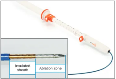
In terms of technical aspects of EUS-RFA, the fine needle is passed through the shortest possible pathway into the normal pancreatic parenchyma and avoiding the bile duct, pancreatic duct, and major vessels. When RFA energy is applied, the real-time EUS visualization of bubbles can be seen on the pancreatic neoplasm, and these results in a hyperechoic lesion at the end of the treatment. After this, RFA electrodes should be repositioned during the same session to ablate the untreated area, and this can be repeated several times. However, during EUS-RFA, it is difficult to accurately estimate the effective area of RFA. Until now, avail-able EUS probes are the Habib EUS-RFA probe (Boston, Marlbor-ough, MA, USA), the EUSRA RF electrode (STARmed, Goyang, Korea) (Fig. 1) and the HybridTherm (ERBE Elektromedizin GmBH, Tübingen, Germany).1 When RFA energy is applied, the amount of
thermal injury depends on different parameters including power (in watts), duration, electrode length, and target temperature. More-over, a heat sink effect that decreases the efficacy of RFA may occur in the area near the blood vessel. Therefore, each safe and effective RFA setting according to the various RFA probes should be presented.
Radiofrequency Ablation Probe
Habib EUS-RFA and EUSRA are monopolar RFA catheters. Habib is a through-the-needle probe and EUSRA is a needle-type catheter. The Habib device is a 1 Fr wire monopolar electrode that can be inserted into a standard 22G needle and connected to a standard electrosurgical unit (ERBE). The EUSRA electrode is an 19G needle connected to a specific VIVA RF generator (STARmed). Typically, this has a pump to cool the EUSRA needle through a chilled saline solution. RFA was stopped when automatically measured impedance exceeded 1,000 Ω. The performances of
100%, and both the devices and the echogenic cloud during the application of RF were clearly visible. At histopathologic analysis, the extent of tissue necrosis was tissue damage ranging from 3.1 ± 0.4 mm (power = 8 W, effect 4, time = 120 sec) to 2.3 ± 0.1 mm (12 W, effect 4, 120 sec) in depth for the Habib probe. Moreover, the ablation depth ranged from 3.6 ± 0.5 mm (power = 30 W, time = 15 sec) to 3.8 ± 0.4 mm (power = 70 W, time = 11 sec) for the EUSRA probe. They showed an effective ablation of pancre-atic tissue about 2.5 mm around the RFA electrode with both de-vices and suggested use of 10 W, 120-second ablation settings for the Habib EUS-RFA probe and 30 W, 15-second settings for the EUSRA when performing EUS-RFA for pancreatic lesions of 5 to 6 mm. Importantly, the ablation of larger lesions should probably require repeated procedures. Interestingly, due to the mechanical properties, each device has its own preferences. Since Habib is a thin device that is used by putting it inside a needle, it is difficult to use repeatedly due to its low durability. On the other hand, EU-SRA is stiffer and technically difficult to handle in some challeng-ing areas of the pancreas.8
Different from the aforementioned pure RFA probes, Hybrid-Therm is a hybrid bipolar needle type probe that is also available with cryogenic cooling using carbon dioxide. The probe has a sharp distal tip with an active part (1.8 mm in diameter and 20 mm in length). The bipolar system has the theoretical advantage of reducing thermal injury; however, its RFA efficacy is limited. In order to overcome the shortcomings, the RFA effect of Hybrid-Therm is augmented by a cryogenic gas, which increases intersti-tial devitalization.9 Indirect comparisons with pure RFA indicate
that a larger ablation zone can be obtained with reduced energy and reduced application time. Preclinical studies have shown the feasibility of the technique and demonstrated a linear correlation between the application time and the size of the ablated tissue, when the risk of necrotizing pancreatitis and other adverse events increased.10 Complication rates were 43% for minor events and a
single case of necrotizing pancreatitis, and all complications oc-curred when applications lasted > 300 seconds. HybridTherm has not been actively used yet, so further research is needed for ap-propriate treatment methods as well as the delivery protocol.
In summary, the limited available clinical experience makes it difficult to draw firm conclusions on the standard protocols for various EUS-RFA methods. Routine antibiotic prophylaxis and administration of rectal nonsteroidal anti-inflammatory drugs are now recommended to reduce the EUS-RFA related morbidity and mortality.11 Furthermore, the proximity to the MPD has raised
some doubts on the possibility of safe ablation of these lesions, and has led to theoretical advocacy for the possibility of prophy-lactic pancreatic stenting in these patients.
Clinical Applications
Pancreatic neuroendocrine neoplasm
PNENs are rare neoplasms that account for approximately 2% to 3% of primary pancreatic malignancies. The incidence of those has increased over the last three decades due to the advancement of diagnostic imaging studies as well as widespread awareness by physicians. The World Health Organization (WHO) 2010 grading system has been proposed to define a new pathologic grade strati-fication, and the system categorized neuroendocrine tumors (NETs) into low-grade (G1), intermediate grade (G2), and high-grade (G3) based on their proliferative rate using the mitotic activity and/or
Insulated
sheath Ablation zone
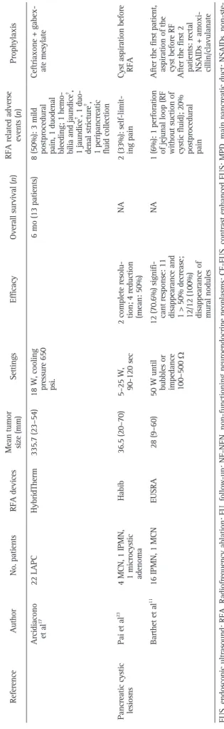
Table 1
Published Data of EUS Guided RFA Treatments in Pancreatic Disease
Reference
Author
No. patients
RFA devices
Mean tumor size (mm)
Settings
Efficacy
Overall survival (
n)
RFA related adverse
events ( n) Prophylaxis Pancreatic neuro -endocrine neoplasm Lakhtakia et al 15 3 insulinomas EUSRA 14–22 50 W
Symptoms relief (100%), persistent at 11 mo FU All patients alive at 11 mo FU
0 NA Choi et al 14 7 NF-NENs, 1 insulinoma EUSRA 20 (8–28) 50 W (10 sec) 6 complete respons
-es, 2 incomplete responses; remis
-sion of hypoglyce
-mic symptoms in insulinoma patient
2 (25%): 1 abdomi
-nal pain, 1 pancre
-atitis
Broad-spectrum antibiotics
Oleinikov et al
16
7 insulinomas, 11 NF-NENs (some multifocal; 2 with treatable metastasis); 27 total lesions
EUSRA
14.4 (4.5–30)
10–50 W, 5–12 sec
26/27 lesions with typical post- ablative changes at CE-EUS; 7 (100%) symptoms resolution in insulinomas; no recurrence after mean 8.7 mo FU
2 (11%) mild pan -creatitis Broad-spectrum antibiotics Barthet et al 11 14 NF-NENs (Grade 1) EUSRA 12 (10–20)
50 W until bubbles or impedance 100–500
Ω
12 (86%) complete disappearance; 2 absence of Dop
-pler at EUS
NA
2 (14%): 1 acute necrotizing pancreatitis (RF without suction of cystic fluid), 1 MPD stenosis; 20% postproce
-dural pain
After the first 2 patients: rectal NSAIDs + amoxi
-cillin/clavulanate
Pancreatic ductal adenocarcinoma
Wang et al 21 3 LAPC Habib 37.3 10–15 W, 2 min
Mean reduction in tumor size: 13.94%
NA 0 NA Song et al 20 4 LAPC + 2 MPC EUSRA 38 (30–90) 20–50 W, 10 sec NA 2 (33%) self-limiting pain Broad-spectrum antibiotics Scopelliti et al 19 10 LAPC EUSRA 25–75
20 W (lesion < 3 cm), 30 W until impedeance 500
Ω
Mean diameter of necrosis at 30 day CT: 30 ± 13 mm
NA
4 (40%): 2 post
-procedural self limiting pain, 2 asymptomatic ascite
Broad-spectrum anti
-biotics + octreotide + LMWH
Crinò et al
18
8 LAPC + 1 metastatic rectal cancer
EUSRA
36 (22–67)
30 W
Ablated area in all patients at 30 day CT: mean diameter 3.75 cm 3 (0.72–
12.6 cm
3 ), 30% of
tumor mass
NA
3 (33%): 3 mild abdominal pain
NA
Yang and Zhang
22
8 unresectable pancreatic cancer
Habib
NA
NA
Ablated area inside th
e tu m or in 1 00 % 8.3 mo 0 NA
fore, in PNEN G1/2, debulking surgery is considered as a primary therapeutic modality as well as a systemic treatment with a soma-tostatin analog or a molecular targeted agent. In particular, since PNEN G1/2 < 2 cm seems to harbor relatively low progressive potential, surveillance is a contemplated strategy to be balanced with surgical resection, whose morbidity and mortality seem un-justified in most cases. For this reason, there have been several studies attempting the local ablative treatment instead of simple observation in PNENs that are cumbersome to operate. Besides, since hyperhormonal symptoms evoked by functional PNENs can be controlled through local ablation therapy, the usefulness of local treatment can be more justified. Previous experiments of 1 to 8 patients have been published showing the feasibility of EUS-RFA in this setting (Table 1).11,14-23
One recent prospective study11 including 14 PNENs (G1, <2
cm) demonstrated a 6-months success rate of 71%, whereas 85.7% of tumors completely disappeared at 12 months, possibly due to the late response related to RFA related immunomodulation. Two RFA related adverse events of acute necrotic pancreatitis and pancreatic duct stricture developed. Another recent study16 also
evaluated the efficacy of EUS-RFA in 18 patients with PNENs of heterogenous prognostic significance (symptomatic insulinomas, nonfunctioning G1, small NETs unwilling surveillance, and PNEN G3 unfit for surgery). The mean tumor size was 14 mm, with five cases having tumors sized between 2 cm and 3 cm. The post-RFA ablation area was found in 96% of lesions with one incomplete ablation due to its proximity to the MPD on postprocedural EUS and computed tomography (CT). Only two cases (11%) of mild acute pancreatitis occurred. In another study15 for symptomatic
PNENs, the complete resolution of hypoglycemia was obtained in all seven insulinomas within 1 hour from the RFA, which represents a valuable and long-lasting treatment for functioning PNENs that are not operable.
Based on the previous results, the appropriate indication for EUS-RFA in PNEN can be presented as follows: (1) PNEN patients required for surgery but unfit (Fig. 2); (2) hyperhormonal symp-tom control in small functioning NETs; and (3) small nonfunc-tioning, G1/2 NETs as an alternative for surveillance.
Pancreatic ductal adenocarcinoma
PDAC is one of the most aggressive malignancies, and a leading cause of cancer related mortality. Surgical resection is the only curative option; however, only 15% to 20% of patients have resectable tumors at initial diagnosis and most patients with locally advanced or metastatic PDAC require systemic chemo-therapy.2 Local ablative therapy including cryotherapy,
irrevers-ible electroporation, stereotactic body radiation therapy, and RFA might become potentially relevant in two major indications. One is palliation of cancer related symptoms and the other is local dis-ease control. Although, local ablative therapy has not been helpful in improving the outcomes in PDAC, those administrable by EUS offer the best combination of excellent real-time visualization and precise localization with minimal invasiveness for selective ablation of the pancreatic lesions. Therefore, EUS-guided RFA has been increasingly employed in experimental and clinical settings in PDAC. Moreover, the role of RFA may go beyond local effects to the immunomodulation of PDAC that has low immunogenic-ity. RFA can alter the stroma and the permeability of vessels, and activate the adaptive immune response.24
To date, clinical efficacy and safety of EUS-RFA have been
Table 1 Continued Reference Author No. patients RFA devices
Mean tumor size (mm)
Settings
Efficacy
Overall survival (
n)
RFA related adverse
events ( n) Prophylaxis Arcidiacono et al 17 22 LAPC HybridTherm 335.7 (23–54)
18 W, cooling pressure 650 psi.
6 mo (13 patients)
8 (50%): 3 mild postprocedural pain, 1 duodenal bleeding; 1 hemo
-bilia and jaundice
† , 1 jaundice †, 1 duo -denal stricture †,
1 peripancreatic fluid collection Ceftriaxone + gabex ate mesylate
Pancreatic cystic lesiosns
Pai et al
23
4 MCN, 1 IPMN, 1 microcystic adenoma
Habib
36.5 (20–70)
5–25 W, 90-120 sec
2 complete resolu
-tion; 4 reduction (mean: 50%)
NA
2 (33%): self-limit
-ing pain
Cyst aspiration before RFA
Barthet et al
11
16 IPMN, 1 MCN
EUSRA
28 (9–60)
50 W until bubbles or impedance 100–500
Ω
12 (70.6%) signifi
-cant response: 11 disappearance and 1 > 50% decrease; 12/12 (100%) disappearance of mural nodules
NA
1 (6%): 1 perforation of jejunal loop (RF without suction of cystic fluid); 20% postprocedural pain After the first patient, aspiration of the cyst before RF After the first 2 patients: rectal NSAIDs + amoxi cillin/clavulanate
EUS, endoscopic ultrasound; RFA, Radiofrequency ablation; FU, follow-up; NF-NEN, non-functioning neuroendocrine neoplasms; CE-E
US, contrast enhanced EUS; MPD, main pancreatic duct; NSAIDs, non-ste
roidal anti-inflammatory drugs; LAPC, locally advanced pancreatic cancer; MPC, metastatic pancreatic cancer; CT, computed tomog
raphy; LMWH, low-molecular-weight heparin; MCN, mucinous cystic neoplasm;
evaluated. EUS-RFA allows both reduced invasiveness and a real-time control of the treatment. Furthermore, it is an easily repeat-able procedure, if necessary. Availrepeat-able experiments of EUS-RFA of PDAC include a few small cohorts and focus mainly on techni-cal feasibility and safety (Table 1).11,14-23 From these researches,
EUS-RFA is technically feasible and has no major procedure-re-lated adverse events such as mortality. Most RFA reprocedure-re-lated adverse events were minor, such as self-limiting postprocedural abdomi-nal pain. As for efficacy, when a 30-day CT was executed,18,19
an ablative necrotic area was identified compared to the original
tumor volume (between 5.7% and 73.5%).18 However, there are
several problems in the interpretation of these results. There is a lack of data on long-term survival, and the differences between the three RFA catheters were not evaluated.
In PDAC, the efficacy of the local treatment has not been demonstrated, except for systemic anti-cancer treatment. For this reason, even if EUS-RFA is minimally invasive and technically feasible, its efficacy must be additionally verified to the validity of this procedure. In order to allow the correct positioning of the lo-cal treatment of EUS-RFA, further efforts should be made to find typical molecular characteristics of PDAC in which local ablation may be effective. In addition, prospective and controlled studies are required in parallel, to compare with systemic chemotherapy for the verification of the efficacy of EUS-RFA in PDAC.
Pancreatic cystic lesions
The PCLs are diagnosed with increasing frequency because of the widespread use of cross-sectional imaging. Although most patients undergo follow-up, in selected cases such as intraductal papillary mucinous neoplasms and mucinous neoplasms, serial morphologic changes pose an indication for surgery. EUS-guided cystic ablation using ethanol and/or an injection of paclitaxel has been suggested as an alternative for unfit for surgery patients with high risk of a malignant transformation. To date, EUS-RFA for PCLs has been proposed, and could provide a better control of the ablative area without the risk of fluid spread.
Unlike PDAC and PNEN, previous experiments on PCL are rel-atively few (Table 1).11,14-23 A prospective study including 17 PCLs
(mean size, 28 mm; range, 9–60 mm) of 16 intraductal papillary mucinous neoplasms with worrisome features and one mucinous cystic adenoma unfit for surgery demonstrated a 71% significant response rate at 12 months. (11 complete disappearances and one partial response in which the diameter was decreased by > 50%). In terms of the EUS-RFA technique, they experienced one case of jejunal perforation adjacent to a cyst treated without aspirating the fluid. EUS-RFA was applied after suction of the liquid from the cyst until a thin layer of film remained, to reduce damage to collateral structures. After this measure, no further complication was experienced. In the entire cohort of 30 patients, also includ-ing NETs, only 6 (20%) experienced minor events such as mild
A
B
Fig. 2. Endoscopic ultrasound and Doppler flow images. (A) The tip of EUSRA needle was placed into the pancreatic neuroendocrine neoplasm (ar-rows) in head, (B) echogenic cloud around the needle during radiofrequency ablation (19 gauge EUSRA with 10 mm electrode, power 30 W, dura-tion 15 seconds) (arrows).
in PCL has been reported, consensus has not been reached. Since Intraductal pancreatic mucinous neoplasm, irrespective of who underwent cystic ablation, or required surveillance of the remain-ing pancreas, there has been a fundamental question of whether the treatment of PCLs is necessary. However, in some high-risk groups where surgery is difficult, EUS-RFA may be possibly used as an alternative solution; more well-designed studies to evaluate the safety and long-term efficacy are necessary.
Celiac ganglia neurolysis
Because pancreatic cancer is commonly associated with in-tense and refractory pain, non-pharmacologic therapies are ad-ministered with the aim of improving pain control and quality of life, while reducing the risks of opioid-induced side effects. Celiac plexus neurolysis and block can be performed percutaneously, surgically, or under EUS guidance. Among them, EUS-guided celiac plexus/ganglia neurolysis using bupivacaine, followed by alcohol injection is one of the most established; however, there is a possibility of severe adverse effects such as arterial embolism or spinal cord infarction due to the migration of ethanol. Recently, EUS-RFA has been proposed as a palliative treatment option, which allows the accurate control of the ablation zone and there-fore produces more specific results without severe adverse events. A recent randomized controlled trial (RCT)25 comparing
EUS-guid-ed ethanol injection versus RFA for the celiac plexus/ganglia neu-rolysis using 1 Fr monopolar probe passed via a 19G FNA needle. They concluded that EUS-RFA provided more pain relief and less severe GI symptoms in patients with pancreatic cancer. However, some doubts have arisen on the problem of small sample size and risk of the procedures.
Conclusions and Future Perspectives
RF ablation has been regarded as an established technique in various disease. Although the application of RFA in the pan-creas has been limited due to the inpan-creased risk of adverse events, EUS-RFA has generated interest as a novel minimally invasive treatment that combines real-time visualization with precise lo-calization of the treatment procedure. For over a decade, several experiments which support the safety and feasibility regarding EUS-RFA have been reported, but the evidence to suggest the ap-propriate indications and to describe the long-term therapeutic ef-fects is still lacking. For this reason, various multicenter prospec-tive studies (NCT0234369262, NCT0369032349, NCT0233667250) are being conducted in order to involve EUS-RFA as a part of multimodal treatments for the cure of pancreatic malignant and premalignant lesions.
In conclusion, EUS-RFA is a technically feasible, safe and minimally invasive ablation treatment in selected patients with PNEN, PDAC, and PCL. For the verification of each indication of EUS-RFA according to various pancreatic neoplasms, future pro-spective and well-designed controlled studies with longer follow-up are warranted. Furthermore, while evidence-based answers are generated, EUS-RFA should be included within research protocols, and centralized in high-volume EUS-centers which have multidis-ciplinary support.
Conflicts of Interest
No potential conflict of interest relevant to this article was
re-ORCID
Jae Hee Cho, https://orcid.org/0000-0003-4174-0091 Sung Ill Jang, https://orcid.org/0000-0003-4937-6167 Dong Ki Lee, https://orcid.org/0000-0002-0048-9112
References
1. Vanella G, Capurso G, Arcidiacono PG. Endosonography-guided radiofrequency ablation in pancreatic diseases: time to fill the gap between evidence and enthusi-asm. J Clin Gastroenterol. 2020;54:591-601.
2. Kim EJ, Kim YJ, Lee HI, Jeong SH, Nam HJ, Cho JH. NRF2 knockdown resensitizes 5-fluorouracil-resistant pancreatic cancer cells by suppressing HO-1 and ABCG2 expression. Int J Mol Sci. 2020;21:4646.
3. Lee YN, Jeong S, Choi HJ, Cho JH, Cheon YK, Park SW, et al. The safety of newly developed automatic temperature-controlled endobiliary radiofrequency ablation system for malignant biliary strictures: A prospective multicenter study. J Gastro-enterol Hepatol. 2019;34:1454-9.
4. Kim EJ, Cho JH, Kim YJ, Lee TH, Kim JM, Jeong S, et al. Intraductal temperature-controlled radiofrequency ablation in malignant hilar obstruction: a preliminary study in animals and initial human experience. Endosc Int Open. 2019;7:E1293-300.
5. Kim EJ, Chung DH, Kim YJ, Kim YS, Park YH, Kim KK, et al. Endobiliary radiofre-quency ablation for distal extrahepatic cholangiocarcinoma: a clinicopathological study. PLoS One. 2018;13:e0206694.
6. Cho JH, Lee KH, Kim JM, Kim YS, Lee DH, Jeong S. Safety and effectiveness of endobiliary radiofrequency ablation according to the different power and target temperature in a swine model. J Gastroenterol Hepatol. 2017;32:521-6.
7. Cho JH, Jeong S, Kim EJ, Kim JM, Kim YS, Lee DH. Long-term results of temper-ature-controlled endobiliary radiofrequency ablation in a normal swine model. Gastrointest Endosc. 2018;87:1147-50.
8. Barret M, Leblanc S, Rouquette A, Chaussade S, Terris B, Prat F. EUS-guided pan-creatic radiofrequency ablation: preclinical comparison of two currently available devices in a pig model. Endosc Int Open. 2019;7:E138-43.
9. Carrara S, Arcidiacono PG, Albarello L, Addis A, Enderle MD, Boemo C, et al. Endoscopic ultrasound-guided application of a new hybrid cryotherm probe in porcine pancreas: a preliminary study. Endoscopy. 2008;40:321-6.
10. Petrone MC, Arcidiacono PG, Carrara S, Albarello L, Enderle MD, Neugebauer A, et al. US-guided application of a new hybrid probe in human pancreatic adeno-carcinoma: an ex vivo study. Gastrointest Endosc. 2010;71:1294-7.
11. Barthet M, Giovannini M, Lesavre N, Boustiere C, Napoleon B, Koch S, et al. Endo-scopic ultrasound-guided radiofrequency ablation for pancreatic neuroendocrine tumors and pancreatic cystic neoplasms: a prospective multicenter study. Endos-copy. 2019;51:836-42.
12. Lee KJ, Cho JH, Lee SH, Song SY, Lee KH, Jeong S, et al. Clinical outcomes of everolimus in patients with advanced, nonfunctioning pancreatic neuroendocrine tumors: a multicenter study in Korea. Cancer Chemother Pharmacol. 2017;80:799-805.
13. Cho JH, Ryu JK, Song SY, Hwang JH, Lee DK, Woo SM, et al. Prognostic validity of the American Joint Committee on Cancer and the European Neuroendocrine Tumors staging classifications for pancreatic neuroendocrine tumors: a retrospec-tive nationwide multicenter study in South Korea. Pancreas. 2016;45:941-6. 14. Choi JH, Seo DW, Song TJ, Park DH, Lee SS, Lee SK, et al. Endoscopic
ultrasound-guided radiofrequency ablation for management of benign solid pancreatic tu-mors. Endoscopy. 2018;50:1099-104.
15. Lakhtakia S, Ramchandani M, Galasso D, Gupta R, Venugopal S, Kalpala R, et al. EUS-guided radiofrequency ablation for management of pancreatic insulinoma by using a novel needle electrode (with videos). Gastrointest Endosc. 2016;83:234-9. 16. Oleinikov K, Dancour A, Epshtein J, Benson A, Mazeh H, Tal I, et al. Endoscopic
ultrasound-guided radiofrequency ablation: a new therapeutic approach for pan-creatic neuroendocrine tumors. J Clin Endocrinol Metab. 2019;104:2637-47. 17. Arcidiacono PG, Carrara S, Reni M, Petrone MC, Cappio S, Balzano G, et al.
Fea-sibility and safety of EUS-guided cryothermal ablation in patients with locally advanced pancreatic cancer. Gastrointest Endosc. 2012;76:1142-51.
18. Crinò SF, D'Onofrio M, Bernardoni L, Frulloni L, Iannelli M, Malleo G, et al. EUS-guided radiofrequency ablation (EUS-RFA) of solid pancreatic neoplasm using an 18-gauge needle electrode: feasibility, safety, and technical success. J Gastrointes-tin Liver Dis. 2018;27:67-72.
19. Scopelliti F, Pea A, Conigliaro R, Butturini G, Frigerio I, Regi P, et al. Technique, safety, and feasibility of EUS-guided radiofrequency ablation in unresectable pan-creatic cancer. Surg Endosc. 2018;32:4022-8.
20. Song TJ, Seo DW, Lakhtakia S, Reddy N, Oh DW, Park DH, et al. Initial experience of EUS-guided radiofrequency ablation of unresectable pancreatic cancer. Gastro-intest Endosc. 2016;83:440-3.
21. Wang D, Jin Z, Lei W, Leung JW, Li Z. Mo1524 endoscopic ultrasound guided radiofrequency ablation for the treatment of advanced pancreatic carcinoma. Gas-trointest Endosc. 2013;77:AB414.
ablation in unresectable pancreatic cancer. Gastrointest Endosc. 2019;89:AB588-9. 23. Pai M, Habib N, Senturk H, Lakhtakia S, Reddy N, Cicinnati VR, et al. Endoscopic
ultrasound guided radiofrequency ablation, for pancreatic cystic neoplasms and neuroendocrine tumors. World J Gastrointest Surg. 2015;7:52-9.
24. Giardino A, Innamorati G, Ugel S, Perbellini O, Girelli R, Frigerio I, et al. Immuno-modulation after radiofrequency ablation of locally advanced pancreatic cancer by
monitoring the immune response in 10 patients. Pancreatology. 2017;17:962-6. 25. Bang JY, Sutton B, Hawes RH, Varadarajulu S. EUS-guided celiac ganglion
radio-frequency ablation versus celiac plexus neurolysis for palliation of pain in pancre-atic cancer: a randomized controlled trial (with videos). Gastrointest Endosc. 2019; 89:58-66.e3.