A Comparison of Ultrasound-Guided Fine Needle Aspiration versus Core Needle Biopsy for Thyroid Nodules: Pain, Tolerability, and Complications
Eun Ji Jeong1,*, Sae Rom Chung1,*, Jung Hwan Baek1, Young Jun Choi1, Jae Kyun Kim2, Jeong Hyun Lee1
1Department of Radiology and Research Institute of Radiology, Asan Medical Center, University of Ulsan College of Medicine;
2Department of Radiology, Chung-Ang University College of Medicine, Seoul, Korea
Background: To compare pain, tolerability, and complications associated with fine needle aspiration (FNA) versus core needle bi- opsy (CNB).
Methods: FNAs were performed using 23-gauge needles and CNBs were performed using 18-gauge double-action spring-activated needles in 100 patients for each procedure. Patients were asked to record a pain score using a 10-cm visual analog scale and proce- dure tolerability. Complications and number of biopsies were recorded.
Results: The median pain scores were similar for the FNA and CNB approaches during and 20 minutes after the biopsy procedures (3.7 vs. 3.6, P=0.454; 0.9 vs. 1.1, P=0.296, respectively). The procedure was tolerable in all 100 FNA patients and in 97 CNB pa- tients (P=0.246). The mean number of biopsies was fewer in the CNB group (1.4 vs. 1.2, P=0.002). By subgroup analysis (staff vs.
non-staff), no significant difference was detected in any parameter. There were no major complications in either group, but three pa- tients who underwent CNB had minor complications (P=0.246).
Conclusion: FNA and CNB show no significant differences for diagnosing thyroid nodules in terms of pain, tolerability, or compli- cations.
Keywords: Thyroid nodule; Biopsy, fine-needle; Biopsy, large-core needle; Safety
INTRODUCTION
Ultrasound (US)-guided fine needle aspiration (FNA) has been suggested to be a cost-effective and safe diagnostic procedure for assessing thyroid nodules and US-guided FNA has been es- tablished as the method of choice for this purpose [1]. However, FNA shows non-diagnostic results in 10% to 42% of cases [2-
4], and atypia of undetermined significance or follicular lesions of undetermined cause in 3% to 18% of the thyroid nodules tested [4-6]. Despite these known limitations of FNA, the opti- mal management of these nodules has not yet been clearly es- tablished.
Recently, core needle biopsy (CNB) has been suggested as an alternative to FNA. CNB has shown efficacy for testing thyroid
Received: 15 December 2017, Revised: 3 January 2018, Accepted: 18 January 2018
Corresponding author: Jung Hwan Baek
Department of Radiology and Research Institute of Radiology, Asan Medical Center, University of Ulsan College of Medicine, 88 Olympic-ro 43-gil, Songpa-gu, Seoul 05505, Korea
Tel: +82-2-3010-4348, Fax: +82-2-476-0090, E-mail: [email protected]
*These authors contributed equally to this work.
Copyright © 2018 Korean Endocrine Society
This is an Open Access article distributed under the terms of the Creative Com- mons Attribution Non-Commercial License (http://creativecommons.org/
licenses/by-nc/4.0/) which permits unrestricted non-commercial use, distribu- tion, and reproduction in any medium, provided the original work is properly cited.
nodules that show non-diagnostic [7], or indeterminate [8,9]
FNA results, and for clinically suspected lymphoma or anaplas- tic carcinoma [10]. Additionally, CNB is effective in the diagno- sis of calcified thyroid nodules [11] and follicular neoplasms [12,13]. Although CNB has been suggested to be an effective procedure [14], assessments of its safety and tolerability in pre- vious publications have been limited [15,16]. On our present study therefore we evaluated the pain, tolerability, and compli- cations associated with CNB in comparison with FNA.
METHODS
Patients
This retrospective study was approved by the Institutional Re- view Board of Asan Medical Center (S2013-1765-0001) and in- formed consent was obtained from all patients before each pro- cedure. From September to November 2013, 100 consecutive patients (male:female, 17:83; mean age, 51.3 years [range, 28 to 78]) who underwent FNA and 100 consecutive patients (male:
female, 13:87; mean age, 49.7 years [range, 26 to 77]) who re- ceived CNB at our hospital (total cohort: male:female, 30:170;
mean age, 50.5 years [range, 26 to 78]) were retrospectively evaluated.
US-guided FNA and CNB procedures
US examinations were performed using one of three systems:
an iU 22 (Philips Medical Systems, Bothell, WA, USA); an EUB-7500 (Hitachi Medical Systems, Tokyo, Japan); or an Aix- plorer (Super Sonic Imagine S.A., Aix-en-Provence, France) equipped with a linear, high frequency probe (5 to 14 MHz). All US examinations and US-guided FNA or CNB procedures were performed by experienced staff, and by non-staff (fellows, or resident radiologists) under the supervision of two experienced thyroid radiologists with 18 and 12 years of thyroid US experi- ence, respectively. Whether to perform CNB or FNA was deter- mined according to the referring physicians’ preference. After evaluation of thyroid and perithyroidal structures, including the vessels or esophagus, we measured the size of the nodule [17].
FNAs were performed using a 10-mL plastic syringe attached to a conventional 23-gauge needle with a combination of capillary and aspiration techniques that were selected according to the nodule characteristics. No local anesthesia was performed be- fore performing FNA. US-guided CNB was performed using a disposable 18-gauge double-action spring-activated needle (1.1- or 1.6-cm excursion; TSK Ace-cut, Create Medic, Yokohama, Japan). Local anesthesia with 1% lidocaine was selectively per-
formed before performing CNB [7,9,18]. Using a freehand technique, the core of the needle was advanced from the isth- mus of the thyroid towards the target nodule using the transisth- mic approach [19]. When the needle tip was advanced into the edge of the nodule, the stylet and cutting cannula of the needle were sequentially fired. A second FNA or CNB was performed when adequate tissue was not obtained based on a visual inspec- tion [7,9,20]. The number of biopsies and types of core needles were recorded in our radiology US-guided biopsy report.
Questionnaire
Immediately after the FNA or CNB procedure, patients were asked by one author to rate their pain on a 10-cm visual analog scale (0 to 10 cm) with “0” representing “no pain” and “10”
representing the “the worst pain imaginable.” After compres- sion of the biopsy site for 20 minutes, patients were asked again to rate their pain on a 10-cm visual analog scale (0 to 10 cm) and also whether the procedure was tolerable or not.
Statistical analyses
Statistical analyses were performed using SPSS version 18.0 for Windows (SPSS Inc., Chicago, IL, USA). We analyzed and compared parameters that included pain score, the number of biopsies, complications, tolerability, and non-diagnostic patho- logical results between the FNA and CNB groups. We also per- formed subgroup analyses between staff and non-staff groups.
The chi-square test or Fisher exact test for categorical variables and the Mann-Whitney U test for quantitative variables were used to compare study groups. Statistical significance was ac- cepted for P<0.05.
RESULTS
All patients underwent FNA or CNB for one nodule respective- ly. All 200 patients also completed questionnaires to evaluate the level of pain associated with FNA or CNB. Both staff and non-staff performed the FNA (75 by non-staff, 25 by staff) or CNB (30 by non-staff, 70 by staff) procedures.
Demographic data for the patients who underwent FNA or CNB are summarized in Table 1. There were no significant dif- ferences in either gender or age between the two groups (P=
0.553 and P=0.318, respectively). The mean size of nodules of FNA group was statistically significantly smaller than that of CNB group (1.2 cm vs. 1.7 cm, P<0.001). However, the num- ber of nodules <1 cm between the FNA and CNB patients showed no significant difference (46 vs. 40, P=0.475).
Table 2 details the questions and patient responses following both FNA and CNB procedures. Regarding the mean pain score during and at 20 minutes after the procedures, there were no significant differences between the two groups (3.7 vs. 3.6, P=
0.454; 0.9 vs. 1.1, P=0.296, respectively). Regarding proce- dure-related complications and tolerability, there was also no difference detected between the groups. Regarding the mean number of biopsies, this was greater in the FNB group (1.4 vs.
1.2, P=0.002). Although we detected a tendency for FNA to be more frequently associated with non-diagnostic pathological re- sults than CNB, this was not significant (5 vs. 0, P=0.059).
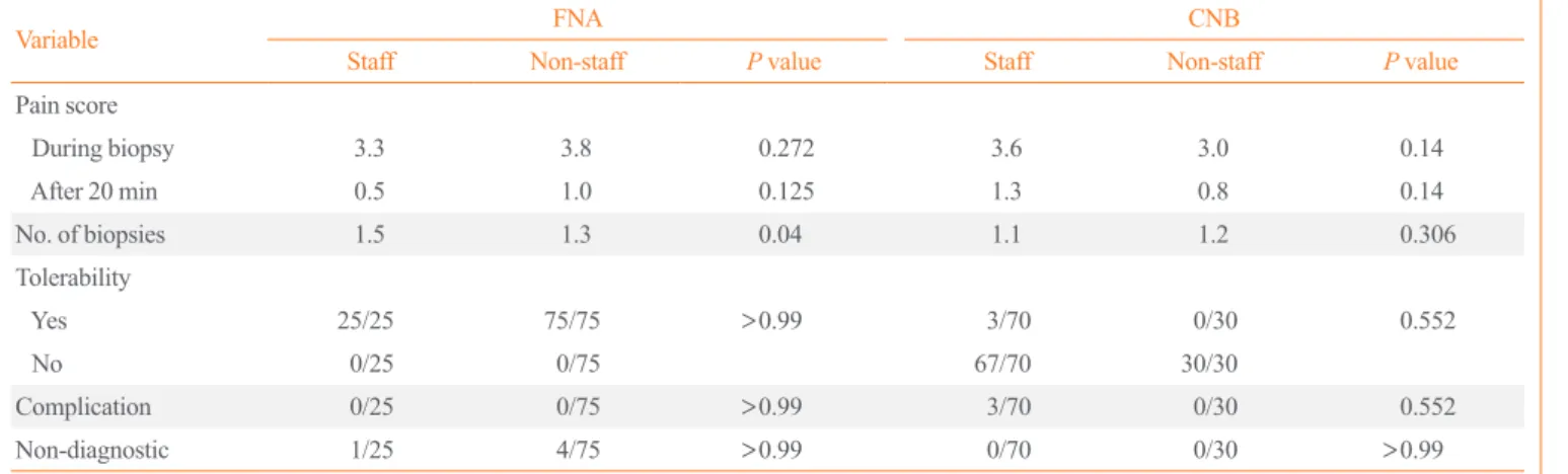
We performed subgroup analysis stratified by staff and non- staff (Table 3). There were no significant differences for any pa- rameter analyzed for the two biopsy methods, including pain score, biopsy number, tolerability, complications, and non-diag- nostic pathological results. However, the number of biopsies for non-staff was less than that for staff in the FNA group (P=0.04).
Complications
There were no major complications in either the FNA or CNB
groups, and no patient required hospital admission or interven- tional treatment. In the case of the CNB group, there were three minor complications, including perithyroidal hemorrhage and parenchyma edema; however, there were no significant differ- ences with regard to this parameter between the groups (P=
0.246). In the patients with hemorrhage and parenchymal ede- ma, the symptoms were relieved following manual compression from 30 minutes to 2 hours. After compression, US evaluations showed the nearly complete disappearance of the hematoma or edema. There have been no delayed complications reported dur- ing the follow-up period in our current patient series.
DISCUSSION
We observed no significant differences in terms of pain, tolera-
Table 1. Demographic Data for the FNA and CNB Patients Characteristic FNA (n=100) CNB (n=100) P value
Sex, male/female 17/83 13/87 0.553
Age, yr 51.3±11.3 (28–78) 49.7±10.9 (26–77) 0.318 Nodule size, cm 1.2±0.7 (0.3–4.1) 1.7±1.2 (0.3–5.1) <0.001 No. of nodule,
size <1 cm 46 40 0.475
Values are expressed as mean±SD (range).
FNA, fine needle aspiration; CNB, core needle biopsy.
Table 2. Comparison of the Indicated Parameters between the FNA and CNB Groups
Variable FNA (n=100) CNB (n=100) P value Pain score
During biopsy 3.7 (0–10) 3.6 (0–8) 0.454 After 20 min 0.9 (0–8) 1.1 (0–8) 0.296 No. of biopsies 1.4 (1–3) 1.2 (1–3) 0.002
Tolerability 0.246
Yes 100 97
No 0 0
Complication 0 3 0.246
Non-diagnostic 5 0 0.059
Values are expressed as median (range).
FNA, fine needle aspiration; CNB, core needle biopsy.
Table 3. Comparison of Staff vs. Non-Staff Subjects
Variable FNA CNB
Staff Non-staff P value Staff Non-staff P value
Pain score
During biopsy 3.3 3.8 0.272 3.6 3.0 0.14
After 20 min 0.5 1.0 0.125 1.3 0.8 0.14
No. of biopsies 1.5 1.3 0.04 1.1 1.2 0.306
Tolerability
Yes 25/25 75/75 >0.99 3/70 0/30 0.552
No 0/25 0/75 67/70 30/30
Complication 0/25 0/75 >0.99 3/70 0/30 0.552
Non-diagnostic 1/25 4/75 >0.99 0/70 0/30 >0.99
FNA, fine needle aspiration; CNB, core needle biopsy.
bility, or complications between the FNA and CNB procedures.
The rate of non-diagnostic pathologic result was lower in CNB, even though CNB achieved a fewer number of biopsies than FNA. In our subgroup analysis (staff vs. non-staff), we detected no significant difference in any parameter in terms of efficacy or safety. Our present findings thus suggest that FNA and CNB are equivalent in terms of pain, tolerability, and possible com- plications.
Two previous studies have also compared the tolerability of FNA versus CNB [15,16] and suggested that the two procedures are similar in terms of tolerability and pain. Nasrollah et al. [16]
found that the occurrence of pain during the first few minutes
following CNB was significantly higher than FNA, although there was no significant difference in pain detected at later time points for either procedure. The patients in that study were asked to evaluate the degree of tolerability for both procedures, and no significant difference was reported. Stangierski et al. [15]
reported that CNB was slightly more painful than FNA but re- mained tolerable for most patients. In our present study, we found no differences in either pain or tolerability for either pro- cedure during and at 20 minutes after the biopsy. We further found that CNB required fewer numbers of biopsies than FNA.
Considering the different experience level of the staff versus non-staff who performed these procedures, we also performed
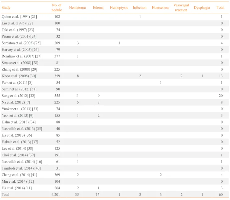
Table 4. Previously Reported Complications for Ultrasound-Guided Core Needle Biopsy
Study No. of
nodule Hematoma Edema Hemoptysis Infection Hoarseness Vasovagalreaction Dysphagia Total
Quinn et al. (1994) [21] 102 1 1
Liu et al. (1995) [22] 100 0
Taki et al. (1997) [23] 74 0
Pisani et al. (2001) [24] 32 0
Screaton et al. (2003) [25] 209 3 1 4
Harvey et al. (2005) [26] 79 0
Renshaw et al. (2007) [27] 377 1 1
Strauss et al. (2008) [28] 81 0
Zhang et al. (2008) [29] 225 0
Khoo et al. (2008) [30] 359 8 2 2 1 13
Park et al. (2011) [8] 54 1 1
Samir et al. (2012) [31] 90 0
Sung et al. (2012) [32] 555 11 9 20
Na et al. (2012) [7] 225 5 3 8
Yunker et al. (2013) [33] 74 0
Yeon et al. (2013) [9] 155 1 2 3
Hahn et al. (2013) [34] 88 0
Nasrollah et al. (2013) [35] 40 0
Ha et al. (2013) [36] 85 0
Hakala et al. (2013) [37] 52 0
Lee et al. (2014) [38] 125 0
Choi et al. (2014) [39] 191 1 1
Nasrollah et al. (2014) [16] 61 1 1
Trimboli et al. (2014) [40] 31 0
Zhang et al. (2014) [41] 369 2 2 4
Min et al. (2014) [12] 104 0
Ha et al. (2014) [11] 264 2 1 3
Total 4,201 35 15 1 3 3 2 1 60
subgroup analysis, but again found no significant differences in terms of pain, tolerability, or complications for both procedures.
In recent studies, CNB using 18 to 22 gauges cutting needles with US guidance has allowed operators to obtain thyroid tissue samples without major complications. From 1994 to 2014, many CNB studies have reported associated complications (Ta- ble 4) [7-9,11,12,16,21-41]. The total number of nodules report- ed in that period was 4,201, for which there were 60 recorded complications (1.43%) including hematoma, parenchyma ede- ma, hemoptysis, infection, hoarseness, vasovagal reaction, and dysphagia. Hematoma was the most common complication, which was reported in 35/4,201 cases (0.83%). There were three patients described with minor soft tissue infections after CNB that were successfully managed by orally administered antibiot- ics. In our present study, only three of the cases in the CNB se- ries (3%) experienced minor hematoma after the procedure and there was no significant difference in the complication rate be- tween the FNA and CNB groups. All of the reported symptoms among our current study subjects were mild and recovery oc- curred in all cases with simple compression within 2 hours. Fur- thermore, additional treatments or hospitalization were unneces- sary in these cases. Although CNB showed no major complica- tions and similar pain or discomfort when compared with FNA in our study, it remains important for investigators to understand the anatomy of the thyroid and perithyroidal areas to minimize adverse events after either procedure [19].
In terms of the advantages of CNB, it provides a larger tissue sample and can facilitate a more precise histological diagnosis.
At the microscopic level, the core of the CNB nodule sample of- fers the possibility of evaluating the general architecture of the lesion, alterations of the follicular structures, and the integrity of the capsule of the nodule along with its relationship with adja- cent tissues. Specifically, CNB core samples allow for the dif- ferentiation of cancer in up to 98% of thyroid nodules that showed previously indeterminate FNA results [8]. Additionally, Na et al. [7] reported that CNB is more useful for reducing the number of inconclusive diagnoses than FNA, and that this meth- od will serve a useful complementary diagnostic role for the op- timal management of thyroid nodules with previous non-diag- nostic findings or atypia from FNA readings. The devices and techniques associated CNB have been developed over the past 20 years. Unlike the old technique of palpation-guided large needle biopsy, US-guided CNB, using a modern spring-activat- ed biopsy needle, has been reported to be a safe and well tolerat- ed procedure [16,21,22]. Furthermore, the guidelines of the Na- tional Cancer Institute [14] also suggest that CNB is safe, well
tolerated, and associated with a low incidence of complications.
Our study had several limitations of note. First, it was retro- spective in design, which could have introduced an inherent bias regarding patient selection. However, all patients were con- secutively enrolled. Second, multiple operators with different levels of experience were involved. However, the inclusion of multiple operators reflects the real practice in our hospital and we performed subgroup analysis between staff and non-staff.
Third, we used relatively small sample size to assess the low in- cidence of complication of FNA and CNB. Although CNB has been known as a safe procedure [42-44], a further prospective investigation with a larger sample size is necessary to verify the results of our study. Finally, we performed local anesthesia with 2% lidocaine when performing the CNB procedure, but not with FNA procedure. It can be influence the pain score of the patients who underwent CNB procedure. However, 70% of pa- tients, who underwent CNB by staff, did not undergo local an- esthesia. Thus majority of patients who underwent CNB actual- ly did not have received local anesthesia.
In conclusion, FNA and CNB show no significant differences in terms of pain, tolerability, or complications for diagnosing thyroid nodules.
CONFLICTS OF INTEREST
No potential conflict of interest relevant to this article was re- ported.
AUTHOR CONTRIBUTIONS
Conception or design: J.H.B., E.J.J., Y.J.C., J.H.L., J.K.K., S.R.C. Acquisition, analysis, or interpretation of data: J.H.B., E.J.J., S.R.C. Drafting the work or revising: J.H.B., E.J.J., S.R.C. Final approval of the manuscript: J.H.B., E.J.J., Y.J.C., J.H.L., J.K.K., S.R.C. Statistical analysis: J.K.K.
ORCID
Sae Rom Chung http://orcid.org/0000-0003-4219-7166 Jung Hwan Baek http://orcid.org/0000-0003-0480-4754 Young Jun Choi http://orcid.org/0000-0001-7098-5042 Jeong Hyun Lee http://orcid.org/0000-0002-0021-4477
REFERENCES
1. Lee YH, Baek JH, Jung SL, Kwak JY, Kim JH, Shin JH, et
al. Ultrasound-guided fine needle aspiration of thyroid nod- ules: a consensus statement by the Korean Society of Thy- roid Radiology. Korean J Radiol 2015;16:391-401.
2. Alexander EK, Heering JP, Benson CB, Frates MC, Doubi- let PM, Cibas ES, et al. Assessment of nondiagnostic ultra- sound-guided fine needle aspirations of thyroid nodules. J Clin Endocrinol Metab 2002;87:4924-7.
3. Orija IB, Pineyro M, Biscotti C, Reddy SS, Hamrahian AH.
Value of repeating a nondiagnostic thyroid fine-needle aspi- ration biopsy. Endocr Pract 2007;13:735-42.
4. Yang J, Schnadig V, Logrono R, Wasserman PG. Fine-nee- dle aspiration of thyroid nodules: a study of 4703 patients with histologic and clinical correlations. Cancer 2007;111:
306-15.
5. Nayar R, Ivanovic M. The indeterminate thyroid fine-needle aspiration: experience from an academic center using termi- nology similar to that proposed in the 2007 National Cancer Institute Thyroid Fine Needle Aspiration State of the Sci- ence Conference. Cancer 2009;117:195-202.
6. Yassa L, Cibas ES, Benson CB, Frates MC, Doubilet PM, Gawande AA, et al. Long-term assessment of a multidisci- plinary approach to thyroid nodule diagnostic evaluation.
Cancer 2007;111:508-16.
7. Na DG, Kim JH, Sung JY, Baek JH, Jung KC, Lee H, et al.
Core-needle biopsy is more useful than repeat fine-needle aspiration in thyroid nodules read as nondiagnostic or atypia of undetermined significance by the Bethesda system for re- porting thyroid cytopathology. Thyroid 2012;22:468-75.
8. Park KT, Ahn SH, Mo JH, Park YJ, Park DJ, Choi SI, et al.
Role of core needle biopsy and ultrasonographic finding in management of indeterminate thyroid nodules. Head Neck 2011;33:160-5.
9. Yeon JS, Baek JH, Lim HK, Ha EJ, Kim JK, Song DE, et al.
Thyroid nodules with initially nondiagnostic cytologic re- sults: the role of core-needle biopsy. Radiology 2013;268:
274-80.
10. Ha EJ, Baek JH, Lee JH, Kim JK, Song DE, Kim WB, et al.
Core needle biopsy could reduce diagnostic surgery in pa- tients with anaplastic thyroid cancer or thyroid lymphoma.
Eur Radiol 2016;26:1031-6.
11. Ha EJ, Baek JH, Lee JH, Kim JK, Kim JK, Lim HK, et al.
Core needle biopsy can minimise the non-diagnostic results and need for diagnostic surgery in patients with calcified thyroid nodules. Eur Radiol 2014;24:1403-9.
12. Min HS, Kim JH, Ryoo I, Jung SL, Jung CK. The role of core needle biopsy in the preoperative diagnosis of follicular
neoplasm of the thyroid. APMIS 2014;122:993-1000.
13. Yoon RG, Baek JH, Lee JH, Choi YJ, Hong MJ, Song DE, et al. Diagnosis of thyroid follicular neoplasm: fine-needle aspiration versus core-needle biopsy. Thyroid 2014;24:1612- 7.
14. Baloch ZW, Cibas ES, Clark DP, Layfield LJ, Ljung BM, Pitman MB, et al. The National Cancer Institute thyroid fine needle aspiration state of the science conference: a summa- tion. Cytojournal 2008;5:6.
15. Stangierski A, Wolinski K, Martin K, Leitgeber O, Ruchala M. Core needle biopsy of thyroid nodules: evaluation of di- agnostic utility and pain experience. Neuro Endocrinol Lett 2013;34:798-801.
16. Nasrollah N, Trimboli P, Rossi F, Amendola S, Guidobaldi L, Ventura C, et al. Patient’s comfort with and tolerability of thyroid core needle biopsy. Endocrine 2014;45:79-83.
17. Choi YJ, Baek JH, Hong MJ, Lee JH. Inter-observer varia- tion in ultrasound measurement of the volume and diameter of thyroid nodules. Korean J Radiol 2015;16:560-5.
18. Shin JH, Baek JH, Chung J, Ha EJ, Kim JH, Lee YH, et al.
Ultrasonography diagnosis and imaging-based management of thyroid nodules: revised Korean Society of Thyroid Radi- ology consensus statement and recommendations. Korean J Radiol 2016;17:370-95.
19. Ha EJ, Baek JH, Lee JH. Ultrasonography-based thyroidal and perithyroidal anatomy and its clinical significance. Ko- rean J Radiol 2015;16:749-66.
20. Moon WJ, Baek JH, Choi JW, Kim YJ, Ha EJ, Lim HK, et al. The value of gross visual assessment of specimen ade- quacy for liquid-based cytology during ultrasound-guided, fine-needle aspiration of thyroid nodules. Endocr Pract 2015;21:1219-26.
21. Quinn SF, Nelson HA, Demlow TA. Thyroid biopsies: fine- needle aspiration biopsy versus spring-activated core biopsy needle in 102 patients. J Vasc Interv Radiol 1994;5:619-23.
22. Liu Q, Castelli M, Gattuso P, Prinz RA. Simultaneous fine- needle aspiration and core-needle biopsy of thyroid nodules.
Am Surg 1995;61:628-32.
23. Taki S, Kakuda K, Kakuma K, Annen Y, Katada S, Ya- mashita R, et al. Thyroid nodules: evaluation with US-guid- ed core biopsy with an automated biopsy gun. Radiology 1997;202:874-7.
24. Pisani T, Bononi M, Nagar C, Angelini M, Bezzi M, Vecchi- one A. Fine needle aspiration and core needle biopsy tech- niques in the diagnosis of nodular thyroid pathologies. Anti- cancer Res 2000;20(5C):3843-7.
25. Screaton NJ, Berman LH, Grant JW. US-guided core-needle biopsy of the thyroid gland. Radiology 2003;226:827-32.
26. Harvey JN, Parker D, De P, Shrimali RK, Otter M. Sono- graphically guided core biopsy in the assessment of thyroid nodules. J Clin Ultrasound 2005;33:57-62.
27. Renshaw AA, Pinnar N. Comparison of thyroid fine-needle aspiration and core needle biopsy. Am J Clin Pathol 2007;
128:370-4.
28. Strauss EB, Iovino A, Upender S. Simultaneous fine-needle aspiration and core biopsy of thyroid nodules and other su- perficial head and neck masses using sonographic guidance.
AJR Am J Roentgenol 2008;190:1697-9.
29. Zhang S, Ivanovic M, Nemcek AA Jr, Defrias DV, Lucas E, Nayar R. Thin core needle biopsy crush preparations in con- junction with fine-needle aspiration for the evaluation of thyroid nodules: a complementary approach. Cancer 2008;
114:512-8.
30. Khoo TK, Baker CH, Hallanger-Johnson J, Tom AM, Grant CS, Reading CC, et al. Comparison of ultrasound-guided fine-needle aspiration biopsy with core-needle biopsy in the evaluation of thyroid nodules. Endocr Pract 2008;14:426- 31.
31. Samir AE, Vij A, Seale MK, Desai G, Halpern E, Faquin WC, et al. Ultrasound-guided percutaneous thyroid nodule core biopsy: clinical utility in patients with prior nondiag- nostic fine-needle aspirate. Thyroid 2012;22:461-7.
32. Sung JY, Na DG, Kim KS, Yoo H, Lee H, Kim JH, et al. Di- agnostic accuracy of fine-needle aspiration versus core-nee- dle biopsy for the diagnosis of thyroid malignancy in a clini- cal cohort. Eur Radiol 2012;22:1564-72.
33. Yunker WK, Hassan SF, Ferrell LB, Hicks MJ, Giannoni CM, Wesson DE, et al. Needle core biopsy in the diagnosis of pediatric thyroid neoplasms: a single institution retro- spective review. Pediatr Surg Int 2013;29:437-43.
34. Hahn SY, Shin JH, Han BK, Ko EY, Ko ES. Ultrasonogra- phy-guided core needle biopsy for the thyroid nodule: does the procedure hold any benefit for the diagnosis when fine- needle aspiration cytology analysis shows inconclusive re- sults? Br J Radiol 2013;86:20130007.
35. Nasrollah N, Trimboli P, Guidobaldi L, Cicciarella Modica
DD, Ventura C, Ramacciato G, et al. Thin core biopsy should help to discriminate thyroid nodules cytologically classified as indeterminate. A new sampling technique. Endocrine 2013;43:659-65.
36. Ha EJ, Baek JH, Lee JH, Song DE, Kim JK, Shong YK, et al. Sonographically suspicious thyroid nodules with initially benign cytologic results: the role of a core needle biopsy.
Thyroid 2013;23:703-8.
37. Hakala T, Kholova I, Sand J, Saaristo R, Kellokumpu- Lehtinen P. A core needle biopsy provides more malignan- cy-specific results than fine-needle aspiration biopsy in thy- roid nodules suspicious for malignancy. J Clin Pathol 2013;
66:1046-50.
38. Lee SH, Kim MH, Bae JS, Lim DJ, Jung SL, Jung CK.
Clinical outcomes in patients with non-diagnostic thyroid fine needle aspiration cytology: usefulness of the thyroid core needle biopsy. Ann Surg Oncol 2014;21:1870-7.
39. Choi YJ, Baek JH, Ha EJ, Lim HK, Lee JH, Kim JK, et al.
Differences in risk of malignancy and management recom- mendations in subcategories of thyroid nodules with atypia of undetermined significance or follicular lesion of undeter- mined significance: the role of ultrasound-guided core-nee- dle biopsy. Thyroid 2014;24:494-501.
40. Trimboli P, Nasrollah N, Guidobaldi L, Taccogna S, Cicci- arella Modica DD, Amendola S, et al. The use of core needle biopsy as first-line in diagnosis of thyroid nodules reduces false negative and inconclusive data reported by fine-needle aspiration. World J Surg Oncol 2014;12:61.
41. Zhang M, Zhang Y, Fu S, Lv F, Tang J. Thyroid nodules with suspicious ultrasound findings: the role of ultrasound- guided core needle biopsy. Clin Imaging 2014;38:434-8.
42. Jung CK, Baek JH. Recent advances in core needle biopsy for thyroid nodules. Endocrinol Metab (Seoul) 2017;32:407- 12.
43. Na DG, Baek JH, Jung SL, Kim JH, Sung JY, Kim KS, et al.
Core needle biopsy of the thyroid: 2016 consensus state- ment and recommendations from Korean Society of Thyroid Radiology. Korean J Radiol 2017;18:217-37.
44. Baek JH. Current status of core needle biopsy of the thyroid.
Ultrasonography 2017;36:83-5.