Anaplastic Thyroid Cancer: Ultrasonographic Findings and the
Role of Ultrasonography-Guided Fine Needle Aspiration Biopsy
Hee Jung Suh,
1,2Hee Jung Moon,
1Jin Young Kwak,
1Ji Soo Choi,
1,3and Eun-Kyung Kim
11Department of Radiology, Research Institute of Radiological Science, Yonsei University College of Medicine, Seoul; 2Department of Cancer Prevention & Detection Center; 3Department of Radiology, National Cancer Center, Ilsan, Korea.
Received: November 28, 2012 Revised: February 5, 2013 Accepted: February 5, 2013
Corresponding author: Dr. Eun-Kyung Kim, Department of Radiology,
Research Institute of Radiological Science, Yonsei University College of Medicine, 50 Yonsei-ro, Seodaemun-gu, Seoul 120-752, Korea.
Tel: 82-2-2228-7400, Fax: 82-2-393-3035 E-mail: [email protected]
∙ The authors have no financial conflicts of interest.
© Copyright:
Yonsei University College of Medicine 2013
This is an Open Access article distributed under the terms of the Creative Commons Attribution Non-Commercial License (http://creativecommons.org/ licenses/by-nc/3.0) which permits unrestricted non-commercial use, distribution, and reproduction in any medium, provided the original work is properly cited.
Purpose: To investigate the ultrasonographic (US) features of anaplastic thyroid cancer (ATC) and the diagnostic performance of US-guided fine needle aspiration biopsy (FNAB) therein. Materials and Methods: Eighteen cases of ATC diag-nosed between January 2001 and May 2011 were included. FNAB was performed in all cases. Initial FNAB results were divided into three groups: 1) the cytological ATC group, cytological diagnosis of ATC; 2) the underestimated group, cytological diagnoses of malignancy other than ATC; and 3) the false negative group, cytologi-cal diagnoses of atypicytologi-cal, benign and non-diagnostic lesions. We retrospectively re-viewed US findings and compared treatment modalities between each group. Re-sults: Among the 18 patients, there were nine in the initially cytological ATC group, four in the underestimated group and five in the false negative group. The most common US features of ATC were a solid (64.7%) and irregular shaped (88.2%) mass with lymph node involvement (76.4%). However, except for lymph node in-volvement (p=0.003), US findings for each group were not statistically different. The initial cytological diagnostic accuracy of ATC was 50% (9/18). Surgery was performed less in the ATC group (11%) and the false negative group (20%) than the underestimated group (75%). Conclusion: The US features of ATC were not espe-cially different from other types of aggressive thyroid cancer. A correct diagnosis of ATC by initial US-FNAB was made in 50% of the patients, which is significant in that therapeutic surgery can be undertaken in lower numbers if correctly diagnosed.
Key Words: Anaplastic thyroid cancer, fine needle aspiration biopsy, ultrasonog-raphy
INTRODUCTION
Anaplastic thyroid cancer (ATC) is the most aggressive form of thyroid cancer, with median survival on the order of 3 to 5 months following diagnosis.1-4 Even though
less than 1-3% of all thyroid cancers are ATC, it contributes to 14-50% of the annu-al mortannu-ality associated with thyroid cancer.5-7 The treatment for ATC has not been
standardized because therapy has not been conclusively proven to be effective in prolonging survival. Until now, surgery has played an important role in survival for patients with intrathyroidal tumors.8-10 However, many patients have also presented
of six radiologists with 1 to 14 years of experience in thy-roid imaging. US-FNAB was performed with a 23-gauge needle attached to a 20-mL disposable plastic syringe and aspirator or a 2-mL syringe disposable plastic syringe. As-pirated materials were expelled on glass slides and immedi-ately fixed in 95% alcohol for Papanicolaou staining. The remaining material in the syringe was rinsed in normal sa-line for cell block processing. US-guided core biopsy was performed using a freehand technique, and each procedure was performed with a 18-gauge dual-action semiautomatic core biopsy needle (Stericut with coaxial; TSK Laboratory, Tochigi, Japan). A cytopathologist was not on site during the aspiration procedure. Additional special staining was performed according to the requirements set by the cytolo-gist. From 2001 to December 2009, cytology reports were divided into the following categories: 1) nondiagnostic 2) benign 3) indeterminate 4) suspicious for malignancy and 5) malignant. Afterwards, the Bethesda classification was used to classify cytology results at our institution.20
Data analysis
Each US image was reviewed retrospectively by two radi-ologists in consensus. US images were reviewed according to size, composition, echogenicity, margin, presence of cal-cification, shape and presence of lymph node involvement. Lymph node involvement was considered pathologic if any one of the following suspicious features were present: round shape, increased echogenicity, cystic change, or presence of microcalcifications.21
To evaluate cytological diagnostic accuracy for ATC, FNAB results were divided into three groups: 1) the cyto-logical ATC group, cytocyto-logical diagnosis of ATC; 2) the un-derestimated group, cytological diagnoses of malignancy rather than ATC, such as suspicious for papillary ma, papillary carcinoma, and poorly differentiated carcino-ma; and 3) the false negative group, cytological diagnoses of atypical, benign and non-diagnostic lesions. We included atypia in the false negative group, because the Bethesda sys-tem recommends follow-up FNAB for patients with atypia instead of surgery.20 We compared US findings between
these three groups.
For statistical analysis, the SPSS statistical package, ver-sion 11.0, for Windows (SPSS Inc., Chicago, IL, USA) was used. Pearson chi square tests and Fisher’s exact tests were used to study categorical variables and the Kruskal-Wallis test was used to compare continuous variables. p-values < 0.005 were considered statistically significant.
with inoperable disease; macroscopic complete resection is only possible in up to one-third of patients at presenta-tion.11-14 The best survival results have been observed in
in-operable patients who received primary chemotherapy and radiation therapy rather than primary surgical resection.10,15
Therefore, an accurate preoperative diagnosis of ATC may help avoid unnecessary surgery and allow treatment to pro-ceed directly to medical therapy.4,16
A diagnosis of ATC is usually suspected on clinical ex-amination and ultrasonography (US), which can be con-firmed by fine needle aspiration biopsy (FNAB), core biop-sy or surgery. US and the following FNAB are the first di-agnostic modalities in the evaluation of a palpable thyroid mass.17 Several studies have focused on the treatment
mo-dalities or outcomes of ATC,1,4,9,11,14,15,18,19 but there have
been few reports about US features and the role of preoper-ative US-guided biopsy in ATC.
The purpose of this study was to investigate the US fea-tures of ATC and the diagnostic performance of US-guided FNAB.
MATERIALS AND METHODS
This retrospective study was conducted with institutional re-view board approval and patient informed consent was waived.
Study population
From January 2001 to May 2011, a total 18 patients were diagnosed as having ATC by US-FNAB, core biopsy and surgery at our institution and were included in this study.
Initially, all patients had undergone US-FNAB of thyroid masses. According to the initial FNAB results, patients un-derwent follow up FNAB (n=2), core biopsy (n=3), diag-nostic surgery (n=1) and other treatment options including medical treatment or therapeutic surgery (n=3). Among the 18 patients, 5 were men and 13 were women, with ages ranging from 54-81 years (mean age 70.5 years). All pa-tients complained of having a palpable neck mass.
Imaging and diagnostic processes
US was performed before FNAB in all 18 patients with a 7-15 MHz linear array transducer (HDI 3000 or 5000; Phil-ips Medical Systems, Bothell, WA, USA) or 5-12 MHz lin-ear array transducer (Iu22; Philips Medical Systems). Real-time US and subsequent US-FNAB was performed by one
and it increased to 72% (13/18) if the underestimated group was regarded as part of the correct diagnostic group. Among the 4 patients in the underestimated group, one case of sus-picious papillary carcinoma was diagnosed as ATC by fol-low-up core biopsy, and three cases of poorly differentiated thyroid carcinoma (PDTC) that underwent surgery were fi-nally diagnosed as ATC. Among the 5 patients in the false negative group, one atypia case and one non-diagnostic case were diagnosed as ATC by follow-up core biopsy; two be-nign cases were diagnosed as ATC by repeat FNAB and one benign case was determined to be ATC after surgery (Fig. 1). Accordingly, a preoperative diagnosis of ATC was made in 14 out of 18 cases (77.8%). The mean number of days from first visit to confirmative diagnosis of ATC was 10 days for the ATC group, 33.5 days for the underestimate group and 104.5 days for the false negative group.
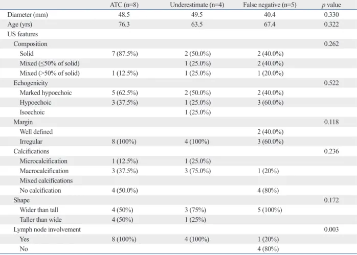
US images were available in 17 out of 18 patients. US features of the 17 ATC patients according to the three groups above are summarized in Table 1. Mean age and mass size were not statistically different between each group. The com-mon US features of ATCs were solid (11/17, 64.7%), marked
RESULTS
Among the 18 patients, there were nine in the initially cyto-logical ATC group; four in the underestimated group, poor-ly differentiated cancer (n=3) and suspicious papillary car-cinoma (n=1); and five in the false negative group, atypia (n=1), benign (n=3), and non-diagnostic (n=1). The initial cytological diagnostic accuracy of ATC was 50% (9/18)
Fig. 1. Preoperative diagnostic methods of 18 anaplastic ATC. Underestimated
group: poorly differentiated cancer and suspicious papillary carcinoma. False negative group: atypia, benign and non-diagnostic. bx, biopsy; ATC, anaplastic thyroid cancer; FNAB, fine needle aspiration biopsy.
Table 1. Ultrasonographic Findings of Anaplastic Thyroid Cancers According to Three Groups
ATC (n=8) Underestimate (n=4) False negative (n=5) p value
Diameter (mm) 48.5 49.5 40.4 0.330 Age (yrs) 76.3 63.5 67.4 0.322 US features Composition 0.262 Solid 7 (87.5%) 2 (50.0%) 2 (40.0%) Mixed (≤50% of solid) 1 (25.0%) 2 (40.0%) Mixed (>50% of solid) 1 (12.5%) 1 (25.0%) 1 (20.0%) Echogenicity 0.522 Marked hypoechoic 5 (62.5%) 2 (50.0%) 2 (40.0%) Hypoechoic 3 (37.5%) 1 (25.0%) 3 (60.0%) Isoechoic 1 (25.0%) Margin 0.118 Well defined 2 (40.0%) Irregular 8 (100%) 4 (100%) 3 (60.0%) Calcifications 0.236 Microcalcification 1 (12.5%) 1 (25.0%) Macrocalcification 3 (37.5%) 3 (75.0%) 1 (20%) Mixed calcifications No calcification 4 (50.0%) 4 (80%) Shape 0.172
Wider than tall 4 (50%) 3 (75%) 5 (100%)
Taller than wide 4 (50%) 1 (25%)
Lymph node involvement 0.003
Yes 8 (100%) 4 (100%) 1 (20%)
No 4 (80%)
ATC, anaplastic thyroid cancer; US, ultrasonographic. Initial FNAB (n=18) Underestimated (n=4) Core bx. (n=1) ATC
diagnosed by: Surgery(n=3) (n=2)FNA Core bx.(n=2) Surgery(n=1) ATC (n=9)
False negative (n=5)
less on patients in the initially confirmed ATC (11%) and false negative groups (20%) than those in the underestimat-ed group (75%).
DISCUSSION
ATC is an extremely aggressive solid tumor that resists most therapeutic efforts and is always fatal.1-4,9,10 It contrasts with
well differentiated thyroid cancer (WDTC), which accounts for most thyroid malignancies, with an indolent course and good prognosis regardless of the type of treatment.2 The
in-cidence of ATC is decreasing, presumably due to earlier de-tection of antecedent disease.22 Several studies have revealed
that potentially curative surgery is the only discriminating variable that retains a significant association with prolonged hypoechogenicity (9/17, 52.9%) irregular margin (15/17,
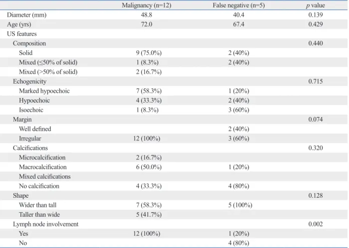
88.2%), internal calcification (9/17, 52.9%), wider than tall shape (12/17, 70.5%), and presence of cervical lymph node involvement (13/17, 76.4%). The US findings for each group were not statistically different, except for lymph node in-volvement (p=0.003). Mixed composition was more fre-quently seen in the underestimated (50%) and false nega-tive groups (60%), compared with the ATC group (12.5%), but this was not statistically different (p=0.262). We also compared US findings between those in the initially diag-nosed malignancy group (the cytological ATC and underes-timated groups) and those in the false negative group, but similarly, lymph node involvement was the only statistical-ly factor that differed between the two groups (Table 2).
Differences in treatment modalities between the three groups are summarized in Table 3. Surgery was performed
Table 2. Ultrasonographic Findings of Anaplastic Thyroid Cancers According to Two Groups
Malignancy (n=12) False negative (n=5) p value
Diameter (mm) 48.8 40.4 0.139 Age (yrs) 72.0 67.4 0.429 US features Composition 0.440 Solid 9 (75.0%) 2 (40%) Mixed (≤50% of solid) 1 (8.3%) 2 (40%) Mixed (>50% of solid) 2 (16.7%) Echogenicity 0.715 Marked hypoechoic 7 (58.3%) 1 (20%) Hypoechoic 4 (33.3%) 2 (40%) Isoechoic 1 (8.3%) 3 (60%) Margin 0.074 Well defined 2 (40%) Irregular 12 (100%) 3 (60%) Calcifications 0.320 Microcalcification 2 (16.7%) Macrocalcification 6 (50.0%) 1 (20%) Mixed calcifications No calcification 4 (33.3%) 4 (80%) Shape 0.128
Wider than tall 7 (58.3%) 5 (100%)
Taller than wide 5 (41.7%)
Lymph node involvement 0.002
Yes 12 (100%) 1 (20%)
No 4 (80%)
Table 3. Differences between Treatment Modalities Based on the Diagnosis of Initial FNAB
Treatment Modality ATC (n=9) Underestimated (n=4) False negative (n=5)
Surgery 1 (11%) 3 (75%) 1 (20%)
Non-surgical treatments 8 (89%) 1 (25%) 4 (80%)
cluded. The diagnostic performance in our study was lower than others and this may be due to a different study design and sample size. Previously, Us-Krasovec, et al.29 reported
the FNAB results of 113 ATCs for 20 years. They reported that 107 patients (107/113, 94.7%) were diagnosed as ma-lignant and 96 (96/107, 89.7%) were diagnosed as ATC through reexamination. If we had reevaluated the pathology slides retrospectively perhaps there would have been an in-crease in diagnostic accuracy. The reason for failures in ob-taining an accurate diagnosis on FNAB might have been due to sampling in areas that consisted of foci of WDTC or in areas burdened with necrosis, fibrosis, or hemorrhage.2,29
In the present study, among the five false negative cases, 60% (3/5) had both solid and cystic portions on US, which contrasted with the 12.5% (1/8) in the initially cytological ATC group. It is difficult to obtain adequate cells for diag-nosis from masses containing both solid and cystic por-tions,34,35 which could explain why false negative diagnosis
was frequent in mixed composition.29 According to Luze, et
al.,31 12% of samples of finally confirmed ATCs contained
only necrotic material. In order to obtain a representative sample, large tumor masses must be aspirated at two or three different sites. This is particularly important in differ-entiated tumors undergoing transformation to ATC, in which only small foci of ATC components may be present and both well differentiated carcinoma and ATC coexist.1,2,29
Multimodality treatment with combination surgery, radi-ation and chemotherapy has been reported to improve out-comes for ATC.2,3,5,18 If tumors can be managed by
aggres-sive surgery with preservation of organ function, adjuvant chemotherapy and radiotherapy may be the best option for now.36 The majority of patients with ATCs die from
aggres-sive local regional disease, primarily upper airway respirato-ry failure. For this reason, aggressive local therapy is indicat-ed in all patients who can tolerate it. Yet complete resection of ATC is rarely performed, which gives the best chance of long term control and disease free survival. Accordingly, more than two-thirds of advanced ATC patients are not ex-pected to show survival benefits from initial surgery.37 In
our study, 92.8% (13/14) of pre-op diagnosed ATC cases did not undergo surgical treatment. Among the 9 patients in the cytological ATC group, only 1 patient underwent surgery, whereas 3 out of 4 patients in the underestimated group un-derwent surgery even though similar US findings were ob-served. If the patients had been diagnosed with ATCs pre-operatively, surgery would not have been performed.
Among the 9 patients in the underestimate and false neg-survival.8,9,18 But, it was seen in previous studies that
sys-temic metastases were present in 46% of ATC patients at presentation, and 68% ATC of patients had metastases di-agnosed during the course of their illness.19 At this stage,
complete resection is not meaningful, so patients with inop-erable disease at diagnosis are considered for combined chemotherapy and radiotherapy.10 Thus it is important to
accurately diagnose ATC before surgery and treatment.16
Grossly, ATC is a nonencapsulated, tan-white, fleshy tu-mor with a direct extension into the surrounding soft tissue of the neck and has regions of necrosis and hemorrhage.23
The imaging features of ATC reflect these gross findings. Large, solid and ill defined masses accompanied by necro-sis, nodular calcification and cervical lymph node involve-ment are the common image features of ATC.24 In our study,
common US features of ATCs included solid (11/17, 64.7%), marked hypoechogenicity (9/17, 52.9%), irregular margin (15/17, 88.2%), internal calcification (9/17, 52.9%), wider than tall shape (12/17, 70.5%) and presence of cervical lymph node involvement (13/17, 76.4%). The image features of ATCs are not that different from other aggressive forms of thyroid cancer, but the prognosis of ATC is significantly worse than PDTC. The 5-year disease free survival and 5-year cause-specific survival of ATC are both 0%, but those of PDTC are 51% and 70%.25 PDTC patients survive
longer, with medial survival being 3.2 years for PDTC and 3.1 months for ATC. In a previous study, one third of ATC patients died of local disease, whereas all patients who died of PDTC had distant metastases.26 ATC more frequently
in-vades the surrounding structures and more often involves regional lymph nodes; moreover, half the patients with ATC present with distant metastases.23
Considering the limitations of US evaluation in studying larger masses, CT is much more useful in defining the local extent of the disease and detecting lymph node metastases. MRI has further added value over CT scan for detecting in-vasion of vascular, airway and bony structures.23,27 CT or
MR information on the extent and location of tumor necro-sis and site of calcification in the tumor is expected to lower false negative diagnoses by appreciating indication sites for biopsy.28 However, when ATC is clinically suspected,
US-guided cytologic confirmation is initially performed and CT or MRI is performed after diagnosis.
The cytological diagnosis accuracy of ATC reported by previous reports was 78.7% to 90%.29-33 In the present study,
the accuracy of initial US-FNAB was 50% (9/18) and creased to 72% (12/18) if the underestimated group was
in-Cancer Data Base report on 53,856 cases of thyroid carcinoma treated in the U.S., 1985-1995 [see commetns]. Cancer 1998;83: 2638-48.
7. Kebebew E, Greenspan FS, Clark OH, Woeber KA, McMillan A. Anaplastic thyroid carcinoma. Treatment outcome and prognostic factors. Cancer 2005;103:1330-5.
8. Sugino K, Ito K, Mimura T, Nagahama M, Fukunari N, Kubo A, et al. The important role of operations in the management of ana-plastic thyroid carcinoma. Surgery 2002;131:245-8.
9. Akaishi J, Sugino K, Kitagawa W, Nagahama M, Kameyama K, Shimizu K, et al. Prognostic factors and treatment outcomes of 100 cases of anaplastic thyroid carcinoma. Thyroid 2011;21:1183-9. 10. Pudney D, Lau H, Ruether JD, Falck V. Clinical experience of the
multimodality management of anaplastic thyroid cancer and litera-ture review. Thyroid 2007;17:1243-50.
11. Heron DE, Karimpour S, Grigsby PW. Anaplastic thyroid carci-noma: comparison of conventional radiotherapy and hyperfrac-tionation chemoradiotherapy in two groups. Am J Clin Oncol 2002;25:442-6.
12. Haigh PI. Anaplastic thyroid carcinoma. Curr Treat Options Oncol 2000;1:353-7.
13. Mitchell G, Huddart R, Harmer C. Phase II evaluation of high dose accelerated radiotherapy for anaplastic thyroid carcinoma. Radiother Oncol 1999;50:33-8.
14. McIver B, Hay ID, Giuffrida DF, Dvorak CE, Grant CS, Thomp-son GB, et al. Anaplastic thyroid carcinoma: a 50-year experience at a single institution. Surgery 2001;130:1028-34.
15. Besic N, Auersperg M, Us-Krasovec M, Golouh R, Frkovic-Grazio S, Vodnik A. Effect of primary treatment on survival in an-aplastic thyroid carcinoma. Eur J Surg Oncol 2001;27:260-4. 16. Greenblatt DY, Woltman T, Harter J, Starling J, Mack E, Chen H.
Fine-needle aspiration optimizes surgical management in patients with thyroid cancer. Ann Surg Oncol 2006;13:859-63.
17. American Thyroid Association (ATA) Guidelines Taskforce on Thyroid Nodules and Differentiated Thyroid Cancer, Cooper DS, Doherty GM, Haugen BR, Kloos RT, Lee SL, et al. Revised American Thyroid Association management guidelines for pa-tients with thyroid nodules and differentiated thyroid cancer. Thy-roid 2009;19:1167-214.
18. Haigh PI, Ituarte PH, Wu HS, Treseler PA, Posner MD, Quivey JM, et al. Completely resected anaplastic thyroid carcinoma com-bined with adjuvant chemotherapy and irradiation is associated with prolonged survival. Cancer 2001;91:2335-42.
19. Lam KY, Lo CY, Chan KW, Wan KY. Insular and anaplastic car-cinoma of the thyroid: a 45-year comparative study at a single in-stitution and a review of the significance of p53 and p21. Ann Surg 2000;231:329-38.
20. Cibas ES, Ali SZ. The Bethesda System for Reporting Thyroid Cytopathology. Thyroid 2009;19:1159-65.
21. Rashid OM, Takabe K. The evolution of the role of surgery in the management of breast cancer lung metastasis. J Thorac Dis 2012; 4:420-4.
22. Boerner SL, Asa SL. Biopsy interpretation of the thyroid. 1st ed. Philadelphia (PA): Lippincott Williams & Wilkins, a Wolters Klu-wer business; 2010.
23. Pasieka JL. Anaplastic thyroid cancer. Curr Opin Oncol 2003;15: 78-83.
24. Lee JW, Yoon DY, Choi CS, Chang SK, Yun EJ, Seo YL, et al. Anaplastic thyroid carcinoma: computed tomographic differentia-tion from other thyroid masses. Acta Radiol 2008;49:321-7.
ative groups, 5 patients were diagnosed as having ATCs through repeat FNAB or core biopsy. Repeated FNAB and core biopsy including immunohistochemical staining and flow cytometry analysis2,19,22 were valuable methods when
suspicions of clinical and imaging findings remained high and initial FNAB was inadequate or benign.
There are some limitations to this study. First, this study was based on a retrospective design, and the initial cytology results were not reviewed. More over as this study had a small sample size, we did not compare the mean survival pe-riod of each group by different treatment modalities. So we did not analyze the survival rate of each group. Second, al-most all cases of ATC in our study were of large size and ad-vanced cases. Recently, the wide use of high resolution US has provided early detection of thyroid malignancy, and this might contribute to an earlier detection of ATC. Han, et al.38
reported time trends of tumor size and characteristics of ATC. They reported that the mean tumor size of ATC de-creased significantly and the frequencies of coexistent dif-ferentiated thyroid cancer (DTC) increased over time. The mean tumor size of long term survivors was significant smaller and the proportion of cases with coexistent DTC was much higher in long term survivors than short term survivors. So smaller ATC and cases with coexistent DTC were increasingly detected, and accordingly, therapeutic ap-proaches have evolved with growing expectations of long term survival for ATC patients.38
In conclusion, in the present study, the most common US features of ATC were a large, solid, irregular shaped mass with lymph node involvement. A correct diagnosis of ATC by initial US-FNAB was made in 50% of the patients, which is significant in that therapeutic surgery can be undertaken in lower numbers if correctly diagnosed.
REFERENCES
1. Are C, Shaha AR. Anaplastic thyroid carcinoma: biology, patho-genesis, prognostic factors, and treatment approaches. Ann Surg Oncol 2006;13:453-64.
2. Patel KN, Shaha AR. Poorly differentiated and anaplastic thyroid cancer. Cancer Control 2006;13:119-28.
3. Nagaiah G, Hossain A, Mooney CJ, Parmentier J, Remick SC. Anaplastic thyroid cancer: a review of epidemiology, pathogene-sis, and treatment. J Oncol 2011;2011:542358.
4. Ain KB. Anaplastic thyroid carcinoma: a therapeutic challenge. Semin Surg Oncol 1999;16:64-9.
5. Jemal A, Siegel R, Ward E, Murray T, Xu J, Thun MJ. Cancer sta-tistics, 2007. CA Cancer J Clin 2007;57:43-66.
thyroid carcinoma: review of 24 cases, with emphasis on cytodi-agnosis and leukocytosis. Taiwan Yi Xue Hui Za Zhi 1989;88: 551-6.
33. Lee MJ, Hong SW, Chung WY, Kwak JY, Kim MJ, Kim EK. Cy-tological results of ultrasound-guided fine-needle aspiration cytol-ogy for thyroid nodules: emphasis on correlation with sonographic findings. Yonsei Med J 2011;52:838-44.
34. Moon HJ, Kwak JY, Kim EK, Kim MJ. Ultrasonographic charac-teristics predictive of nondiagnostic results for fine-needle aspira-tion biopsies of thyroid nodules. Ultrasound Med Biol 2011;37: 549-55.
35. Sohn YM, Yoon JH, Moon HJ, Kim EK, Kwak JY. Mixed echoic thyroid nodules on ultrasound: approach to management. Yonsei Med J 2012;53:812-9.
36. Rinaldi P, Costantini M, Belli P, Giuliani M, Bufi E, Fubelli R, et al. Extra-mammary findings in breast MRI. Eur Radiol 2011;21: 2268-76.
37. Rausch DR. Spectrum of extra-mammary findings on breast MRI: a pictorial review. Breast J 2008;14:592-4.
38. Han JM, Bae Kim W, Kim TY, Ryu JS, Gong G, Hong SJ, et al. Time trend in tumour size and characteristics of anaplastic thyroid carcinoma. Clin Endocrinol (Oxf) 2012;77:459-64.
25. Wreesmann VB, Ghossein RA, Patel SG, Harris CP, Schnaser EA, Shaha AR, et al. Genome-wide appraisal of thyroid cancer pro-gression. Am J Pathol 2002;161:1549-56.
26. Siironen P, Hagström J, Mäenpää HO, Louhimo J, Heikkilä A, Heiskanen I, et al. Anaplastic and poorly differentiated thyroid carcinoma: therapeutic strategies and treatment outcome of 52 consecutive patients. Oncology 2010;79:400-8.
27. Green LD, Mack L, Pasieka JL. Anaplastic thyroid cancer and pri-mary thyroid lymphoma: a review of these rare thyroid malignan-cies. J Surg Oncol 2006;94:725-36.
28. Takashima S, Morimoto S, Ikezoe J, Takai S, Kobayashi T, Koya-ma H, et al. CT evaluation of anaplastic thyroid carcinoKoya-ma. AJR Am J Roentgenol 1990;154:1079-85.
29. Us-Krasovec M, Golouh R, Auersperg M, Besic N, Ruparcic-Oblak L. Anaplastic thyroid carcinoma in fine needle aspirates. Acta Cytol 1996;40:953-8.
30. Cornillot M, Cappelaere P, Granier AM, Houcke M, Triplet I. [Cy-tologic diagnosis of thyroid cancer]. Lille Med 1977;22:366-71. 31. Luze T, Tötsch M, Bangerl I, Hittmair A, Sandbichler P, Ladurner
D, et al. Fine needle aspiration cytodiagnosis of anaplastic carci-noma and malignant haemangioendothelioma of the thyroid in an endemic goitre area. Cytopathology 1990;1:305-10.