CASE REPORT
내시경초음파 유도하 세침흡인술로 진단한 폐암으로 오인된 복강 내 폐흡충증 1예
오초롱, 김미진, 이광혁
성균관대학교 의과대학 삼성서울병원 내과학교실 소화기내과
A Case of Intra-abdominal Paragonimiasis Mimicking Metastasis of Lung Cancer Diagnosed by Endoscopic Ultrasound-guided Fine Needle Aspiration
Cho Rong Oh, Mi-Jin Kim, and Kwang Hyuck Lee
Division of Gastroenterology, Department of Internal Medicine, Samsung Medical Center, Sungkyunkwan University School of Medicine, Seoul, Korea
Paragonimiasis has been continuously decreasing in Korea. However, it still occurs by ingesting raw or incompletely cooked fresh water crab or crayfish. The diagnosis of paragonimiasis is challenging because of its rarity. It may be confused with other inflammatory disease or carcinomatosis. Endoscopic ultrasound-guided fine needle aspiration (EUS-FNA) has lower risk of complications such as bleeding, perforation than percutaneous fine needle aspiration. EUS-FNA is more accurate and popular method to find mucosal or submucosal tumors and the lesions of several organs. Benign and malignant tumors, infectious diseases have been diagnosed by EUS-FNA, but there was no report describing the use of EUS-FNA for diagnosing paragonimiasis.
Herein, we present a 47-year-old male patient with paragonimiasis diagnosed by EUS-FNA. Imaging studies revealed mass lesions in the lung and peritoneal cavity, which was eventually confirmed as paragonimiasis using EUS-FNA. (Korean J Gastroenterol 2015;66:41-45)
Key Words: Paragonimiasis; Endoscopic ultrasound-guided fine needle aspiration
Received November 3, 2014. Revised December 26, 2014. Accepted January 6, 2015.
CC This is an open access article distributed under the terms of the Creative Commons Attribution Non-Commercial License (http://creativecommons.org/licenses/
by-nc/4.0) which permits unrestricted non-commercial use, distribution, and reproduction in any medium, provided the original work is properly cited.
Copyright © 2015. Korean Society of Gastroenterology.
교신저자: 이광혁, 135-710, 서울시 강남구 일원로 81, 삼성서울병원 소화기내과
Correspondence to: Kwang Hyuck Lee, Division of Gastroenterology, Department of Internal Medicine, Samsung Medical Center, 81 Irwon-ro, Gangnam-gu, Seoul 135-710, Korea. Tel: +82-2-3410-3409, Fax: +82-2-3410-6983, E-mail: [email protected]
Financial support: None. Conflict of interest: None.
서 론
폐흡충증은 폐흡충(Paragonimus westermani)에 의해 발 생하는 기생충 질환으로 과거보다 감소하였지만 민물게장 섭 취 등으로 인해 아직도 국내에서 산발적으로 발생하고 있다.
피낭유충(metacercaria)의 복잡한 생활사 때문에 폐 이외의 여러 조직에서도 발견될 수 있는데, 복강 내 폐흡충증은 흔히 폐암의 전이나 결핵 같은 질환과 혼동되기도 한다. 이는 폐흡 충증 자체가 드물어 처음 환자가 방문 시 의심하기가 쉽지 않으며 임상양상, 방사선 특징도 폐암, 결핵과 비슷한 부분이
많기 때문이다.1,2
내시경초음파 유도하 세침흡인술(endoscopic ultrasound- guided fine needle aspiration, EUS-FNA)은 상부위장관에 인접한 복강 내 병변을 진단하는 정확하고 안전하며 간편한 시술이다. EUS-FNA로 진단된 다양한 양성, 악성종양, 감염 질환이 보고된 바 있으나 현재까지 폐흡충증을 진단하여 보고 한 증례는 없었다.3,4
저자들은 객혈을 주소로 내원하여 시행한 검사에서 폐와 복강 내 종괴가 발견되어 폐암의 복강 내 전이를 의심하고 EUS-FNA로 조직검사 후에 폐흡충증으로 최종 진단된 증례
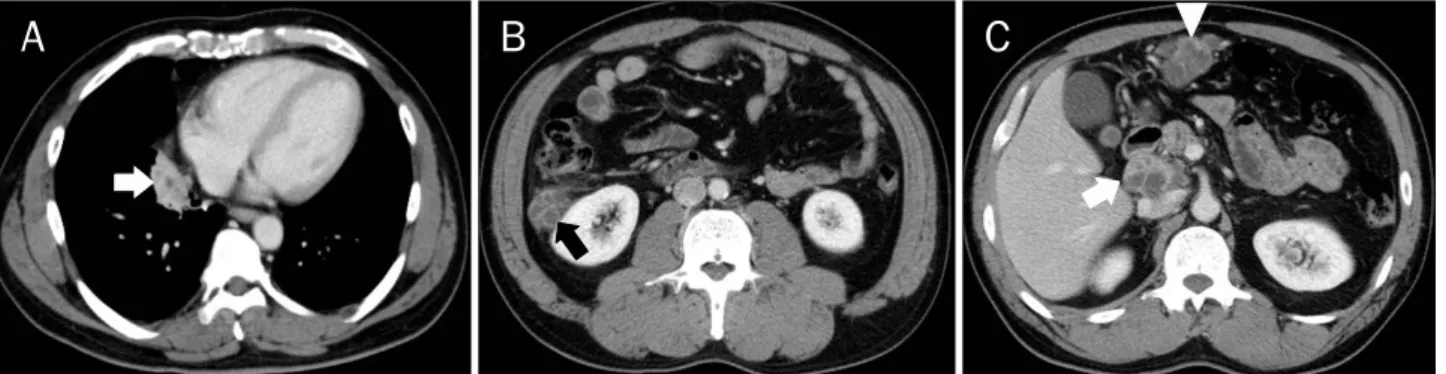
Fig. 1. CT scans of chest and abdomen at admission. (A) At medial basal segment of right lower lobe, there is a 4.0 cm sized soft tissue density mass (arrow) harboring multifocal low attenuation areas. (B, C) On upper abdomen, there are three soft tissue density masses containing multiple necrotic low attenuation areas of bunch-of-grapes shape. A 3.5 cm sized mass (black arrow) is observed at right peri-renal space, a 4.5 cm sized mass (white arrowhead) at greater curvature side of stomach lower body along gastroepipoloic vessel, and a 4.6 cm sized mass (white arrow) at portocaval space.
Fig. 2. PET scan images. (A) PET scan reveals an ovoid nodular opacity (arrow) with mild F-18 fluorodeoxyglucose (FDG) uptake in the right lower lobe of lung (maximal standardized uptake value [SUV] 3.2). (B-D) PET scan reveals three low attenuation lesions (arrows) with increased FDG uptakes in the portocaval space, gastroepiploic area and right peri-renal space (maximal SUV 5.1).
를 경험하였기에 보고하는 바이다.
증 례
47세 남자가 4개월 전부터 시작된 주 2-3회의 객혈을 주소 로 내원하였다. 환자는 13년 전 중이염으로 수술받은 과거력
이 있었고, 25갑년의 흡연자였으나 4년 전부터 금연 상태였 다. 환자의 어머니는 67세에 위암으로 사망하였다.
내원 시 생체징후는 안정적이었고, 호흡음을 포함한 이학적 검사는 정상이었다. 말초혈액 검사에서 적혈구침강속도 23 mm/hr (0-15 mm/hr)로 상승되어 있었고, 백혈구 7,050/μL (호산구 5.2%), 혈색소 14.9 g/dL, 암배아항원 0.9 ng/mL로
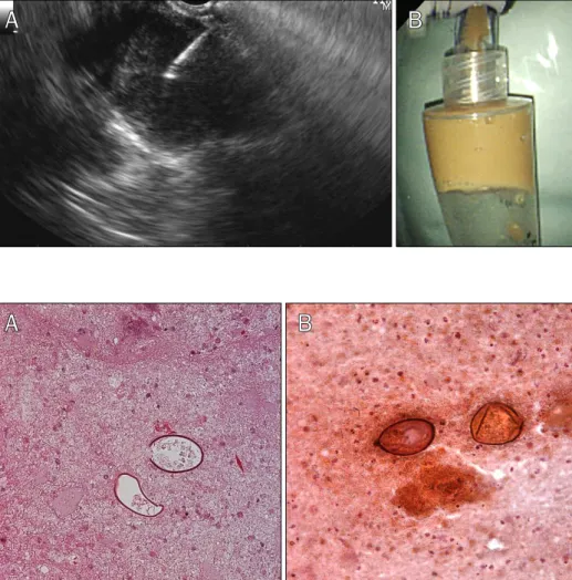
Fig. 3. Endoscopic ultrasonography- guided fine needle aspiration findings.
(A) On portocaval space, there was a heterogeneous hypo-echoic 3 cm sized cystic mass with much debris. (B) After fine needle aspiration, muddy colored pus was obtained.
Fig. 4. Eggs of Paragonimus wester- mani obtained by endoscopic ultra- sonography-guided fine needle aspi- ration from tissue (A) and pus (B). The eggs are ovoid, with a thick shell and often asymmetrical with one end slightly flattened (H&E stain, ×400).
정상 범위였다. 객담 결핵균 항산성 세균 염색 결과 음성이었다.
흉부 전산화단층촬영에서 폐 우하엽에 괴사성 저음영 부분 을 내부에 포함한 4.0 cm 크기의 연조직음영 종괴가 관찰되 었다(Fig. 1A). 복부 전산화단층촬영에서 복강 내에 세 개의 종괴가 관찰되었는데, 내부에 포도송이 모양으로 뭉쳐진 괴사 성 저음영 부분을 포함한 3.5-4.6 cm 크기의 연조직음영 종괴 가 오른쪽 신장에 인접한 부분, 위와 복벽 사이, 문맥 대정맥 부위에 있었다(Fig. 1B, C). 양전자방출 단층촬영 검사에서 폐 의 종괴는 약하게, 복강 내 종괴는 세 개 모두 F-18 fluo- rodeoxyglucose (FDG) 섭취가 증가되어 악성의 가능성을 의 심하게 했다(Fig. 2).
내시경 초음파(GF-UCT240P-AL5; Olympus, Center Valley, PA, USA)를 이용하여 십이지장 구부에서 관찰하였을 때, 문 맥 대정맥 부위에 3 cm 크기의 종괴가 있었다. 종괴 내부에 많은 부유물이 있는 불균일한 저음영의 낭성 종괴였으며 주변 혈관이나 인접 장기로의 침범 소견은 없었다(Fig. 3A). 세침 (echo-22; Cook, Winston-salem, NC, USA)을 이용하여 흡 인하자 황토색의 농이 나왔다(Fig. 3B). 채취한 조직과 농에서 타원형 모양에 두꺼운 껍질을 가진 기생충 충란이 관찰되어
폐흡충증으로 진단하였다(Fig. 4). 추가로 시행한 혈청 폐흡충 증 특이 항체 검사(enzyme-linked immunosorbent assay, ELISA)에서도 양성 소견을 보였다.
환자에게 확인해보니 전라도 섬진강 인근에 거주하면서 근 처에서 구입하거나 직접 잡아서 담근 손바닥만 한 크기의 민 물게장을 즐겨 먹었다고 하였다. 환자는 25 mg/kg의 prazi- quantel을 하루 3회, 이틀간 복용 후 증상과 영상의학 소견이 호전되어(Fig. 5), 외래 경과 관찰 중이다.
고 찰
폐흡충증은 흡충류인 폐흡충의 피낭유충을 섭취하여 발생 하는 기생충 질환이다. 피낭유충이 십이지장에서 탈낭한 후 십이지장벽을 뚫고 복강으로 이동하여 복벽 근육을 거쳐 다시 복강에서 횡경막과 흉막을 뚫고 폐조직으로 이동하게 된다.
이동 경로가 복잡하기 때문에 폐 이외에 뇌척수, 장벽, 간, 비장, 신장, 복강, 피하조직, 복벽 등 여러 장기에서 발견될 수 있다.
감염된 환자는 대부분 무증상이지만 침범된 장기에 따라 다양 한 증상이 나타난다. 폐흡충증은 객혈, 기침, 가래, 흉통, 호흡
Fig. 5. Follow-up CT scans of chest and abdomen. (A-D) Fifteen months later after chemotherapy, chest and abdomen CT scans reveal much decreased mass lesions (black and white arrows and arrowhead).
곤란, 발열 등의 증상을 보이고, 복강 내 이소성 폐흡충증은 복통, 간농양, 경직, 설사, 압통 등의 증상을 보일 수 있다.2,5
폐흡충의 영상의학적 소견은 감염 단계에 따라 다르다. 감 염 초기에는 기흉, 수성기흉(hydropneumothorax), 국소 기 실 경화(focal air space consolidation), 선상음영 소견을, 후 기에는 낭성 병변, 단단한 종괴, 결절, 기관지확장증 등 다양 한 소견을 보인다. 육아종성 병변을 만들기 때문에 전산화단 층촬영에서 괴사성 저음영을 포함한 뭉쳐진 결절 또는 다발성 의 뭉쳐진 낭성 병변으로 관찰되고, 괴사성 폐결절과 이어진 국소적 흉막비후와 흉막하 선상 결절 소견을 보인다. 이번 증 례에서는 보이지 않았으나 폐결절과 이어진 국소적 흉막 비후 는 폐흡충 진단에 중요한 소견이다.5-7
양전자방출 단층촬영 검사는 알려져 있다시피 암뿐만 아니 라 염증반응이나 기생충을 포함한 감염증과 같이 대사율이 증 가하는 여러 상황에서 FDG 섭취율 증가를 보일 수 있고, 폐 흡충증에서 섭취율이 증가된 예도 보고되고 있기 때문에 암과 의 감별을 위한 진단도구로 사용하기 어렵다.8
혈청학적으로는 ELISA로 진단할 수 있는데 ELISA 검사는 감염 초기에도 진단이 되며 높은 민감도(92%)와 특이도를 보 여 유용하다.2,8 흉부 방사선 검사에서 이상소견을 보이면서 기왕력에서 민물게장, 가재를 자주 섭취하였거나 혈액검사에 서 호산구 증가증이 보이는 환자의 경우, 폐흡충증의 가능성 을 염두에 두고 ELISA를 실시하면 비교적 간단하며 정확하 게 진단할 수 있다. 불필요한 침습적 시술 또는 수술을 피하려 면 가능성이 있는 환자에서 폐흡충증을 의심하고 ELISA를 확인하는 것이 진단에 중요하다.
객담이나 대변, 또는 병변에서 직접 얻은 조직에서 특징적 인 충란을 발견하면 확진을 할 수 있다. 감염 초기에는 객담에 서 충란이 검출되지 않고 이후에도 충란 검출률이 28-38%로 낮기 때문에 주의해야 한다. 조직학적 검사는 병변의 위치에 따라 다양한 방법을 고려할 수 있다. 폐만 침범하였을 경우, 비수술적 방법으로 전산화단층촬영 또는 초음파 유도하 경피 적 세침흡인술, 고식적 기관지경술을 이용한 경기관지 폐생검 등으로 진단할 수 있다. 최근에는 초음파 기관지 내시경 유도
하 경기관지 폐생검(endobronchial ultrasound-guided trans bronchial needle aspiration)으로 진단한 예도 보고되었다.9 복강 내 폐흡충증의 경우에는 수술이나 초음파 유도하 경피적 세침흡인술로 진단한 예가 많다.1,6 이번 증례에서는 EUS-FNA 로 진단하였기에 소개하고자 한다.
1980년대에 개발된 방사형 내시경초음파는 내시경 종축에 수직방향 영상을 볼 수 있어 천자침이 점상 에코로만 보였다.
1991년 내시경 종축방향으로 초음파 주사방향이 진행되는 선 형배열 내시경초음파가 개발되면서 천자침의 끝부분 위치를 볼 수 있게 되어 조직검사가 가능해졌다.10 EUS-FNA는 췌장 과 림프절 조직검사에 가장 많이 쓰이나, 위장관 점막 또는 점막하병변과 부신, 담낭, 담도, 간, 신장, 폐 등 위장관과 인 접한 다양한 장기에서 조직을 얻어 진단하는 등 널리 이용되 고 있다.4 위장관 벽만을 통과하면 되고 도플러를 이용하여 혈관을 피할 수 있으며 병변을 관찰하면서 실시간으로 조직검 사가 가능하다. Eloubeidi와 Tamhane11의 보고에 의하면, 540명의 환자에서 다양한 병변에 대해 시행한 EUS-FNA 656건 의 민감도, 특이도, 양성예측도, 음성예측도는 각각 91.7%, 97.1%, 98.1%, 87.7%를 보였다. 연구에 따라 0.3-2.2%에서 감염, 출혈, 천공 등의 합병증이 발생할 수 있다고 보고되었
다.12,13 기존의 경피적 세침흡인술에 비해 혈관이나 주변장기에
출혈, 천공 같은 합병증 발생 확률은 낮으면서도 더 정확한 검 사가 가능하다.
위장관 주변의 다양한 병변을 진단하는 데 EUS-FNA가 이 용되면서 EUS-FNA로 진단된 여러 양성 및 악성종양과 감염 질환이 보고되고 있다. 기생충 질환으로는 췌장의 종괴에 EUS-FNA로 람블편모증이 진단된 증례는 있었으나14 폐흡충 증이 보고된 예는 이번 증례가 처음이다.
저자들은 폐암의 전이로 의심되었으나 EUS-FNA로 조직 검사 결과 복강 내 폐흡충증으로 진단된 1예를 경험하였기에 EUS-FNA의 유용성과 함께 보고하는 바이다.
REFERENCES
1. Lee CH, Kim JH, Moon WS, Lee MR. Paragonimiasis in the ab- dominal cavity and subcutaneous tissue: report of 3 cases.
Korean J Parasitol 2012;50:345-347.
2. Choi DW. Paragonimus and paragonimiasis in Korea.
Kisaengchunghak Chapchi 1990;28 Suppl:79-102.
3. Catalano MF, Sial S, Chak A, et al. EUS-guided fine needle aspi- ration of idiopathic abdominal masses. Gastrointest Endosc 2002;55:854-858.
4. Costache MI, Iordache S, Karstensen JG, Săftoiu A, Vilmann P.
Endoscopic ultrasound-guided fine needle aspiration: from the past to the future. Endosc Ultrasound 2013;2:77-85.
5. Jeon K, Koh WJ, Kim H, et al. Clinical features of recently diag- nosed pulmonary paragonimiasis in Korea. Chest 2005;128:
1423-1430.
6. Kim AY. Heterotopic paragonimiasis presented by intra-ab- dominal masses. Korean J Gastroenterol 2013;61:351-353.
7. Kim TS, Han J, Shim SS, et al. Pleuropulmonary para- gonimiasis: CT findings in 31 patients. AJR Am J Roentgenol 2005;185:616-621.
8. Kim KU, Lee K, Park HK, Jeong YJ, Yu HS, Lee MK. A pulmonary paragonimiasis case mimicking metastatic pulmonary tumor.
Korean J Parasitol 2011;49:69-72.
9. Lee JJ, Choi CM, Kwon HH, et al. A case of pulmonary para- gonimiasis mimicking lung cancer diagnosed by EBUS-TBNA.
Korean J Med 2013;84:423-427.
10. Kim EY. Linear array endoscopic ultrasonography 1. Korean J Gastrointest Endosc 2009;38:1-8.
11. Eloubeidi MA, Tamhane A. Prospective assessment of diag- nostic utility and complications of endoscopic ultrasound- guided fine needle aspiration. Results from a newly developed academic endoscopic ultrasound program. Dig Dis 2008;26:
356-363.
12. Jenssen C, Dietrich CF. Endoscopic ultrasound-guided finenee- dle aspiration biopsy and trucut biopsy in gastroenterology - An overview. Best Pract Res Clin Gastroenterol 2009;23:743-759.
13. O'Toole D, Palazzo L, Arotçarena R, et al. Assessment of compli- cations of EUS-guided fine-needle aspiration. Gastrointest Endosc 2001;53:470-474.
14. Carter JE, Nelson JJ, Eves M, Boudreaux C. Giardia lamblia in- fection diagnosed by endoscopic ultrasound-guided fine-nee- dle aspiration. Diagn Cytopathol 2007;35:363-365.