288
http://dx.doi.org/10.4046/trd.2012.73.5.288 ISSN: 1738-3536(Print)/2005-6184(Online) Tuberc Respir Dis 2012;73:288-291
CopyrightⒸ2012. The Korean Academy of Tuberculosis and Respiratory Diseases. All rights reserved.
A Case of Typhlitis Developed after Chemotherapy with Irinotecan and Cisplatin in a Patient with Small Cell Lung Carcinoma
Eun Hye Ji, M.D., Young Min Kim, M.D., Soo Jeong Kim, M.D., Soo Jeong Yeom, M.D., Sung Eun Ha, M.D., Hyeon Hui Kang, M.D., Ji Young Kang, M.D., Sang Haak Lee, M.D., Hwa Sik Moon, M.D
Department of Internal Medicine, The Catholic University of Korea College of Medicine, Seoul, Korea
Typhlitis is a necrotizing colitis that usually occurs in neutropenic patients and develops most often in patients with hematologic malignancies such as leukemia and lymphoma. Typhlitis may proceed to bowel perforation, peritonitis and sepsis, which requires immediate treatment. Irinotecan is a semisynthetic analogue of the natural alkaloid camptothecin which prevents DNA from unwinding by inhibition of topoisomerase I. It is mainly used in colon cancer and small cell lung carcinoma (SCLC), of which the most common adverse effects are gastrointestinal toxicities. To the best of our knowledge, no case of typhlitis after chemotherapy with a standard dose of irinotecan in a solid tumor has been reported in the literature. We, herein, report the first case of typhlitis developed after chemotherapy combining irinotecan and cisplatin in a patient with SCLC.
Key Words: Typhlitis; Irinotecan; Small Cell Lung Carcinoma
Address for correspondence: Sang Haak Lee, M.D.
Department of Internal Medicine, St. Paul's Hospital, The Catholic University of Korea College of Medicine, 620-56, Jeonnong 1-dong, Dongdaemun-gu, Seoul 130-709, Korea Phone: 82-2-958-2114, Fax: 82-2-968-7250
E-mail: [email protected] Received: Aug. 13, 2012 Revised: Aug. 21, 2012 Accepted: Oct. 23, 2012
CCIt is identical to the Creative Commons Attribution Non-Commercial License (http://creativecommons.org/licenses/by-nc/3.0/).
Introduction
Irinotecan (camptothecin-11) is a semisynthetic ana- logue of the natural alkaloid camptothecin, which can be used as the first-line chemotherapy of colorectal can- cer and small cell lung carcinoma (SCLC). In a random- ized, phase III study involving 154 patients with ex- tensive small cell lung carcinoma, irinotecan-plus-cispla- tin therapy was compared with etoposide-plus-cisplatin therapy resulting in more effective outcome with higher overall response rate (84.4% vs. 67.5%) and median sur- vival (12.8 months vs. 9.4 months)
1. Gastrointestinal toxicities are common side effects in the patients treated with irrinotecan, such as abdominal pain, nausea, and
vomiting. Especially, diarrhea is the most common and serious complication, reporting incidence rate of more than 80%
2. Typhlitis, neutropenic enterocolitis, is most commonly reported in leukemias while it is rare in solid tumors. We report a case of typhlitis in a patient with SCLC after treatment with standard dose of irinotecan and cisplatin with a review of the literature.
Case Report
A 73-year-old man was admitted to our hospital with a complaint of diffuse abdominal pain that had lasted for 2 days. He was diagnosed as extensive stage SCLC of the left lung with multiple metastases of brain, right adrenal gland, pancreas and lumbar spine, 2 months ago, and had received radiation therapy for the meta- static brain tumor. He had been taking hydro- chlorothiazide for 5 years under the diagnosis of hyper- tension, and had a history of 35 pack-year smoking.
Ten days before admission, chemotherapy consisting of irinotecan 100 mg (60 mg/m
2days 1 and 8) and cis- platin 100 mg (60 mg/m
2day 1) was started. On admis- sion day, vital signs were blood pressure 130/70 mm
Case Report
Tuberculosis and Respiratory Diseases Vol. 73. No. 5, Nov. 2012
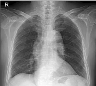
289 Figure 1. Chest posteroanterior view shows mass on the
left hilum and infrahilar area.
Figure 2. Abdominal X-ray shows nonspecific gas dis- tension in the colon.
Figure 3. Abdominal com- puted tomography shows long segmental edematous wall thickening in the ce- cum (white arrow) and ter- minal ileum (black arrow).
Hg, heart rate 76/min, respiration rate 20/min, and body temperature 36.6
oC. Bowel sound was hyperactive, and there was mild tenderness on the right lower quadrant without rebound tenderness or palpable mass. Labor- atory data showed white blood cell (WBC) count of 4,000/mm
3with absolute neutrophil 3,600/mm
3, hemo- globin 10.6 g/dL, platelet 210,000/mm
3, high sensitivity C-reactive protein was 8.10 mg/dL. His liver and renal function panel were unremarkable. Chest X-ray films showed left hilar mass without interval change com- pared with the one taken at the time of cancer diagnosis (Figure 1). On the abdominal X-ray films, there was a nonspecific gaseous distension (Figure 2). He was treat-
ed with antispasmodics, symptomatically.
On the day 3 of hospitalization (day 13 of chemo-
therapy), despite supportive care, he complained of ag-
gravated abdominal pain and developed fever of
37.6
oC. Laboratory data revealed a hematological tox-
icity, hemoglobin decreased to 9.2 mg/dL, WBC count
to 1,400/mm
3, absolute neutrophil count to 1,064/mm
3,
and platelets to 161,000/mm
3. An enhanced abdomen
computed tomography (CT) scan revealed bowel wall
thickening in the cecum and terminal ileum, and sur-
rounding mesenteric inflammation, consistent with ty-
phlitis (Figure 3). He was treated with empirical anti-
biotics and supportive therapy of bowel rest, intra-
EH Ji et al: Typhlitis induced by irinotecan
290
venous fluids, and nutritional support.
During the next 4 days, his abdominal pain improved slowly. On the day 7 of hospitalization, oral intake was available and absolute neutrophil count increased to 1,704/mm
3. Follow-up ultrasonography performed on the day 16 of hospitalization showed resolution of bow- el wall thickening, and he was discharged on hospital day 29. After discharge, he refused additional chemo- therapy and expired 3 months later due to progression of SCLC.
Discussion
Typhlitis, a necrotizing enterocolitis, is usually re- ported in neutropenic patients during chemotherapy, which mainly involves cecum, ileum, and the ascending colon. Typhlitis can be a life-threatening complication in the neutropenic patients, that may progress to in- testinal necrosis, hemorrhage, perforation, peritonitis, and eventually sepsis
3. Mechanism of chemotherapy re- lated typhlitis is as follows. Anticancer agents cause di- rect mucosal cytotoxicity of the gastrointestinal tract and neutropenia leading to enteral microorganism prolifer- ation. Bacterial endotoxins result in intestinal mucosal ischemia, necrosis, and a breakdown of intestinal mcos- al barrier
4. Cecum is the most common site of typhlitis, because of its poor blood supply, lymphatic drainage and direct exposure to colonic bacteria, making it more liable to infection
4,5.
The typical clinical manifestations of typhlitis include abdominal pain and fever in a patient during chemo- therapy, as in this case. Early diagnosis is the most im- portant in the treatment of typhlitis. Abdominal CT, ul- trasonography, and barium enema can be used as diag- nostic tools. CT scan is a valuable tool for early diagnosis. Findings include an edematous colon, sym- metric mucosal thickening more than 5 mm, and in- flammation of the pericolonic tissues
3,5,6, as shown in Figure 3. Ultrasonography is also a good diagnostic tool, which may reveal a target sign encircling mural thickening as a result of mucosal edema in the ileum and cecum
6. For this reason, we performed abdominal
ultrasonography to follow him up.
Typhlitis usually occurs in leukemia patients receiving chemotherapy. Recently, as high-dose chemotherapy became popular, the incidence of typhlitis is increasing in patients with solid tumors
7. In particular, several cas- es of typhlitis have been reported in lung cancer and pemetrexed, paclitaxel, docetaxel, 5-fluorouracil and vi- norelbine were suggested as causative agents
8. Irinotecan is a derivatives of the natural cytototoxic compound camptothecin which produces anticancer ef- fects through inhibition of DNA topoisomerase I
9. The most common complication of irinotecan is gastro- intestinal mucosal toxicities, in particular, more than 80% of patients receiving irinotecan therapy experience diarrhea
2.
Although five cases of typhlitis have been reported in phase I study determining the maximum tolerated dose of camtothecin analogues (topotecan and irinote- can)
9, no case has been reported in patients treated with standard dose. It is unknown how typhlitis occurs after chemotherapy with irinotecan. The following character- istics of irinotecan may be helpful in the comprehension of the irinotecan induced typhlitis. Irinotecan has a stronger gastrointestinal mucosal cytotoxicity than any other anticancer drug. In addition, it is well known that patients with UGT1A1 genetic mutation tend to develop severe side effects
10. In this case, due to the lack of his genetic study at that time, we were not able to com- pletely exclude the possibility of the genetic mutation.
We presume that he might have had a UGT1A1 poly- morphism, because irinotecan induced typhlitis was de- veloped under not severe neutropenia.
It is still controversial whether medical or surgical
treatment is more proper. In the past, surgical treatment
was preferred
3,5. However, because of poor prognosis
due to underlying diseases, and high mortality due to
severe postoperative complications, priority is shifting to
conservative treatment including broad spectrum anti-
biotics, bowel rest, intravenous fluids, and parenteral
nutritional support
5. On the other hand, surgical treat-
ment takes priority in patients with gastrointestinal hem-
orrhage, bowel perforation, and sepsis
4,11. In this case,
Tuberculosis and Respiratory Diseases Vol. 73. No. 5, Nov. 2012