Kyoung Lee , Sung-Ryong Ko and Ja-Young Moon*
1Department of Bio Health Sciences, College of Natural Sciences, Changwon National University, Changwon, Gyungnam 51140, Korea
2Bureau of General Affairs, The Korean Society of Ginseng, Seoul 06732, Korea Received July 22, 2016 /Revised August 8, 2016 /Accepted August 16, 2016
This study was performed to investigate the modulatory effects of two prototypes of Panax ginseng saponin fractions, 20(S)-protopanaxadiol saponins (PDS) and 20(S)-protopanaxatriol saponins (PTS), on the induction of inflammatory mediators in lipopolysaccharide (LPS)-treated RAW264.7 murine macro- phage cells. For this purpose, RAW264.7 cells were treated with LPS (10 μg/ml) before, after, or si- multaneously with PDS or PTS (150 μg/ml), and the released level of nitric oxide (NO) and expression levels of inducible nitric oxide synthase (iNOS) and cyclooxygenase-2 (COX-2) were evaluated. When RAW264.7 cells were treated with LPS and ginseng saponin fractions simultaneously for 24 hr, PTS, compared to PDS, more strongly attenuated the NO production induced by LPS treatment. When the cells were pretreated with LPS for 2 hr followed by PDS or PTS treatment for 24 hr, both ginseng saponins strongly reduced NO release. The pretreatment of RAW264.7 cells with PDS or PTS for 2 hr followed by LPS treatment for 24 hr significantly attenuated the LPS-induced production of NO.
PTS showed stronger inhibitory potency to NO generation than PDS. Our western blot experiment showed that both PDS and PTS (150 μg/ml) also significantly down-regulated the expressions of iNOS and COX-2 induced by LPS treatment. Our results suggest that both PDS and PTS possess strong pro- tective effects against LPS-stimulated inflammation and that their protective effects are mediated by the suppression of NO synthesis via down-regulation of pro-inflammatory enzymes, iNOS, and COX-2 in the RAW264.7 cells.
Key words : COX-2, iNOS, lipopolysaccharide, 20(S)-protopanaxadiol, 20(S)-protopanaxatriol
*Corresponding author
*Tel : +82-55-213-3552 , Fax : +82-55-213-3550
*E-mail : [email protected]
This is an Open-Access article distributed under the terms of the Creative Commons Attribution Non-Commercial License (http://creativecommons.org/licenses/by-nc/3.0) which permits unrestricted non-commercial use, distribution, and reproduction in any medium, provided the original work is properly cited.
Introduction
Macrophages play a central role in inflammatory proc- esses through the release of pro-inflammatory cytokines, chemokines and chemoattractants as well as cytotoxic and inflammatory molecules such as nitric oxide (NO), reactive oxygen species (ROS), and prostaglandin E2 (PGE2) [20, 24].
Nitric oxide and ROS are representative inflammatory medi- ators produced by macrophages under inflammatory con- ditions [20, 24]. These molecules are generated by the activa- tion of inducible nitric oxide synthase (iNOS) and cyclo- oxygenase-2 (COX-2) [16, 21].
Korean ginseng (Panax ginseng C.A. Meyer) is one of the
most widely used medicinal plants, particularly in tradi- tional oriental medicine. The root of Panax ginseng C. A.
Meyer is a widely used as a valuable herbal medicine for preventive and therapeutic purposes for several thousands of years in East Asian countries such as Korea, China, and Japan. Panax ginseng has been empirically used as a psychic energizer and as a general tonic in traditional medicine to increase vitality, health and longevity and for cancer-pre- venting potential. Because it has been used for a long time without showing any toxic properties, Korean Red Ginseng (KRG, steamed root of Panax ginseng Meyer, Araliaceae) is considered more beneficial for human health. In fact, several studies suggest that Korean ginseng takes its pharmaco- logical effects mostly by multiple active constituents includ- ing ginsenosides and acid polysaccharides [1, 6, 11, 14].
The main components of Panax ginseng are known to be the ginsenosides that are classified structurally into two types, 20(S)-protopanaxadiol-type ginsenosides such as Ra, Rb, Rc, Rd, Rg3, and Rh2 and 20(S)-protopanaxatriol-type ginsenosides such as Re, Rf, Rg1, Rg2, and Rh1 [26] (Fig.
A B
Fig. 1. Chemical structures of Panax ginseng (A) 20(S)-protopanax- adiol saponins (PDS) and (B) 20(S)-protopanaxatriol saponins (PTS), and distribution ratios of ginsenosides in the two pro- totypes of ginseng saponins.
1). Ginsenosides are generally recognized as the principle bioactive ingredients in Panax ginseng and reported to have a wide variety of physiological and pharmacological effects [3]. At present, it is also extensively used as an ingredients for formulation of herbal supplements. Recently, many stud- ies on anti-inflammatory effects of traditional medicines have been reported [15, 30]. However, mechanisms for the inhibitory effects of the two proto-types of Panax ginseng saponin fractions, 20(S)-protopanaxadiol saponins (PDS) and 20(S)-protopanaxatriol saponins (PTS) on NO production system in the pro-inflammatory conditions have not yet been fully demonstrated. Thus, underlying mechanisms for the immunomodulatory effects of PDS and PTS remained to be discovered.
In this study, we investigated anti-inflammatory activities of the two Panax ginseng saponins, PDS and PTS at the cel- lular level in relation to NO generation system in the lip- opolysaccharide (LPS)-induced RAW264.7 murine macro- phage cells, which have been used as a model of inflamma- tory macrophages.
Materials and Methods
Materials
Panax ginseng saponin fractions of PDS and PTS were kindly provided by the Research Institute of Technology, Korea Ginseng Corporation (Daejeon, Korea). PDS are a mix- ture of ginsenosides containing higher amounts of Ra, Rb1, Rb2, Rc, and Rd. PTS are a mixture of ginsenosides contain- ing higher amounts of Re, Rf, Rg1, Rg2, and Rh1. The con- centration of stock solution were 20 mg/ml in dimethyl sulf- oxide (DMSO) and stored at -80℃. Dulbecco’s modified Eagle’s medium (DMEM), fetal bovine serum (FBS), and oth-
er tissue culture reagents were purchased from Gibco BRL Co. (Grand Island, NY, USA). Final concentrations of PDS and PTS used for experiments were prepared by diluting the stock solution with DMEM immediately before use.
RAW264.7 cells were purchased from the Korean Cell Line Bank (Seoul, Korea). LPS (Escherichia coli, serotype O55:B5) was purchased from Sigma-Aldrich (St. Louis, MO, USA) and was used to induce an inflammatory response. Anti- iNOS mouse monoclonal, anti-COX-2 goat polyclonal and anti-β-actin mouse monoclonal antibodies were purchased from Santa Cruz Biotechnology (Santa Cruz, CA, USA).
Donkey anti-goat IgG and anti-mouse IgG conjugated to horseradish-peroxidase were purchased from Santa Cruz Biotechnology and Cell Signaling Technology (Danvers, MA, USA), respectively. The BCA Protein Assay Kit was pur- chased from PIERCE (Rockford, IL, USA). All other reagents were obtained from Sigma-Aldrich unless indicated.
Cell culture and treatment
Murine RAW264.7 macrophage-like cell line was obtained from the Korean Cell Line Bank (Seoul, Korea) and routinely cultured in Dulbecco’s modified Eagle medium (Life Tech- nology, Inc.) supplemented with 10% fetal bovine serum, 100 units/ml penicillin, and 100 μg/ml streptomycin at 37℃ and 5% CO2 in humidified air. Cells were plated and cultured in plastic culture dishes and cells reached to 60% confluent were exposed to LPS. Cells were treated with LPS (10 μg/ml) and PDS or PTS at the indicated concentrations and in- cubated for the indicated time periods. Following in- cubation, the cells were dissociated from dishes by scraping.
Dissociated cells were collected by centrifugation (500× g, 5 min), washed twice with ice-cold PBS, and lysed in an EBC buffer (50mM Tris–HCl, pH 8.0, 120mM sodium chlor-
Cell viability assay
Cell viability was determined colorimetrically by measur- ing the reduction of the tetrazolium salt, 3-[4,5-dimethylthia- zol-2-yl]-2,5-diphenyl tetrazolium bromide (MTT) to for- mazan. Briefly, murine RAW264.7 cells were seeded at a density of 1x10⁵ cells/well in 96-well plates (Falcon, Ger- many) and cultured for 24 hr. After cell attachment, culture media were freshly changed, and various doses of PDS or PTS were added. Cells were additionally cultured for 24 hr and then MTT solution (10 μl, 5 mg/ml in PBS) was added to the wells. After 3 hr incubation, the medium was re- moved, and DMSO was then added to dissolve the formazan produced by the cells. The optical density of formazan sol- ution was measured with a microplate reader at 570 nm.
Assay of NO production
NO production from activated macrophage cells was monitored by measuring the nitrite content in culture medium. In brief, an aliquot (100 μl) of the conditioned me- dium was mixed with an equal volume of Griess reagent (1% sulfanilamide, 0.1% N-1-naphthylenediamine dihydro- chloride in 5% phosphoric acid). Absorbance was measured at 540 nm after incubation for 10 min. Sodium nitrite, diluted in culture medium at concentrations of 10~100 μM, was used as a standard to calculate NO2-
concentrations.
Immunoblot analysis
Cells were harvested, washed twice with ice-cold PBS, and collected in the cell lysis buffer using Ez RIPA Lysis kit (ATTO, Japan) for 20 min on ice. Lysates were then cen- trifuged at 9,000× g for 15 min to remove insoluble material.
Protein concentrations were determined by using the BCA protein assay kit (Pierce, Rockford, USA). Bovine serum al- bumin (BSA) was used as a standard. Protein samples (70 μg for each) were separated by 12% SDS–PAGE and trans- ferred onto polyvinylidene difluoride (PVDF, Bio-Rad) membranes for 2 hr at 90 V. The membrane was blocked with 5% nonfat dry milk in TBST solution for 3 hr. The blots were incubated with primary antibodies, anti-iNOS (1:200
ature and the blots were developed by the ECL (enhanced chemiluminescence) detection system (Amersham Bioscien- ces, Piscataway, NJ, USA). Monoclonal sheep anti-mouse IgG, or donkey anti-goat IgG horseradish peroxidase-con- jugated secondary antibodies were used at 1:2,000 dilutions in TBST. Images were pictured using the Amersham Imager 600 (GE Healthcare Life Sciences) and densitometric data were calculated using the analysis program provided by the Amersham Imager 600.
Statistical analysis
Student’s t-test and a one-way ANOVA were used to de- termine the statistical significance of the difference between values for the various experimental and control groups.
Experimental data are expressed as means ± standard devia- tion (SD), and the results were obtained from at least three independent experiments performed in triplicate. The im- ages of western blot are a representative of three in- dependent experiments (n = 3). A p-value of 0.05 or less was considered statistically significant.
Results
The cytotoxicity of PDS and PTS on RAW264.7 cells
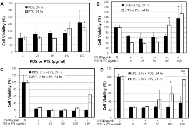
As a first step, in order to identify non-cytotoxic dose ranges of PDS and PTS, RAW264.7 cell viability was ana- lyzed after exposure to the ginseng ingredients with or with- out LPS treatment. When cultured for 24 hr, both PDS and PTS itself up to 150 μg/ml had no cytotoxic effect on RAW 264.7 cells (Fig. 2A). Even though statistically not significant, the viability was rather increased as both PDS and PTS con- centrations increased. As shown in Fig. 2B, when RAW264.7 cells were cultured for 24 hr after co-treatment of LPS with PDS or PTS, PDS at 25, 50 and 100 μg/ml did not have any attenuating effect of the cytotoxicity induced by LPS treat- ment. However, PDS at 150 μg/ml also showed a strong potency to attenuate LPS-stimulated cytotoxicity (Fig. 2B).
Fig.2 B also showed that PTS possessed much stronger at- tenuation potency to the cytotoxicity induced by LPS treat-
A B
C D
Fig. 2. Effect of PDS or PTS on the viability of RAW264.7 cells stimulated by LPS. (A) RAW264.7 cells (1×10⁵ cells) were incubated at 37℃ with increasing concentrations of PDS or PTS (0, 25, 50, 100, and 150 μg/ml) for 24 hr. (B) RAW264.7 cells were treated with both LPS (10 μg/ml) and PDS or PTS simultaneously for 24 hr. (C) RAW264.7 cells were pretreated with indicated concentrations of PDS or PTS for 2 hr prior to incubation with LPS (10 μg/ml) for 24 hr. (D) RAW264.7 cells were pretreated with LPS (10 μg/ml) for 2 hr prior to incubation with indicated concentrations of PDS or PTS for 24 hr. Cytotoxicity of the two types of ginsenosides was measured using a MTT assay system, as described in Materials and methods. Cells from nonconfluent dishes were harvested by treating with trypsin and resuspended in 1 ml PBS. An equal volume (1 ml) of trypan blue was then added and gently mixed. After 2 min, cells were counted. Results are expressed as percentages of the control value and data shown are means ± SD of three replicate experiments. **p<0.01, *p<0.05, significantly different when compared with LPS-stimulated RAW264.7 cells.
ment rather than that PDS did in the RAW264.7 cells. Even though PTS at two low concentrations, 25 and 50 μg/ml, showed low attenuating effect on LPS-stimulated cytotox- icity, PTS at two high concentrations, 100 and 150 μg/ml, strongly and completely attenuated the LPS-stimulated cyto- toxicity (Fig. 2B). When the cells pretreated with four differ- ent concentrations of PDS or PTS for 2 hr were treated with LPS (10 μg/ml) for 24 hr, viability of the RAW264.7 cells was not recovered at the PDS concentrations applied (Fig.
2C). This result suggests that either PDS or PTS does not possess any preventative potency to the cytotoxicity induced by LPS treatment. When RAW264.7 cells were pretreated with LPS for 2 hr, followed by treated with PTS for 24 hr, cytotoxicity induced by LPS treatment was dose-depend- ently attenuated (Fig. 2D). This result suggests that PTS, at 150 μg/ml concentration applied, possessed the protective effects against LPS-stimulated cytotoxicity. However, PDS,
at the concentration ranges applied in this study, did not possess any protective effects against LPS-stimulated cyto- toxicity. These results suggest that both PDS and PTS pos- sess strong protective potencies to the LPS-induced cytotox- icity rather than preventive effects. Further studies are neces- sary to investigate its protective effect mechanistically. Based on these results, the concentrations at 150 μg/ml of PDS or PTS were applied for other experiments in this study.
Suppression effects of both PDS and PTS on the release of nitric oxide in LPS-stimulated RAW264.7 cells
To investigate the anti-inflammatory effects of PDS or PTS, RAW264.7 cells were treated with LPS (10 μg/ml) for 2 hr or 24 hr before, after or even simultaneous treatment with PDS or PTS, and the level of NO production was evaluated. When treated with LPS simultaneously to the
A B
C
Fig. 3. Effect of PDS or PTS on NO production in LPS-stimulated RAW264.7 cells. (A) RAW264.7 cells were treated with both LPS and PDS (150 μg/ml) or PTS (150 μg/ml) simulta- neously for 24 hr. (B) Cells were pretreated with LPS (10 μg/ml) for 2 hr, followed by treatment with PDS or PTS for 24 hr. (C) Cells were pretreated with PDS (150 μg/ml) or PTS (150 μg/ml) for 2 hr, followed by treatment with LPS (10 μg/ml) for 24 hr. Levels of NO were determined from culture medium of RAW264.7 cells by using Griess reagent. **p<0.01, significantly different when compared with LPS-stimulated RAW264.7 cells. 1removed by chang- ing with fresh medium after 2 hr incubation; 2not removed after 2 hr incubation.
24 hr, PDS and PTS strongly reduced NO release in LPS- stimulated RAW264.7 cells (Fig. 3B). Particularly, PTS com- pletely reduced LPS-stimulated NO production regardless of removal of LPS. When RAW264.7 cells were pretreated with PDS or PTS for 2 hr, followed by treated with LPS for 24 hr, the pretreatment with PDS or PTS significantly and statistically attenuated the LPS-induced production of NO (Fig. 3C). The inhibitory potency to the NO generation was much stronger in the presence of PTS than PDS. However, when the RAW264.7 cells following removal of PTS pre- treated for 2 hr were treated with LPS, the reduction of NO produced by LPS was not occurred.
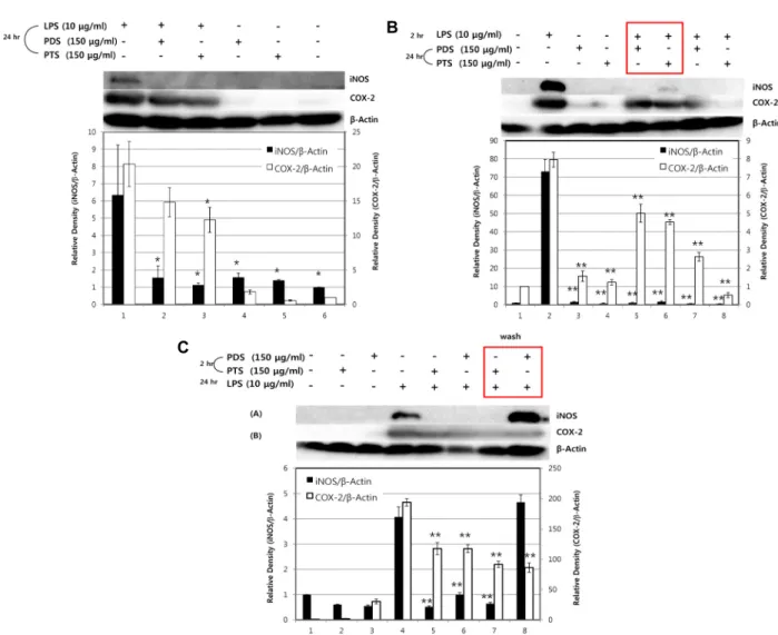
Inhibitory effects of PDS or PTS on the expression of iNOS and COX-2 in LPS-stimulated RAW264.7 cells
Since the production of NO in macrophage is regulated primarily by the expression of inflammatory enzymes, iNOS and COX-2, western blot analysis was performed to de-
cells were simultaneously treated with LPS and PDS or with LPS and PTS for 24 hr, both PDS and PTS showed strong inhibition of the expression of iNOS and COX-2 proteins (Fig. 4A). As shown in Fig. 4, when the cells were stimulated with LPS in the absence of PDS or PTS, the expression of COX-2 and iNOS proteins was significantly induced. How- ever, regardless of the exposure conditions of LPS and PDS or PTS to the RAW264.7 cells, both PDS and PTS potently inhibited the expression of these enzymes. PTS, compared to PDS, showed the stronger inhibitory potency to the ex- pression of iNOS and COX-2 induced by LPS. Even when PDS or PTS was removed from the medium after 2 hr in- cubation, the expression of the two enzymes by the stim- ulation with LPS for 24 hr was attenuated.
Discussion
In this study, we evaluated the inhibitory effects of two Panax ginseng saponins, PDS and PTS, on the production
A B
C
Fig. 4. The effects of PDS or PTS on the expression of iNOS or COX-2 in LPS-stimulated RAW264.7 cells. RAW264.7 cells were incubated with LPS (10 μg/ml) in the presence or absence of PDS (150 μg/ml) or PTS (150 μg/ml) for indicated periods, and then expression levels of iNOS and COX-2 were determined by immunoblot analysis. (A) RAW264.7 cells were treated with both LPS (10 μg/ml) and PDS (150 μg/ml) or PTS (150 μg/ml) simultaneously for 24 hr. (B) RAW264.7 cells were pretreated with LPS (10 μg/ml) for 2 hr, followed by treatment with PDS or PTS for 24 hr. Rectangular box indicates that LPS was removed out by changing with fresh medium after 2 hr incubation. (C) RAW264.7 cells were pretreated with PDS (150 μg/ml) or PTS (150 μg/ml) for 2 hr, followed by treatment with LPS (10 μg/ml) for 24 hr. Rectangular box indicates that PDS or PTS was removed out by changing with fresh medium after 2 hr incubation. **p<0.01, *p<0.05, significantly different when compared with LPS-stimulated RAW264.7 cells.
of inflammatory mediators including nitric oxide and on the expressions of iNOS and COX-2 in LPS-stimulated RAW 264.7 murine macrophage cells. We found that both PDS and PTS inhibited LPS-induced NO generation accompanied by a reduction of iNOS level in RAW264.7 cells. We also found that the two ginseng saponins inhibited COX-2 expression in the LPS-treated macrophage cells. These data suggest that the protective effects of PDS or PTS against LPS-induced in- flammation may be mediated by the suppression of NO syn- thesis via down-regulation of pro-inflammatory enzymes, iNOS and COX-2, in the macrophages.
Inflammation is a complex biological response against harmful stimuli and plays a critical role in immune defense under various external and internal pathogens [23]. Harmful stimuli such as lipopolysaccharides, a toxic molecule derived from gram-negative bacteria cell walls, activate macrophages to release various pro-inflammatory molecules such as NO and cytokines and pro-inflammatory enzymes, iNOS and COX-2 [24]. NO is produced by iNOS, which is the major form induced in response to inflammatory stimuli including LPS [21]. Excessive release of NO has a critical role in vari- ous diseases involving the immune system such as athero-
tential strategy for the treatment of inflammatory diseases.
Thus, our data suggest that both PDS and PTS could be use- ful for treatment of the inflammatory diseases. Because of the unique feature of PDS- and PTS-mediated reduction of iNOS and COX-2 expression, further elucidating their under- lying molecular mechanisms will provide a novel insight in- to the understanding of ginseng-mediated anti-inflammatory and/or immunomodulatory actions.
Ginseng saponin fractions of PDS and PTS used in this study are two characteristic types of triterpenoid saponins in ginseng ginsenosides. As mentioned in introduction, PDS and PTS are mixtures of many structurally different types of ginsenosides. Our results showing that PTS possessed stronger inhibitory potency to NO production than PDS might be due to compositional difference in the ginseng saponins. Our results were supported from the previous re- port that ginsenoside Rd, one component of PDS, per se in- duces COX-2 expression in RAW264.7 macrophages [10]. In the conformational structure, PTS contains one more hydrox- yl group compared to PDS, which might contribute to the differential inhibitory potency to NO generation system.
Structure-activity relation studies are required for further elucidation of this differential potency to the modulation of pro-inflammatory enzymes by using purified single ginseno- sides of PDS such as Ra, Rb, Rc, Rd, Rg3, Rh2 and PTS such as Re, Rf, Rg1, Rg2 and Rh1.
Our finding that both PDS and PTS can suppress ex- pressions of LPS-induced iNOS and COX-2 is especially of clinical interest. In fact, iNOS and COX-2 are important regu- lators of inflammatory responses in various tissues and or- gans, and play important roles in the pathogenesis of human disease [8, 18, 25]. Emerging evidence has also revealed that a delicate cross-talk between NOSs and COXs plays a critical role in the control of inflammation [4, 17]. Although several signaling pathways contribute to LPS-induced iNOS and COX-2 expression [5, 9, 13, 29], both PDS and PTS might interfere with any specific signaling cascades in macro- phages, thereby regulating iNOS and COX-2 expression. In this context, the unique modulatory effect of PDS and PTS on the control of iNOS and COX-2 expression might repre-
It is known that NO generated by the activation of iNOS is primarily controlled by transcriptional and translational regulation by surface receptors such as Toll like receptors (TLR) and its counter adaptor and signaling molecules such as TANK binding kinase (TBK), Toll-interleukin-1 receptor- domain-containing adaptor-inducing interferon-β (TRIF), and TRIF-related adaptor molecule (TRAM) [12]. Particular- ly, TLR-4 is a pattern recognition receptor that responds to LPS and triggers activation of the acquired immune response [7]. Thus, TLR-4 is activated in various inflammatory dis- eases induced by LPS [2]. However, the inhibitory effect of PDS or PTS on TLR-4 production in LPS-stimulated macro- phages has not yet been fully reported. Therefore, further studies are necessary for elucidation of the molecular mecha- nism whether the reduction effects of PDS and PTS on the NO-generation system in LPS-stimulated RAW264.7 cells are involved in the inhibition of LPS binding to Toll-like re- ceptor(s) by the two ginseng saponins.
Inhibition of excessive release of NO, therefore, is consid- ered to be an important therapeutic target against various inflammatory diseases induced by LPS. Elucidation of any other intracellular signaling cascades in response to LPS-in- duced NO production and the modulation by PDS or PTS may also provide additional insights into the molecular basis of its anti-inflammatory effects.
It is known that NO can produce other proinflammatory cytokines and is a harmful stimuli in the cell environment.
Additionally, COX-2 has been shown to trigger sustained ROS production and to be induced by proinflammatory cy- tokine signaling [29]. As shown in Fig. 4, both PDS and PTS at 150 μg/ml decreased the levels of iNOS and COX-2 ex- pression induced by LPS treatment. Various inflammatory diseases involve inflammatory mediators such as NO and PGE2 as well as pro-inflammatory cytokines, such as TNF-α, IL-1β and IL-6, in macrophages [19, 27]. Having shown that the two ginseng saponins facilitate anti-inflammatory effect against LPS, our further study will focus on determination of their inhibitory mechanisms of pro-inflammatory cytokine signaling in macrophages.
In conclusion, from our results that both PDS and PTS
inhibit the release and production of inflammatory media- tors including NO by inhibiting the expression of pro-in- flammatory enzymes, iNOS and COX-2, we suggest that both ginseng saponins may be used as auxiliary substances for treatment of pro-inflammatory diseases.
Acknowledgements
We thank the Research Institute of Technology, Korea Ginseng Corporation (Daejeon, Korea) for generously pro- viding 20(S)-protopanaxadiol saponins and 20(S)-protopan- axatriol saponins for anti-inflammation study. This research is financially supported by Changwon National University in 2015~2016.
References
1. Byeon, S. E., Lee, J., Kim, J. H., Yang, W. S., Kwak, Y. S., Kim, S. Y., Choung, E. S., Rhee, M. H. and Cho, J. Y. 2012.
Molecular mechanism of macrophage activation by red gin- seng acidic polysaccharide from Korean red ginseng.
Mediators Inflamm. 2012, 732860.
2. Cario, E. and Podolsky, D. K. 2000. Differential alteration in intestinal epithelial cell expressionof toll-like receptor 3 (TLR3) and TLR4 in inflammatory bowel disease. Infect.
Immun. 68, 7010-7017.
3. Chu, S. F. and Zhang, J. T. 2009. New achievements in gin- seng research and its future prospects. Chin. J. Integr. Med.
15, 403-408.
4. Cuzzocrea, S. and Salvemini, D. 2007. Molecular mecha- nisms involved in the reciprocal regulation of cyclooxyge- nase and nitric oxide synthase enzymes. Kidney Int. 71, 290- 297.
5. Friedl, R., Moeslinger, T., Kopp, B. and Spieckermann, P.
G. 2001. Stimulation of nitric oxide synthesis by the aqueous extract of Panax ginseng root in RAW264.7 cells. Brit. J.
Pharmacol. 134, 1663-1670.
6. Hasegawa, H. 2004. Proof of the mysterious efficacy of gin- seng: basic and clinical trials: metabolic activation of ginse- noside: deglycosylation by intestinal bacteria and ester- ification with fatty acid. J. Pharmacol. Sci. 95,153-157.
7. Hoshino, K., Takeuchi, O., Kawai, T., Sanjo, H., Ogawa, T.
and Takeda, Y. et al. 1999. Cutting edge: toll-like receptor 4 (TLR4)-deficient mice are hyporesponsive to lipopoly- saccharide: evidence for TLR4 as the LPS gene product. J.
Immunol. 162, 3749-3752.
8. Hui, D. Y. 2008. Intimal hyperplasia in murine models. Curr.
Drug Targets 9, 251-260.
9. Ichikawaa, T., Li, J., Nagarkatti, P., Nagarkatti, M., Hofsethc, L. J., Windust, A. and Cui, T. 2009. American ginseng prefer- entially suppresses STAT/iNOS signaling in activated mac- rophages. J. Ethnopharmacol. 125, 145-150.
10. Jeong, H. G., Pokharel, Y. R., Han, E. H. and Kang, K. W.
2007. Induction of cyclooxygenase-2 by ginsenoside Rd via activation of CCAAT-enhancer binding proteins and cyclic AMP response binding protein. Biochem. Biophys. Res. Com- mun. 359, 51-56.
11. Kim, D. H. 2012. Chemical diversity of Panax ginseng, Panax quinquifolium, and Panax notoginseng. J. Ginseng Res. 36, 1-15.
12. Kim, H. S. and Moon, E. Y. 2009. Reactive oxygen species-in- duced expression of B cell activating factor (BAFF) is in- dependent of Toll-like receptor 4 and myeloid different- iation primary response gene 88. Biomol. Ther. 17, 144-150.
13. Kim, T. W., Joh, E. H., Kim, B. and Kim, D. H. 2012.
Ginsenoside Rg5 ameliorates lung inflammation in mice by inhibiting the binding of LPS to toll-like receptor-4 on macrophages. Int. Immunopharmacol. 12, 110-116.
14. Kwak, Y. S., Kyung, J. S., Kim, J. S., Cho, J. Y. and Rhee, M. H. 2010. Anti-hyperlipidemic effects of red ginseng acid- ic polysaccharide from Korean red ginseng. Biol. Pharm. Bull.
33, 468-472.
15. Lee, K. W., Jung, S. Y., Choi, S. M. and Yang, E. J. 2012.
Effects of ginsenoside Re on LPS-induced inflammatory me- diators in BV2 microglial cells. BMC Complement. Altern.
Med. 12, 196.
16. Lee, S. H., Soyoola, E., Chanmugam, P., Hart, S., Sun, W., Zhong, H., Liou, S., Simmons, D. and Hwang, D. 1992.
Selective expression of mitogen-inducible cyclooxygenase in macrophages stimulated with lipopolysaccharide. J. Biol.
Chem. 267, 25934-25938.
17. Mariotto, S., Suzuki, Y., Persichini, T., Colasanti, M., Suzuki, H. and Cantoni, O. 2007. Cross-talk between NO and arach- idonic acid in inflammation. Curr. Med. Chem. 14, 1940-1944.
18. Mathrani, V. C., Kenyon, N. J., Zeki, A. and Last, J. A. 2007.
Mouse models of asthma: can they give us mechanistic in- sights into the role of nitric oxide? Curr. Med. Chem. 14, 2204- 2213.
19. Moynagh, P. N. 2005. The NF-kappaB pathway. J. Cell Sci.
118, 4589-4592.
20. Nathan, C. 1992. Nitric oxide as a secretory product of mam- malian cells. FASEB J. 6, 3051-3064.
21. Nathan, C. and Xie, Q. W. 1994. Nitric oxide synthases:
roles, tolls, and controls. Cell 78, 915-918.
22. Pacher, P., Beckman, J. S. and Liaudet, L. 2007. Nitric oxide and peroxynitrite in health and disease. Physiol. Rev. 87, 315-424.
23. Ran, S. and Montgomery, K. E. 2012. Macrophage-mediated lymphangiogenesis: the emerging role of macrophages as lymphatic endothelial progenitors. Cancers (Basel) 4, 618- 657.
24. Rossol, M., Heine, H., Meusch, U., Quandt, D., Klein, C., Sweet, M. J. and Hauschildt, S. 2011. LPS-induced cytokine production in human monocytes and macrophages. Crit.
Rev. Immunol. 31, 379-446.
25. Salinas, G., Rangasetty, U. C., Uretsky, B. F. and Birnbaum, Y. 2007. The cycloxygenase 2 (COX-2) story: it’s time to ex- plain, not inflame. J. Cardiovasc. Pharmacol. Ther. 12, 98-111.
초록:RAW264.7 대식세포에서 LPS 매개 iNOS/NO 생성에 대한 protopanaxadiol saponin 및 protopanaxatriol saponin의 억제효과
김진익1․난딘셋세그 나르나투야1․최용원1․강대욱1․김동완1․이 경1․고성룡2․문자영1*
(1창원대학교 생명보건학부, 2고려인삼학회 사무국)
본 연구는 RAW264.7 세포에서 lipopolysaccharide (LPS) 처리에 의한 염증매개인자의 유도에 대한 고려인삼
사포닌 분획인 20(S)-protopanaxadiol saponins (PDS)과 20(S)-protopanaxatriol saponins (PTS)의 조절효능을 탐 구하였다. 이를 위해 RAW264.7 세포에 PDS 또는 PTS를 150 μg/ml의 농도로 LPS (10 μg/ml) 처리 이전이나 처 리 이후 또는 LPS와 동시에 처리하였으며, 처리된 세포에서 nitric oxide (NO)의 방출량, 유도성 nitric oxide syn- thase (iNOS) 및 cyclooxygenase-2 (COX-2)의 발현 량을 분석하였다. PDS에 비하여 PTS는 RAW264.7 세포에 LPS 와 동시에 처리하여 24시간 동안 배양했을 때 LPS 처리에 의해 유도된 NO의 생성을 강하게 감소시켰다. RAW 264.7 세포에 LPS (10 μg/ml)를 2시간 동안 처리한 후에 PDS 또는 PTS를 150 μg/ml 농도로 24시간 동안 처리하 면 두 인삼 사포닌 성분 모두 NO의 생성을 강하게 감소시켰다. RAW264.7 세포에 PDS 또는 PTS를 150 μg/ml농 도로 2시간 동안 처리한 후에 LPS (10 μg/ml)를 24시간 동안 처리했을 경우에도 두 인삼 사포닌 성분 모두 LPS 처리에 의해 유도된 NO 생성을 강하게 감소시켰다. LPS 처리에 의한 NO 생성을 저해하는 효과는 PDS에 비하여 PTS가 더 강하게 나타났다. PDS와 PTS 모두 150 μg/ml 처리농도에서 LPS (10 μg/ml)처리에 의해 유도된 iNOS 와 COX-2의 발현 역시 상당히 감소시켰다. 따라서 본 연구의 결과는 RAW264.7 대식세포에서 PDS와 PTS 두 인 삼 사포닌 성분은 LPS 처리에 의한 염증활성화에 강한 억제효과를 가지고 있음을 의미하며, 전염증성 효소인 iNOS와 COX-2 발현의 감소조절을 통하여 NO의 생성을 억제함으로써 항 염증효과가 나타남을 제시한다.
A. N. 2006. Signaling networks regulating cyclooxygenase-2.
Int. J. Biochem. Cell Biol. 38, 1654-1661.
29. Tsoyi, K., Kim, H. J., Shin, J. S., Kim, D. H., Cho, H. J., Lee,
effects of ginsenosides on NO and TNF-α production by LPS-activated N9 microglia. Int. Immunopharmacol. 7, 313- 320.