해삼의 항위염, 항위궤양 및 항헬리코박터 효과
오홍근1*․문대인1*․김정훈1․강영례1․박정우1․서민영1․박상훈1․강양규2 최충현3․박인선3․김 주3․유강렬3․설으뜸4․김옥진4․이학용1†
1
㈜휴벳,
2(유)해원
3
전주생물소재연구소,
4원광대학교 애완동식물학과
The Effects of Sea Cucumber as an Anti-gastritis, Anti-gastric Ulcer, and Anti-Helicobater
Hong-Geun Oh1*, Dae-In Moon1*, Jung-Hoon Kim1, Young-Rye Kang1, Jung-Woo Park1, Min-Young Seo1, Sang-Hoon Park1, Yang-Gyu Kang2, Chung-Hyeon Choe3, In-Sun Park3, Ju Kim3,
Kang-Yeol Yu3, Eu-Ddeum Seol4, Ok-Jin Kim4, and Hak-Yong Lee1†
1Huvet Co. Ltd., Jeonbuk 570-749, Korea, 2Heawon, Ltd., Jeonbuk 561-739, Korea
3Jeonju Biomaterials Institute, Jeonbuk 561-360, Korea
4Center for Animal Resources Development, Wonkwang University, Jeonbuk 570-749, Korea
Abstract
Sea cucumber, Stichopus japonicus, is used not only as an outstanding tonic food but also as a traditional medicine for the treatment of asthma, hypertension, rheumatism, anemia, and sinus congestion. The purpose of this study was to examine sea cucumber as an anti-gastritis and anti-gastric ulcer in HCl-ethanol-induced gastric and H. pylori-infected animal models. Thirty 7-week-old SD rats and Mongolian gerbils were divided into normal (Nor, n=6), control (Con, 60% HCl-ethanol+water, n=6), groupⅠ (DSCⅠ, 60% HCl-ethanol+sea cucumber 30 mg/kg, n=6), groupⅡ (DSCⅡ, 60% HCl-ethanol+sea cucumber 100 mg/kg, n=6), and group Ⅲ (DSCⅢ, 60% HCl-ethanol+sea cucumber 300 mg/kg, n=6). Sea cucumber significantly suppressed gastric le- sions and ulcers in the 60% HCl-ethanol-induced gastric model. Especially, 100 mg/kg of sea cucumber showed significantly inhibitory effects. In histopathological analysis of the H. pylori model, we found that sea cucumber augmented the eradication rates of H. pylori and attenuated gastric ulcer formation. Our results suggest that sea cucumber has inhibitory effects on gastritis and gastric ulcers. In addition, sea cucumber can be applied for the treatment of H. pylori.
Key words: sea cucumber, gastritis, gastric ulcer, Helicobacter pylori, HCl-ethanol
*
Equally attributed first author
†
Corresponding author. E-mail: [email protected]
†
Phone: 82-63-851-7061, Fax: 82-63-850-7459
서 론
위염(gastritis) 및 위궤양(gastric ulcer)은 하나의 원인에 의해 발병되는 것이 아니라 여러 인자들이 복합적으로 작용 하여 발생한다. 위 점막에 염증이 일어나는 질환인 급성위염 은 술이나 폭식, 불규칙한 식사, 뜨겁거나 차거나 부패한 음 식물의 섭취로 인하여 위 점막에 염증이 야기된 질환으로써, 위부의 불쾌감, 식욕부진, 설사와 구토 증상을 나타내고 전 신 권태를 느끼게 한다. 만성위염은
Helicobacter pylori가 주된 발생 원인체로 위염이나 위궤양뿐만 아니라 암을 유발 시키기도 한다(1-3). 위염의 원인으로는 위장 점막에 대한 공격인자와 방어인자의 평형관계의 균형이 깨어짐으로써 야기된다는 ‘balance theory’가 설득력 있는 이론으로 받아
들여지고 있다(4). 현재 시중에서 판매되고 있는 소화성 궤 양 치료제중 공격인자 억제제로는 위액의 소화력을 억제시 키는 항펩신제 및 위액을 중화시키는 NaHCO
3등의 제산제 와 위액 분비를 억제시키는 항콜린제, 항가스트린제, 점막마 취제, muscarine receptor antoagonist, H
2-receptor antag- onist, H
+K
+-APTase inhibitor 등이 있다(5). 방어인자 강화 제는 위점막 생성을 촉진시키는 목적으로 사용되는 prosta- glandin 제제 등이 있다. 위염에 있어서 점막의 염증 중 가장 중요한 변화는 선조직의 염증이라고 알려져 있으나, 실제로 는 선조직뿐만 아니라 점막하층, 근층, 장막에까지 염증이 미치고 있는 것들을 총괄해서 위염이라 한다.
해삼(
Stichopus japonicus)은 극피동물문(
Phylum Echi- nodermate), 해삼강(
Class Holothuroidea)속으로 항산화,
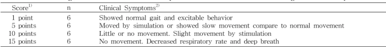
Table 1. Gross finding scores of behavior in the study on effects of dried sea cucumber in gastritis model by HCl-ethanol Score
1)n Clinical Symptoms
2)1 point 5 points 10 points 15 points
6 6 6 6
Showed normal gait and excitable behavior
Moved by simulation or showed slow movement compare to normal movement Little or no movement. Slight movement by stimulation
No movement. Decreased respiratory rate and deep breath
1)
Scores were calculated with the average of three different measurements.
2)
Clinical symptoms were observed by three different persons in each animal for one hour.
항바이러스, 항암, 항응고, 항골다공증 등 많은 효능이 보고되 었다(6-12). 천연물은 비교적 한방 약재로 현재 많이 사용되고 있고 비교적 안전성이 높기 때문에, 강력한 항균효과나 항염 효과, 면역조절 효과 등 다양한 효과가 보고된 해삼은 기능성 식품으로서의 높은 잠재력이 있을 것으로 생각된다(13-15).
따라서 본 연구에서는 60% HCl-ethanol 투여를 통한 급 성 위염모델에서 안전성이 높은 천연물인 해삼 분말을 투여 하여 SD rats의 육안적 위염증상 완화 및 위염, 위궤양 억제 정도를 조직학적인 검경을 통해 관찰하고,
H. pylori감염에 의한 만성 위질환 모델에서 조직병리학적 관찰을 통한 해삼 분말의 항위염, 항위궤양 및 항헬리코박터 효능 평가를 하고 자 하였다.
재료 및 방법
실험재료
㈜해원(전주, 한국)에서 공급받은 해삼 동결건조물(DSC, dried sea cucumber)을 증류수에 혼탁하여 주 1회 체중을 측정하여 각각 30 mg/kg, 100 mg/kg, 300 mg/kg의 농도로 1주일 급여량을 계산하여 조제하고 vortex한 후 냉장 보관 하고, 매일 정해진 시간에 강제 경구투여 하였다.
실험동물
㈜오리엔트(경기, 한국)에서 구입한 Specific-pathogen free(SPF) 상태의 6주령, 체중 150~200 g의 Sprague- Dawley(SD) rats 수컷 40마리와 원광대학교 동원자원개발 연구센터(익산, 한국)에서 공급받은 체중 45~50 g의 Mon- golian gerbil 수컷을 사용하였다. 사육기간 중 온도는 23±2
o
C, 상대습도는 50±5% 환기횟수는 10회/시간, 전배기 방식 및 조명시간은 12시간(7:00~19:00) 조도는 150~300 lux를 유지하였다. 동물 입수 시 모든 동물의 일반 건강상태에 대한 수의학적 검역을 실시하였으며 시험을 실시하는데 적합하 도록 설치류용 고형사료(샘타코, 경기, 한국)와 필터 및 자외 선살균기로 여과 살균된 정제수를 자유롭게 섭취하도록 하 며 약 1주일간의 순화기간을 거쳤다. 본 연구에 사용된 동물 실험에 관련된 모든 실험과정과 절차는 원광대학교 동물실 험윤리위원회의 사전심의와 윤리 규정을 준수하여 수행되 었다(Approval No. WKS 11-005).
군 분리 및 시료투여
60% HCl-ethanol 투여 급성 위염 질환모델 효능평가(SD
rats, n=6)와
H. pylori감염 만성 위염 질환 모델 효능평가 (Mongolian gerbil, n=6)를 위하여 주간 체중에 근거하여 다 음과 같이 군 분리를 하였다. 인체 유효 농도에 대한 근거가 부족하므로 동물을 대상으로 설정된 섭취량을 사람의 외삽 (extrapolation) 계산방법(16)에 따라 정상군(Nor), 대조군 (Con), DSCI(30 mg/kg, 저용량 건해삼), DSCII(100 mg/kg, 중간용량 건해삼), DSCIII(300 mg/kg, 고용량 건해삼)군으 로 총 5군을 각각 임의배정 하여 건해삼을 각 농도별로 강제 경구투여 하였다.
HCl-ethanol 위염 모델 유도 및 관찰
6주령, 체중 250~350 g의 수컷 SD rat 6∼7마리를 한 군 으로 하여 건해삼을 3주간 투여한 후, 위염 유발 물질인 150 mM HCl+60% ethanol을 1 mL씩 단회 경구투여 하여 위염 을 유발하였다. 24시간 이상 절식한 후 Mizui와 Doteuchi (17)의 방법으로 실험하였다. 즉, 실험물질을 경구투여 하고 30분 후에 60% HCl-ethanol용액(60% ethanol에 150 mM HCl을 함유) 1 mL를 경구투여 하여 절식, 절수 하에서 1시간 방치 후 ether로 치사시켰다. 이후 위를 적출하여 유문부를 결찰하고 위내에 10% formalin 용액 10 mL를 주입한 상태로 formalin 용액에 10분간 반응시켜 위 내외를 고정하였다. 염 증의 면적을 측정하기 위하여 대만부를 절개하여 촬영한 후 영상분석 프로그램(Image-Pro
RPlus 7.0, MediaCybernetics
R, Bethesda, MD, USA)으로 분석하여 염증 및 궤양 부위의 면적을 측정하였다.
육안적 측정 및 소견
위염유발 물질(150 mM HCl+60% ethanol)을 투여하고 30분 후에 움직임과 증상을 관찰하여 활동량, 호흡 등 임상 적 증상을 Table 1과 같은 평가기준에 따라 3명의 관찰자가 같은 조건에서 배점하였다. 배점하여 기록하였다. 정상적인 보행에 비슷하며, 텔 고르기 행동이 관찰 시 1점, 자극에 의 하여 움직이거나 정상적인 움직임에 비해 느리거나 감소 시 5점, 움직임이 거의 없으며, 자극에 의하여 약간의 움직임이 관찰 시 10점, 움직임이 없거나 호흡수 저하 및 심호흡 관찰 시 15점으로 배점하였다.
위염의 억제율 계산
위염을 유발시켰을 때 실험약물에 의한 위염의 억제작용
은 다음과 같이 대조군의 지수 및 면적의 억제율(%)로 in-
hibition ratio(%)를 나타내었다.
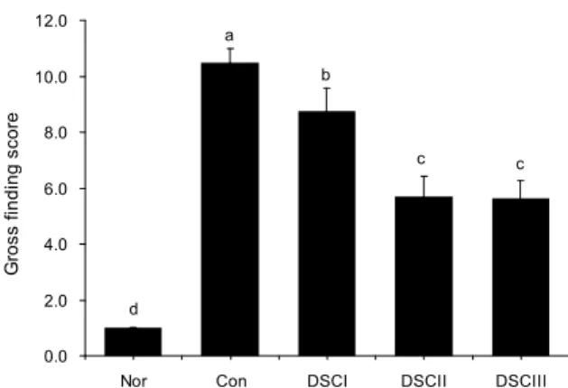
0.0 2.0 4.0 6.0 8.0 10.0 12.0
Nor Con DSCI DSCII DSCIII
G ro s s f in d in g s c o re .
d
a
b
c c
Fig. 1. Effects of dried sea cucumber on gross finding in gas- tritis model by HCl. Data was means±SE (n=7). Bars with dif- ferent letters from the control are significantly different (p<0.05).
Nor: normal; Con: control; DSCⅠ: dried sea cucumber Ⅰ, 30 mg/
kg; DSCⅡ: dried sea cucumber Ⅱ, 100 mg/kg; DSCⅢ: dried sea cucumber Ⅲ, 300 mg/kg.
Inhibition ratio (%)=
lesion length (control)-lesion length (drug) lesion length (control) × 100
H. pylori
균 배양 및 감염유발, 실험물질 투여
H. pylori
(ATCC 43504, American Tissue Culture Col- lection, Rockville, MD, USA) 균주를 10% calf serum이 첨 가된 브루셀라 한천배지에 접종하여 10% CO
2, 37
oC 및 mi- croaerobic 조건에서 3일간 배양하였다. 배양된
H. pylori는 2.0×10
9/mL colony-forming unit(CFU)의 균를 접종하여 배양하였다.
H. pylori
감염 유발
6~7주령 Mongolian gerbil을 12시간 절식시킨 후 2×
10
9/mL CFU의
H. pylori균을 마우스용 존데(sonde)를 이 용하여 0.5 mL씩 이틀 간격으로 3회 경구투여 하여 감염을 유도하였다.
신속요소분해효소 검사
신속요소분해효소검사(rapid urease test)는 campylo- bacter-like organism(CLO) 검사 시약인 Asan Helicobacter Test kit(Asan Pharmaceutical Co., Seoul, Korea)를 이용하 였다. 1주 동안
H. pylori감염 후 4주간 건해삼 투여한 동물 모델의 위 점막의 조직(pyloric region)을 Asan Helicobacter Test kit에 넣고 37
oC로 12시간 배양 후에 판독하였다. 판독 결과 색이 노란색에서 적색으로 바뀐 경우 양성으로 판단하 고, 색깔 변화가 없는 경우를 0점, 약간 붉은색을 나타낼 경 우 1점, 연한 자주색을 나타낼 경우 2점, 진한 자주색을 나타 낼 경우 3점으로 측정하였다.
조직학적 검경
10% formalin 액에서 고정시킨 검체를 위체부에서 육안 손상이 관찰된 부위가 포함되도록 절취하여(trimming) 다 시 10% formalin 액으로 후 고정시킨 후 일반적인 방법에 따라 조직학적 관찰을 실시하였다. 즉, 위조직을 파라핀에 포매 후 5∼7 μm의 두께로 절편하여 세절 후 hematox- ylin-eosin으로 염색하여 광학현미경 하에서 관찰하였다.
통계처리
모든 실험결과는 평균±표준오차(mean±SE)로 계산하였 다. 각 군 간의 통계적 유의성 검정은 ANOVA(one-way analysis of variance test) Duncan 사후검정 비교를 실시하 여 p<0.05일 때 유의한 것으로 판정하였다(SPSS V12., SPSS Inc, Chicago, IL, USA).
결과 및 고찰
HCl-ethanol 모델에서 건해삼의 효과에 대한 육안적 소견 SD rat는 효능평가 및 독성학과 약물학 연구에서 광범위 하게 이용되고 있다. 본 계통의 mice는 풍부한 시험 기초자
료가 축적되어 있어서 시험결과의 해석 및 평가 시에 보다 객관적인 결과를 얻을 수 있다. 이러한 SD rats에 HCl- ethanol을 경구투여하면 위점막층의 지질과산화 증가 및 점 막손상을 유발한다는 보고(18)에 따라 위염 및 위궤양을 유 발하였다. 위염 유발물질인 HCl-ethanol을 투여 30분 후 SD rat의 움직임과 증상을 살펴보고 1시간 동안의 활동량에 따 른 평가를 실시하여 기록하였다. 실험 결과는 Table 1과 같 은 기준으로 평가를 실시하였다(Fig. 1). 정상군에서는 고개 를 가누고 정상적 보행 및 털 고르기, 먹이 섭취 등의 행동을 보였으나 HCl-ethanol와 생리식염수를 투여한 대조군의 육 안적 소견 점수는 10.5±0.50으로 대부분 움직임이 없었으며 자극에 의해서 약간의 움직임을 보였다. 그에 비하여 HCl- ethanol과 함께 건해삼을 농도(30 mg/kg, 100 mg/kg, 300 mg/kg) 의존적으로 투여한 DSCI, Ⅱ, Ⅲ군의 육안적 소견 점수는 각각 8.8±0.82, 5.7±0.71 및 5.6±0.63으로 대조군에 비하여 움직임이 증가하였다. 특히 DSCII와 DSCIII군에서 자극에 의한 움직임 유의적으로 증가하였다(p<0.05). 그러 나 DSCIII군은 DSCII군에 비하여 3배의 고농도의 건해삼을 투여하였음에도 육안적 소견 평가에서 DSCII군과 유사한 결과가 관찰되었다. 이는 건해삼 100 mg/kg 이상의 농도에 서도 위 손상 억제효과는 100 mg/kg과 유사한 효과가 관찰 되었음으로 유효 농도 100 mg/kg에서 위 손상 완화에 효과 가 있음을 간접적으로 시사한다.
HCl-ethanol 위염 모델에서 육안 소견
HCl-ethanol에 의한 위염 및 위궤양은 에탄올이 위 점막 을 직접적으로 자극하여 점막하근층에 부종을 유발하고 국 소적으로 일시적인 허혈상태를 발생시켜 미세혈액순환이 정체되어 급성위염이 유발되고, 또한 HCl이 위 운동을 항진 시켜 급성위염을 더욱 악화시키는 것으로 보고되었다(19).
이 실험모델에서 HCl-ethanol이 위 점막의 방벽을 파괴함으
로써 H
+이온의 역확산(back diffusion)을 유발하여 염증을
악화시키는 것으로 알려져 있다(20). 본 실험에서는 HCl-
Fig. 2. Effects of dried sea cucumber on gross lesion in gas- tritis model by HCl. Nor: normal; Con: control; DSCⅠ: dried sea cucumber Ⅰ, 30 mg/kg; DSCⅡ: dried sea cucumber Ⅱ, 100 mg/ kg; DSCⅢ: dried sea cucumber Ⅲ, 300 mg/kg.
-20 0 20 40 60 80 100
Nor Con DSCI DSCII DSCIII
G a s tr ic d a m a g e in h ib it io n ( % ) .
a
c
c
b b
Fig. 3. Effects of dried sea cucumber on inhibition ratio of gastric damage in gastritis model by HCl. Data was means±
SE (n=7). Bars with different letters from the control are sig- nificantly different (p<0.05). Nor: normal; Con: control; DSCⅠ:
dried sea cucumber Ⅰ, 30 mg/kg; DSCⅡ: dried sea cucumber Ⅱ, 100 mg/ kg; DSCⅢ: dried sea cucumber Ⅲ, 300 mg/kg.
ethanol을 투여하지 않은 정상군에 비하여 HCl-ethanol과 함께 생리식염수만을 투여한 대조군은 HCl-ethanol 투여 후 위체부와 위저부 등에 선명한 점막 손상과 띠 모양으로 선상 의 출혈 소견이 관찰되었다(Fig. 2). 이는 Robert 등(21)이 보고한 HCl-ethanol에 의한 위 손상 병변의 육안소견과 일 치하게 glandular portion의 출혈에 의한 검붉은 선의 lesion 이 발생된 것으로 확인할 수 있었다. 이에 비하여 농도별로 해삼을 투여한 군에서 위 손상이 억제된 것을 관찰할 수 있 었다. 위 손상 면적을 측정하여 억제율을 정량화하였다(Fig 3). 대조군에서는 위 전체 면적대비 25.4±2.50%의 위 손상 이 관찰되었으며 건해삼을 저용량으로 투여한 실험군(DSC
Ⅰ)에서는 19.2±4.05%로 위 손상 억제율이 24.5±15.93%로 관찰되었으나 유의성은 없었다. 그러나 중, 고용량의 건해삼 투여한 실험군(DSCⅡ, Ⅲ)에서는 위 손상 면적이 각각 9.4
± 1.91%와 10.0±1.48%로 위 손상 억제률이 63.1±7.49%와 60.7±5.80%로 대조군에 비하여 유의하게 위 손상이 억제되
었다(p<0.05).
해삼의 구성성분의 조성에 대한 연구에서 동결건조 해삼 분말은 대부분 당단백질과 황산콘드로이틴(chondroitin sul- fate, ChS)으로 이루어져 있으며 화학적인 조성은 sulfate esters 함량이 0.90~1.21%, 다당류 함량이 23.08~26.97%, 구성단당류는 fucose의 함량이 30% 이상을 차지하고 있다 (22,23). 이러한 해삼의 ChS는 D-glucuronic acid, N-ace- tyl-D-galactosamine과 황산기로 결합되어 있는 뮤코다당 (mucopolysaccharide)이다. 점액층을 구성하는 뮤코다당은 위장의 점액층 형성을 도와줌으로써 위장내벽상피세포를 기계적인 손상이나 화학적인 자극으로부터 보호하는 기능 을 한다(24-31).
해삼은 다양한 기능성 성분이 다량 함유되어 있고, 이러한 기능성분들은 HCl-ethanol과 같은 위 손상 물질로부터 위 조 직을 보호할 것으로 생각되며, 본 연구에서 해삼 투여는 HCl-ethanol에 의한 위 손상을 감소시킴으로써 위 보호에 중요한 작용을 할 것으로 생각된다.
H. pylori
감염모델에서의 신속요소분해효소 검사
H. pylori
감염은 만성위염, 소화성 궤양 등 각종 소화기 질환의 주요 발병요인(32)이며 장시간의 감염은 위암의 발 병원인(33)으로 알려져 있다. Mongolian gerbil(
Meriones unguiculatus)에서
H. pylori는 사람의 병태 생리와 유사하 게 만성으로 진행되어 위염, 위궤양, 위암을 유발하는 것으 로 알려져 있으며(34)
H. pyloristrain을 Mongolian gerbil 에 감염시키면 감염율이 높으며, 병리소견 또한 사람과 유사 하다는 보고(35) 이후 현재까지
H. pylori의
in vivo실험은 Mongolian gerbil을 주로 사용하여 수행되고 있다(36). 신속 분해효소 검사는 위 점막에
H. pylori로부터 분비된 요소분 해효소(urease)를 가수분해에 의해 변화된 pH를 측정함으 로써
H. pylori감염유무를 확인하기 위하여 주로 이용된다.
본 연구에서는 1주간
H. pylori를 감염시킨 Mongolian gerbil을 이용하여 4주간 건해삼 투여하여 신속요소분해효 소 검사를 실시하였으며, 결과는 Fig. 4와 같았다.
H. pylori를 감염시키지 않은 정상군에서 모든 개체에서 색의 변화은 없었으나,
H. pylori감염군인 대조군과 실험물질 투여군은 적색을 나타내어 양성반응을 나타내었다. DSCⅢ군의 신속 요소분해효소는 대조군 1.83±0.31점에 비하여 1.33±0.49로 신속요소분해효소 양은 감소한 결과를 나타났으나, 통계적 유의성은 없었다.
신속분해효소 검사는 위점막
H. pylori감염유무를 판정하 는 정성적 자료만 제공하는 목적으로 고안되었지만
H. pylo- ri의 세균 수에 의해 발색 정도의 차이가 나타나며, 발색 정 도의 차이로 간접적인 세균의 수 차이를 비교할 수 있다(37).
본 연구에서는 대조군에 비하여 해삼 투여군의 신속분해
효소 검사 점수가 감소하는 경향은 관찰되었으나 유의성은
없었다. 이는
H. pylori의 5주간의 감염기간으로 짧은 감염
기간으로 인해 통계학적인 유의성 있는 결과를 관찰하지 못
-0.5 0.0 0.5 1.0 1.5 2.0 2.5 3.0
Nor Con DSCI DSCII DSCIII
C L O t s e t s c o re .
aa
a
a
b
Fig. 4. Effects of dried sea cucumber on change of CLO test score in gastritis model by H. pylori infection. Data was means±SE (n=6). Bars with different letters from the control are significantly different (p<0.05). Nor: normal; Con: control; DSCⅠ:
dried sea cucumber Ⅰ, 30 mg/kg; DSCⅡ: dried sea cucumber
Ⅱ, 100 mg/ kg; DSCⅢ: dried sea cucumber Ⅲ, 300 mg/kg.
Table 2. Histopathological lesion scores of the stomach in the study on therapeutic effects of H. pylori infection with several dried sea cucumber (5 week experiment)
Group n Inoculation
1)Histopathological lesion scores
2)H. pylori Treatment 4 Weeks Nor Con
DSCⅠ DSCⅡ DSCⅢ
6 6 6 6 6
No
3)Yes Yes Yes Yes
PBS PBS Low High Med
0.0±0.00 3.0±2.16 2.2±1.47 2.1±1.86 1.3±1.11 Nor: normal; Con: control; DSCⅠ: dried sea cucumber Ⅰ, 30 mg/kg; DSCⅡ: dried sea cucumber Ⅱ, 100 mg/kg; DSCⅢ: dried sea cucumber Ⅲ, 300 mg/kg.
1)
Treatment was conducted daily during 6 weeks after H. pylori inoculation.
2)
Histological lesion scores were calculated with the sum of his- tological grades of 3 tissues in each animal.
3)
The animals of this group were inoculated culture media alone instead of H. pylori.
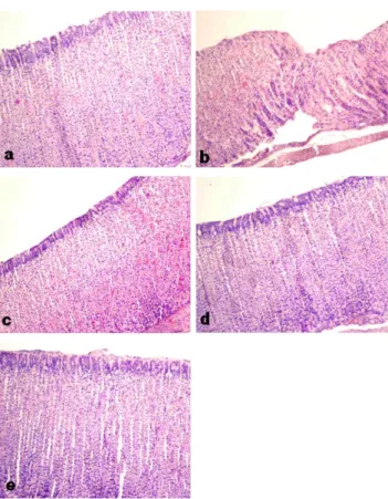
Fig. 5. Histopathological findings of stomach 5 weeks after H. pylori infection with or without extracts. H&E (×100). a:
Normal. No histopathological lesion. b: control. Moderate cellular degeneration and atrophy in the mucous layer. c: dried sea cu- cumber Ⅰ (DSCⅠ), 30 mg/kg. Mild cellular degeneration and atrophy in the mucous layer. Mild cellular degeneration and atro- phy in the mucous layer. d: dried sea cucumber Ⅱ (DSCⅡ), 100 mg/kg. Mild cellular degeneration and atrophy in the mucous layer. e: dried sea cucumber Ⅲ (DSCⅢ), 300 mg/kg. Mild cellular degeneration and atrophy in the mucous layer.
한 것으로 예상되며, 감염기간을 증가 시 통계학적 유의성 있는 결과를 관찰할 것으로 생각된다.
H. pylori
감염 모델 병리조직학적 검사 결과
H. pylori
감염 후 4주간 해삼추출물을 투여 받은 실험군 들의 위 점막의 병리조직학적 검사결과는 Table 2와 같다.
H. pylori
비감염군인 정상군의 모든 개체에서 위 점막에 유의한 병변은 관찰되지 않았다(Fig. 5A). 반면
H. pylori감염군인 대조군에서는 개체에 따라 병변의 정도차이는 있 지만 위점막 상피세포의 손상과 상피세포 위축 소견이 관찰 되었다(Fig. 5B).
H. pylori감염 후 해삼 추출물 저농도 투여 군인 DCSⅠ군, 중간농도 투여군인 DCSⅡ, 고농도 투여군 인 DCSⅢ 모두에서 개체에 따라 가벼운 위점막 상피세포 위축 소견과 염증세포 침윤이 있는 개체가 일부 관찰되었으 나 대조군보다 병변 정도가 개선된 것을 알 수 있었다(Fig.
5C~E).
H. pylori감염 후 해삼 추출물 저농도 투여군인
DCSⅠ군의 병리조직학적 점수(histological lesion scores) 는 2.2±1.47로서 대조군의 병리조직학적 점수인 3.0±2.16 과 비교하여 유의성은 확인되지 않았으나 감소 경향이 있음 을 알 수 있었다.
H. pylori감염 후 해삼 추출물 중간농도 투여군인 DCSⅡ군의 병리조직학적 점수는 2.1±1.86으로서 대조군의 병리조직학적 점수인 3.0±2.16과 비교하여 유의 성은 확인되지 않았으나 감소 경향이 있음을 알 수 있었다.
H. pylori
감염 후 해삼 추출물 고농도 투여군인 DCSⅢ군의
병리조직학적 점수는 1.3±1.11로서 대조군의 병리조직학적
점수인 3.0±2.16과 비교하여 유의성은 확인되지 않았으나
감소 경향이 있음을 알 수 있었다(Table 2). 실험에 사용된
개체수와 점수의 단위의 관계에 의해 통계 분포가 바뀔 수
있음을 감안할 때, 평가방법을 좀 더 다르게 수정한다면 통
계적으로 유의한 결과를 얻을 수도 있을 것이다. 본 연구에
서는 해삼의 항위염, 항위궤양 및 항헬리코박터 효과에 대한
가능성에 초점을 맞춰 연구를 수행하였으며, 항위염, 항위궤
양 및 항헬리코박터 효과에 대한 연구에 있어서 기초자료로
써 그 의미를 둘 수 있을 것으로 생각된다.
요 약
이 연구에서는 SD rat과 Mongolian gerbil에서 실험적으 로 HCl-ethanol(150 mM HCl+60% ethanol)을 투여하여 급 성 위염을 유발하고,
H. pylori감염시켜 만성 위염, 위궤양 을 유발한 후, 건해삼을 투여하여 건해삼에 의한 항위염, 항 위궤양 효과 및
H. pylori감염 경향성 및 치료율을 검토하였 던바 다음과 같은 결과를 얻었다. 7주령 SD rats와
H. pylori에 감염시킨 7주령 Mogolian gerbils를 정상군(Nor, n=6)과 대조군(Con, 60% HCl-ethanol+증류수(vehicle), n=6)에 비 하여 실험군 Ⅰ(DSD Ⅰ, 60% HCl-ethanol+건해삼 30 mg/
kg, n=6), 실험군 Ⅱ(DSD Ⅱ, 60% HCl-ethanol+건해삼 100 mg/kg, n=6), 실험군 Ⅲ(DSD Ⅲ, 60% HCl-ethanol+건 해삼 300 mg/kg, n=6)에서는 건해삼 투여 용량에 따른 항위 염, 항위궤양 및 항헬리코박터 효과를 육안적 소견, CLO test 및 병리조직학적 검사를 통해 확인할 수 있었다. 이상의 결 과를 종합해볼 때 건해삼이 새로운 위염 및 위손상 치료제와 기능성식품의 개발에 활용할 수 있음을 확인하였다.
감사의 글
본 연구는 2010 지식경제부 지역연계기술개발사업(과제 번호: 70007950)의 지원에 의해 이루어진 것이며, 이에 감사 를 드립니다.
문 헌