REVIEW ARTICLE
척수 손상에서의 치료적 중재: 약물, 재활, 이식, 심리 치료
임우택
1,2
ㆍ최봉삼1,2
우송대학교
1
보건복지대학 물리치료학과,2
첨단융합 스포츠재활 연구소Current Therapeutic Approaches in Spinal Cord Injury:
Pharmacological, Rehabilitation, Cell-based, and Psychological Intervention
Wootaek Lim 1,2 , Bongsam Choi 1,2
1
Department of Physical Therapy, College of Health and Welfare,
2Advanced Institute of Convergence Sports Rehabilitation, Woosong University, Daejeon, Korea
Received December 29, 2016 Revised March 10, 2017 Accepted March 10, 2017 Corresponding author Bongsam Choi
Department of Physical Therapy, College of Health and Welfare, Woosong University, 171 Dongdaejeon-ro, Dong-gu, Daejeon 34606, Korea Tel: +82-42-630-4622 Fax: +82-42-630-4611 E-mail: [email protected]
Copyright © 2017 by Stress. All rights reserved.Key messages
이 연구는 현재 척수 손상시 널리 적용되는 치료적 중재에 대한 고찰 및 제한점에 대해 알아보고자 하였다. 척수 손상은 신경학적 손상과 그에 따른 심각한 근육 위축을 초래한다. 손상 초기에는 산화적 스트레스, 지질 과산화 등과 같은 이차적 손상으로 인한 추가 신경 손상을 막기 위해 약물요법이 널리 사용되고 있다. 또한 삶의 질과 관련성이 높은 보행 능력에 대한 임상 전, 임상 연구는 보행 능력의 회복 에 그 촛점을 맞추고 있으며, 이에 대한 예로서, 트레드밀 훈련, 자전거 훈련, 로봇 훈련 등이 시행되고 있다. 최근에는 세포나 조직을 이식하는 이식치료가 많은 관심 속에 연구되고 있으나 안전성을 확보하기 위한 추가적인 연구가 필요하다. 척수손상은 단순히 신체손상뿐아니라 정신건강에도 부정적 영향을 주 는 다면적인 손상을 초래할 수 있기때문에 단독치료 접근방법 보다는 심리인지적 치료를 포함한 다양한 치료가 결합된 형태를 고려해야 한다.
중심단어: 보행 훈련, 외상 후 스트레스 장애, 척수손상, 줄기세포, 스테로이드 Abstract
The purpose of this review is to discuss current therapeutic interventions and their limitations for the optimal treatment outcomes of individuals with spinal cord injury (SCI). SCI leads to neurologic deficits and subsequent severe muscle atrophy. In clinics, despite the controversy, pharmacological therapy using methylprednisolone has widely been accepted to reduce additional neurologic deficits caused by a secondary injury such as oxidative stress and lipid peroxidation. Moreover, it facilitates the recovery process. Since the loss of locomotor function may reduce the quality of life for individuals with SCI, many pre-clinical and clinical studies have focused on the recovery of locomotor function.
Various forms of locomotor training such as treadmill training, cycle training, and robotic-assisted training are currently available for individuals with SCI. Additionally the cell-based interventions have been receiving much attention as one of potential therapeutic interventions which required further clarifications due to the issues of safety. The physical impairment associated with spinal cord injury may cause an adverse effect on mental health. It is now recommended that combined physical and psychological interventions should be considered to maximize the efficacy of therapeutic interventions.
Key Words: Locomotor training, Post-traumatic stress disorder, Spinal cord injury, Stem cell, Steroid
Introduction
In the U.S., it is estimated that 282,000 persons live with spinal cord injury (SCI), with approximately
17,000 new cases each year (
Spinal Cord Injury
, 2016).The most frequent neurologic categories are incomplete, followed by complete (
Spinal Cord Injury
, 2016). Since very few people experience complete neurologic recovery, most patients end up requiring extensiveTable 1. Summary of National Acute Spinal Cord Injury Study I, II, and III
Study NASCIS
aI (1984) NASCIS II (1990) NASCIS III (1997)
Number of patients 330 487 499
Dosing MP
b100 mg + 25 mg every 6 hours for 10 days or MP 1,000 mg + 250 mg every 6 hours for 10 days
MP 30 mg/kg + 5.4 mg/kg/hr for 23
hours MP 5.4 mg/kg for 24 hours or MP 5.4
mg/kg/hr for 48 hours Outcomes No significant differences between two
groups Significant improvement in motor
function Significant improvement in motor
function in individuals treated for 48 hours
a
National Acute Spinal Cord Injury Study,
bmethylprednisolone
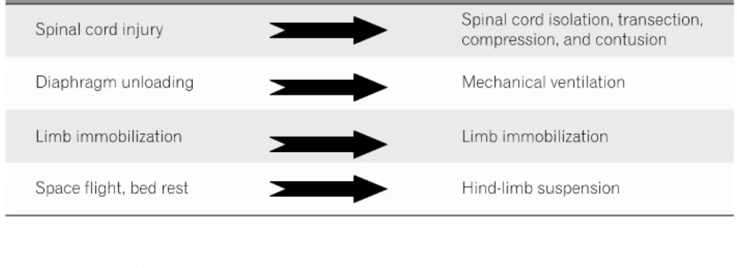
Fig. 1. Human conditions and corres- ponding experimental models.
physical therapy care, for an extended period of time up to many years. Thus, better understanding of SCI is very important for physical therapist to help, guide and establish rehabilitation strategies.
The analysis of clinical data is complicated by the broad diversity consequences, and the population affected resulting from the injuries. Also, data are not easily collected at the early stage post injury in clinics.
For these reasons, experimental models have widely been accepted in preclinical research. The patho- physiological changes in muscles and the preventive strategies for the neurologic deficits following SCI have widely been studied in animal models of SCI (Fig. 1).
The safety, feasibility, and effectiveness of therapeutic interventions strongly support the potential for translation of these experimental models for clinical application.
In traumatic SCI, which is mostly observed in human beings, the overall damage is determined by the combined secondary injury at the molecular and/or cellular levels after primary mechanical insult to the spinal column. The injury caused by external force is inevitable, but it is possible to delay secondary physi- ological and biological changes using pharmacological interventions. In addition, various physical rehabil- itation programs are being applied for rapid physical recovery. Rapid recovery of functional ability can lead to a reduced economic burden of care by shortening hospital length of stay and complications. Finally,
recent advances in medical science and technology, along with the development of cell transplantation therapies, are expected to overcome the therapeutic limitations of conventional approaches.
However, most of the therapeutic approaches currently in use have some limitations, along with the positive effects. This literature review aims to briefly explore and describe current therapeutic interventions and their limitations.
Search Strategy
Electronic searches were conducted using EMBASE, MEDLINE, and PubMed to identify relevant literatures published until February 2017. The keywords used to search were (spinal AND cord AND injury), ((pharma- cological, rehabilitation, OR cell-based) AND (interven- tion OR therapy)). The terms were identified in the title or the abstract of journal articles. The studies written in English were included in this review, while Conference abstracts, Case study, and editorial notes were excluded.
Therapeutic Approaches in Spinal Cord Injury
1. Pharmacological interventions
Because of its anti-inflammatory function, steroids have been considered as one of potential interventions
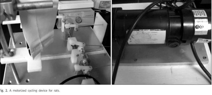
Fig. 2. A motorized cycling device for rats.
in the management of SCI for more than last 40 years.
It is presumed that steroids can inhibit or minimize oxidative stress, inflammation, intracellular calcium accumulation, and vascular abnormalities. The efficacy of steroids in the management of SCI was established by National Acute Spinal Cord Injury Studies (NASCIS) I, II, and III. These studies examined methylpredni- solone (MP) as a pharmacological treatment interven- tion in SCI. In NASCIS II and III, significant improve- ment of sensory and motor function was observed at 6 and 12 months post-injury with MP treatment, which is given within 8 hours of injury (Bracken
et al
., 1992; Brackenet al
., 1990; Brackenet al
., 1997;Bracken
et al
., 1998) (Table 1). However, the findings of these studies are controversial due to issues of statistical analyses and its interpretations. In addition, the potential side effects of methylprednisolone treatment were not taken into consideration while monitoring the effectiveness of steroid therapy in SCI patients (Kwonet al
., 2004).In an attempt to halt the detrimental effects of glutamate accumulation following SCI, antagonists of glutamate receptors were used in several studies to inhibit glutamate toxicity and stop the excitotoxicity cascade. Although these drugs provided neuropro- tection with subsequent improvement in animal behavior following contusion SCI (Mills
et al
., 2002), no improvement was seen in human studies. Additio- nally, calcium- and sodium-channel blockers have been tested as potential treatment interventions in SCI.In animal studies, the calcium channel blockers failed to show any significant improvement (Haghighi
et al
., 1993). In contrast to calcium blockers, significant im- provement in neuroprotective function was observed after treatment with sodium channel blockers in rats (Schwartzet al
., 2001; Tenget al
., 1997). However, similar benefits of treatment with sodium-channel blockers were not observed in few clinical studies.To prevent the programmed cell death following SCI, inhibition of caspases has also been considered as a potential therapeutic intervention. Caspases are proteases involved in apoptosis, necrosis, and inflam- mation (Eldadah
et al
., 2000). After administration of minocycline, a significant decrease in caspase-3 and apoptosis was seen in moderate contusion SCI in rats (Festoffet al
., 2006). Additionally, in another contusion model of SCI in rats, inhibitiory action of oxidative stress was observed following minocycline admini- stration, specifically lipid peroxidation (Sonmezet al
., 2013). Since much more tissue damage is induced by secondary pathophysiological events including oxida- tive stress and lipid peroxidation, the suppression of oxidative stress plays a key role in the amount of secondary damage.Besides the pharmacologic interventions, the use of inhibitors of cyclooxygenase such as Ibuprofen and meclofenamate have been confirmed on improving its function following moderate injury in animal models of SCI (Resnick
et al
., 1998). Recently, other pharma- cologic interventions such as inhibitor of p38, one ofmitogen-activated protein kinases, and fluorocarbon have also been reported to be effective for improving locomotor function (Umezawa
et al
., 2017; Yacoubet al
., 2014). In summary, pharmacologic interventions have primarily been focused on minimizing secondary injuries and enhancing locomotor recovery by inhi- biting inflammation, free radicals, glutamate accumula- tion, apoptosis, and demyelination. These factors may hold promise for future clinical trials.2. Rehabilitation interventions
Significant improvement in muscle properties and function has been reported by implementing activity- based rehabilitation strategies for both incomplete and complete SCI despite the very limited functional recovery in human beings. Strategies of Locomotor training such as treadmill training, cycle training (Fig.
2), and robotic-assisted training take advantage of the central pattern generator to induce movement, and stimulates the spared region of the spinal cord resulting in temporary as well as permanent muscle and spinal cord adaptations. In studies by the use of animal model, even one week of treadmill training after moderately contused SCI have proved that there were increases in muscle fiber CSA and improvements in locomotor function (Stevens
et al
., 2006). Studies using bicycle training by Boseet al
. and Liuet al
. had also confirmed similar effects on muscle recovery or locomotor function compared to treadmill training (Boseet al
., 2012; Liuet al
., 2008). In humans, an increase in muscle CSA was observed in thigh (4.9%) and calf muscle (8.2%) after 12 months of body weight supported treadmill training (BWSTT) (Giangregorioet al
., 2006). Similarly, thigh and lower leg muscle CSA were increased to 4.1-56.9% and 3.8-53.6%, respec- tively after 48 session of BWSTT (Giangregorioet al
., 2005). Additionally, improvement of gait speed (180%) and distance (335%) has been reported after BWSTT (12 months, 144 sessions) (Hickset al
., 2005). Hickset al
. also reported that most subjects with SCI main- tained walking scores even at 8 months post-training.Thus, locomotor training attenuates muscle atrophy, while it enhances muscle activity and gait quality (Behrman
et al
., 2000; Giangregorioet al
., 2006).However, few studies do not support the beneficial effects of physical training after injury. In a study by Fouad
et al
., 2000, no significant difference in kinema- tic and locomotor functional tests was observed in rats with incomplete SCI after early treadmill training(Fouad
et al
., 2000). Similarly, a study by Unget al
., 2010 reported no difference in locomotor function and muscle fiber conversion after treadmill training in complete transection injured mice (Unget al
., 2010).Optimal window of locomotor training with appro- priate intensity and duration might influence the effects of training after injury.
The changes in properties of spinal cord induced by physical activity are relatively less investigated as compared with those caused by other interventions. It has been demonstrated that physical activity in normal adult rat increases brain-derived neurotrophic (BDNF) and nerve growth factor, both which are related to survival of neurons and neural plasticity in various CNS regions (Alderson
et al
., 1990; Neeperet al
., 1996).Further, voluntary exercise increases BDNF and neuro- trophic factors in the lumbar spinal cord (Gomez- Pinilla
et al
., 2001). In rodent SCI, axonal sprouting and regrowth with increased synaptic markers were observed after locomotor training (Goldshmitet al
., 2008) and lesion volume was found to be smaller compared to a sedentary group (Andradeet al
., 2010).Interestingly, the levels of BDNF mRNA in the brain or lumbar spinal cord significantly correlated with the functional activity in normal adult rats (Gomez-Pinilla
et al
., 2001). These findings suggest that physical activity after SCI might induce neuro-regeneration and protection.Recently, increasing attention has been drawn to the approach that involved electrical stimulation on spinal cord. The communication between brain and the spinal cord below the level of injury is disrupted although motor neurons and nervous tissues are still intact at the levels above and below injury. The application of tonic stimulation at the lumbosacral level of spinal cord has been shown to have positive effects on loco- motion and stepping movements, even without sup- raspinal or sensory input. In addition, tonic epidural stimulation of the dorsum of the spinal cord has been shown to induce stepping movements in decerebrated cats (Gerasimenko
et al
., 2009) and in complete spinal rats (Ichiyamaet al
., 2005). The epidural spinal cord stimulation is now known to modulate the excitability of spinal circuits and locomotor function (Alamet al
., 2017; Edgertonet al
., 2012; Gadet al
., 2013).3. Cell-based interventions
In recent years, cell-based interventions have been introduced and advanced rapidly. This rapid progress will continue to grow (Gu
et al
., 2017; Laneet al
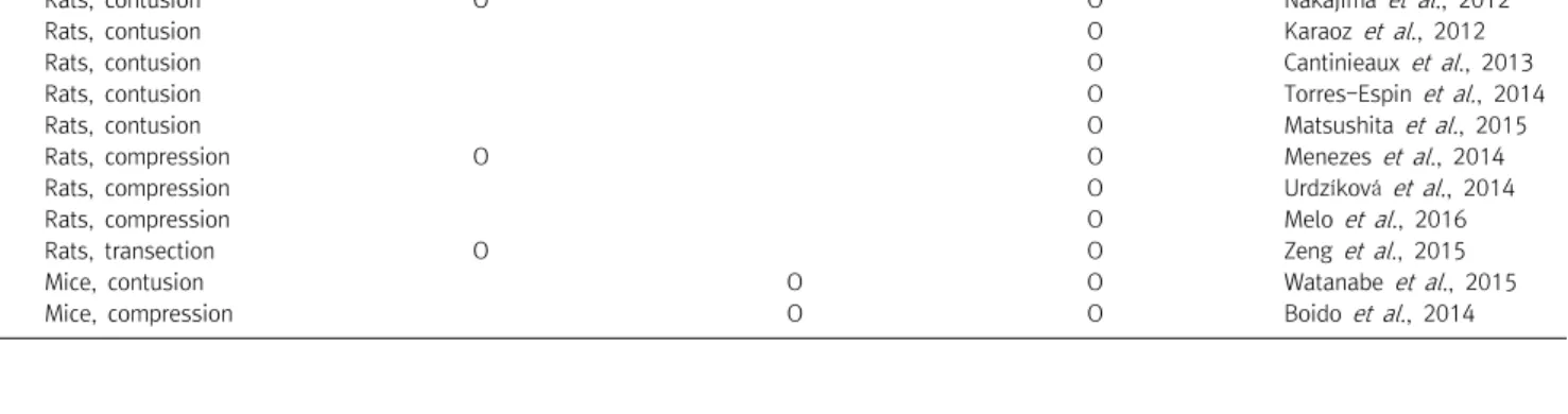
.,Table 2. Mesenchymal stem cell transplantation in an animal model of SCI
SCI models Neuronal or axonal regeneration Sensory improvement Motor improvement References
Rats, contusion O O Nakajima et al ., 2012
Rats, contusion O Karaoz et al ., 2012
Rats, contusion O Cantinieaux et al ., 2013
Rats, contusion O Torres-Espin et al. , 2014
Rats, contusion O Matsushita et al ., 2015
Rats, compression O O Menezes et al ., 2014
Rats, compression O Urdzíková et al ., 2014
Rats, compression O Melo et al ., 2016
Rats, transection O O Zeng et al ., 2015
Mice, contusion O O Watanabe et al ., 2015
Mice, compression O O Boido et al ., 2014
2016; Wang
et al
., 2016; Watzlawicket al
., 2016).Transplantation of cells and tissues focuses on the replacement or reconnection of tissue and neuron because conventional approaches showed a limited effect on axonal regeneration and functional recovery.
Cell-based intervention began with bone marrow transplantation in the late 1950’s and neural stem cells for neurons were successfully isolated
in vitro
from the mammalian neural crest and cultured in the early 1990’s (Mathéet al
., 1959; Stemple & Anderson, 1992). Since then, various stem cellsin vitro
were further studied. However, the findingsin vitro
in the laboratory do not guarantee that the same results can be obtainedin vivo
because studiesin vitro
are conducted in a relatively controlled environment and the complex reactions between cells are simplified outside of organisms. With progress inin vitro
envir- onment, additional experiments are performed inin vivo
and repeated in large animal models of SCI in different species. Cell survival, distribution, migration, and diffe- rentiationin vivo
can be assessed through various tools. In recent years, the tracking of cells in living or- ganisms has become possible to measure non-invasively through cellular magnetic resonance imaging (Jirjiset al
., 2017; Shroff, 2017; Sykovaet al
., 2007). Once the safety and efficacy have been demonstrated inin vitro
andin vivo
, clinical trials for application to individuals with SCI are currently available. In many clinical trials, however, ethical issues may hinder the progress and application of the experiments. In addition, unexpected problems can arise with the different physical and physiological characteristics of each patient. In patients with SCI, chronic patients may be considered as pre- ferentially to subacute patients in the phase I clinical trial due to spontaneous recovery at the subacute stage.Currently, embryonic, neural, and mesenchymal stem cell are being widely investigated and are likely to
expand further in the future. Embryonic stem cells are pluripotent stem cells derived from the inner cell mass of the early embryo (Blair
et al
., 2011). Those cells are known to be capable of self-renew indefinitely and differentiate all cell types (Puriet al
., 2012). The transplant proceedsin vivo
after pre-differentiationin vitro
(Hendrickset al
., 2006). Significant motor recovery and remyelination were commonly reported in a rat model of SCI (Liuet al
., 2000; McDonaldet al
., 1999).In other studies, however, tumor formation was observed after transplantation (Nussbaum
et al
., 2007; Riesset al
., 2007). Neural stem cells are multipotent and used to replace lost tissue after injury. In bothin vitro
andin vivo
, various neurotrophic factors were secreted by neural stem cells (Luet al
., 2003). Improvement of locomotor function, remyelination of axons and normal conduction velocities from the remyelinated axons have been shown (Akiyamaet al
., 2001; Bottaiet al
., 2008), but neuronal differentiation limited as transplanted cells remained undifferentiated and is a potential pro- blem. Lastly, mesenchymal stem cells are also multipotent cells. These cells can be easily isolated from bone marrow and adipose. In particular, bone marrow which can be obtained by iliac crest puncture is preferred.Therefore, it is relatively free from ethical issues compared to others. Differentiation in vitro into the neurons, astrocytes, myoblasts, and osteoblasts have been observed (Jiang
et al
., 2002; Pittengeret al
., 1999). Interestingly, neurotropic factors, when combined with transplantation of fetal tissue, were more beneficial as shown by improvement in axonal growth, length of the projection, and number of fiber branches (Bregmanet al
., 1997). Another beneficial factor of mesenchymal stem cells is an anti-inflammatory effect. The upre- gulation of transforming growth factor beta 1, an anti- inflammatory factor that controls cell proliferation and apoptosis, has been observed and significantly reducedoxidative stress (Chen
et al
., 2011; Hawryluket al
., 2012; Kempet al
., 2010; Whoneet al
., 2012). Unlike others, there are almost no allergic reaction in mesen- chymal stem cells. It is known not to cause hypersensi- tivity reactions (Carradeet al
., 2011; Kramperaet al
., 2003).However, unlike animal studies (Table 2), the efficacy has been reported to be somewhat limited in clinical trials (Syková
et al
., 2006; Yoonet al
., 2007). In conclusion, many studies based on cell transplantation showed signi- ficant and valuable changes of neural tissue and motor function, while an understanding of the exact mechanism of recovery or regeneration requires further studies because much still remains unclear and unexpected adverse effects may also occur.4. Psychological interventions
Although many pre-and clinical studies with human and animal models have been widely conducted for the treatment of spinal cord injury, little is known about the psychological intervention. Spinal cord injury does not simply end with physical impairment, but carries an adverse effect on human mental health. Human mental health can directly affect patients’ outcome of treatments.
For instance, depression following SCI reduces the effectiveness of treatment and increases the length of hospital stay and treatment (Malec
et al
., 1983; Tateet al
., 1994). In 2011, Fann and colleagues reported 23%of patients with SCI were suffering from depression and only about 29% to 11% of them were on medi- cation or receiving psychotherapy (Fann
et al
., 2011).Post-traumatic stress disorder (PTSD) is another issue in SCI. Individuals with SCI have been known to ex- perience PTSD at a higher rate. In a study by Radnitz, the current and lifetime prevalence of PTSD were 12%
and 29% (Radnitz
et al
., 1998). Unfortunately, many of them were accompanied by pain (Ullrichet al
., 2013).The psychological treatments may have only moderate effects in treating depression (Perkes
et al
., 2014).Although there is a lack of evidence for the effect on PTSD, psychological treatment may be of some help in relieving pain in patients with the trauma(Mohta
et al
., 2003). Rather than performing the psychological therapy alone, it may be better to combine cognitive behavioral therapy with psychoeducation (Perkeset al
., 2014).Conclusion
Various therapeutic interventions are effectively and selectively being performed in clinical practice. Each of
them has some limitations and it may require attention to application. Incorrect use of hi-dose steroids can be detrimental and aggravate neuronal damage (Hall
et al
., 2004). It is widely accepted that locomotor training after SCI assists in the qualitative, quantitative, and functional recovery of muscles. However, early loco- motor training may not be beneficial, but disa- dvantageous. The detrimental effects of locomotor training on the spinal cord and muscle may suggest the importance of the optimal time window and appro- priate mode of locomotor training by a physical therapist following SCI. Additionally, the cell-based approach is beyond the limits of existing conventional interventions, but unregulated stem cell transplant has the potential risk of tumor formation. It will be important to establish the safety of transplantation strategies before translating the preclinical findings to human beings. In order to maximize the effectiveness of treatment in SCI and ensure safety and feasibility, it is necessary to thoroughly understand each intervention as well as psychological treatment with its limitations.References