INTRODUCTION
The pathophysiology of acute spinal cord injury (SCI) is characterized by an initial, mechanical injury (primary injury), followed by a series of secondary injury events including ischemia, calcium- and sodium-mediated cellular injury, ex- citotoxic cell death, inflammation, and apoptosis. A variety of promising substances have been tested in animal models of acute SCI, but few have had potential application to human SCI patients. Methylprednisolone (MP) and GM-1 ganglio- side have met rigorous criteria in laboratory testing and initial human investigations. Although the National Acute Spinal Cord Injury Studies (NASCIS) II and III advocated the clin- ical use of MP for acute SCI, critical reviews and contradict- ing studies have cast doubts on its efficacy (1-3). After NASCIS II study, many clinical researches have shown that MP admin- istered according to the NASCIS regimen failed to improve outcome at least in moderate-to-severe cord injuries (4-8).
Thus, the beneficial effect of MP may not be great enough to advocate it as the standard of care for acute SCI.
Recent study has shown that MP depresses the production of growth-promoting factors such as brain-derived neurotroph- ic factor (BDNF) and neurotrophin-3 (NT3) following acute SCI (9). This suggests the possibility that, while MP prevents
secondary injury in the acute stage of SCI through various mechanisms, including inhibition of lipid peroxidation, calci- um influx, ischemia, axonal dieback, calpain-mediated cyto- skeletal degradation, and anti-inflammatory effects, it may hinder functional recovery after the acute stage, especially in severe spinal cord injuries. Thus, the combination of MP with other pharmacological agents that can promote functional recovery is desirable. Some studies have tested combination treatments but have focused on secondary injury reactions or on promoting regeneration rather than influencing both the acute and chronic stage following SCI.
The ability of growth factors to promote axonal regenera- tion in injured spinal cord has been extensively studied. BDNF is one of the best-characterized of the neurotrophic factors.
The neurotrophic activity of BDNF has been studied in var- ious models of central nervous system (CNS) injury, such as spinal cord trauma, contusion injury, and spinal cord hemi- section and axonal lesion. It has been shown to enhance sur- vival and to stabilize both sensory and motor neurons (10, 11).
Recent study has shown functional evidence that BDNF is an anterograde neuronal trophic factor in the CNS of the adult rat (12).
Adjunct neurotrophic factor, such as BDNF, combined with initial MP treatment may enhance functional outcome
Daniel H. Kim, Tae-Ahn Jahng*
Department of Neurosurgery, Stanford University School of Medicine, *Department of Neurosurgery, Seoul National University Hospital, Seoul, Korea
Address for correspondence Tae-Ahn Jahng, M.D.
Department of Neurosurgery, Seoul National University Hospital, 28 Yongon-dong, Chongno-gu, Seoul 110-744, Korea
Tel : +82.2-760-2356 , Fax : +82.2-744-8459 E-mail : [email protected]
113
Continuous Brain-derived Neurotrophic Factor (BDNF) Infusion After Methylprednisolone Treatment in Severe Spinal Cord Injury
Although methylprednisolone (MP) is the standard of care in acute spinal cord injury (SCI), its functional outcome varies in clinical situation. Recent report demonstrated that MP depresses the expression of growth-promoting neurotrophic factors after acute SCI. The present study was designed to investigate whether continuous infu- sion of brain-derived neurotrophic factor (BDNF) after MP treatment promotes func- tional recovery in severe SCI. Contusion injury was produced at the T10 vertebral level of the spinal cord in adult rats. The rats received MP intravenously immediately after the injury and BDNF was infused intrathecally using an osmotic mini-pump for six weeks. Immunohistochemical methods were used to detect ED-1, Growth asso- ciated protein-43 (GAP-43), neurofilament (NF), and choline acethyl transferase (ChAT) levels. BDNF did not alter the effect of MP on hematogenous inflammatory cellular infiltration. MP treatment with BDNF infusion resulted in greater axonal sur- vival and regeneration compared to MP treatment alone, as indicated by increases in NF and GAP-43 gene expression. Adjunctive BDNF infusion resulted in better locomotor test scores using the Basso-Beattie-Bresnahan (BBB) test. This study demonstrated that continuous infusion of BDNF after initial MP treatment improved functional recovery after severe spinal cord injury without dampening the acute effect of MP.
Key Words : Regeneration; Brain-Derived Neurotrophic Factor; Methylprednisolone; Spinal Cord Injuries
Received : 21 May 2003 Accepted : 9 October 2003
by preventing secondary injuries and by promoting neuronal regeneration. This study was designed to test this possibility in a contusion-injury model in the rat spinal cord. Combina- tion therapy was compared to no other treatment but to MP treatment alone.
MATERIALS AND METHODS Spinal Cord Contusion Injury
Forty-five adult female Sprague-Dawley rats weighing 200- 250 g were maintained under standard conditions at the Uni- versity Animal Care System. Spinal cord contusion injuries were performed using the UCSF spinal cord injury model.
Spinal cord injury model was tested several times with patho- logic examination, as well as neurological deficit to properly induce severe spinal cord injury. Rats were anesthetized with ketamine (60 mg/kg) and xylazine (10 mg/kg) injected int- raperitoneally. The lamina and spinous process of T10 were removed to expose a circular region of dura measured at app- roximately 2.8 mm in diameter. The spinous processes of T9 and T11 were clamped with tissue forceps, which in turn were held by Kopf rat spinal clamps, to stabilize the spinal column. The clamps were then raised so the thoracic region of the rat was suspended by the spinous processes. The animals were positioned under a 10-g weight device, which consisted of a Teflon impounder loosely fitted over the end of a brass rod and attached to a three-dimensional stereotaxic frame. A brass rod was inserted through the weight, which was secured by a removable pin. The impounder was centered over the laminectomy and was lowered onto the dura. The weight dropped by 5 cm when the pin was pulled to make contusion injury onto T10 level. Five seconds later, the entire rod assem- bly was raised off the dura. After sustaining the contusion injury, the muscles of the rat were sutured over the laminec- tomy site, and the skin was closed with wound clips. Post- operatively, the rats received a 5-mL subcutaneous injection of saline and an intramuscular injection of gentocin (2.5 mg) daily, and their bladders were expressed twice daily until reflex- es returned. The rats were maintained in pairs in separate cages.
Twenty-seven rats (3 rats per group for 1, 3, 7 days after injury respectively) were used for RT-PCR and eighteen rats (6 rats per group) were used for functional assessment.
MP Treatment and BDNF Infusion
The rats were divided into control, MP only (+ placebo infusion), and MP+BDNF infusion groups. Control rats were not treated after spinal cord contusion injury. For MP treat- ment, rats received 60 mg/kg MP intravenously immediately after SCI, and four hours later they received 30 mg/kg MP intravenously. For MP+BDNF treatment, BDNF (Alomone Co., Israel) was dissolved in an artificial cerebrospinal fluid
(CSF) solution (Na+150 mM, K+3 mM, Ca2+1.4 mM, Mg2+
0.8 mM, phosphate 1 mM, Cl-155 mM) at a concentration of 0.33 mg/mL. Each rat was implanted with an osmotic minipump-brain infusion assembly for continuous infusion (placebo or BDNF) of the injured site (Alzet, Palo Alto, CA, U.S.A.; Model 2004, mean pumping rate=6 L/day). The brain infusion assembly consisted of a catheter tube (2.5-cm length) and a stainless steel cannula with adjustment spacers to obtain the correct stereotaxic depth. The cannula was filled with the appropriate solution and connected to the pump.
The entire device was incubated in sterile saline at 37℃for at least 40 hr prior to surgery in order to ensure immediate flow of the solution once the pump was inserted in the rats.
Following the contusion injury, the cannula was inserted intrathecally at the injury site under surgical magnification using loupe and was secured by sutures. The osmotic pump was housed in a subcutaneous pocket in the back of the rat and was infused maximally for six weeks. Rats were subse- quently sacrificed and evaluated at the interval of one, three, and seven days, and then at ten weeks of post injury.
Immunohistochemical Analysis
The anesthetized rats were perfused with cold 0.1 M, pH 7.2 PBS containing heparin, and then perfused with 4%
buffered paraformaldehyde. Spinal cord from the contused site was cut and fixed in a 10% formalin solution for two hours, embedded in paraffin, then cut in 4- m sections for immunohistochemical staining. Briefly, the sections were treated with 0.3% H2O2in methanol to inactivate the endoge- nous peroxidase, and nonspecific staining was blocked with a solution of 3% normal serum. Primary antibodies raised against ED-1 (rabbit anti-ED-1, Serotech, U.K., 1:1,000);
NF (mouse anti-Neurofilament SMI31, Sternberger Mono- clonals, Baltimore, MD, U.S.A., 1:1,000); growth associated protein-43 (GAP-43, Chemicon, 1:1,000); and choline acetyl transferase (goat anti-ChAT; Chemicon, 1:500) were diluted in 1% normal serum, applied to the tissue sections for one hour at 37℃, and then applied to the tissue sections overnight at 4℃. Sections were incubated with an appropriate biotiny- lated secondary antibody (1:200, Vector laboratories, Burlin- game, CA, U.S.A.) followed by ABC complex for 45 min at room temperature. Diaminobenzidine (DAB-brown color;
Sigma Chemical Co., St. Louis, MO, U.S.A.) was used as the chromatogen. The sections were counterstained with hema- toxylin, dehydrated in graded alcohols, and mounted. Sections were also stained with appropriate normal serum for control comparison. Omission of primary antibodies resulted in only weak, nonspecific labeling of the blood vessels. Quantification of the proportional area at the center of the lesion at a mag- nification level of 40× was performed by using a computer- assisted digital image analysis system (Image Measure/IP Ver.
4.0-NIH image program), and by manually setting the rel- ative optical density threshold to identify positive stained
properties within a defined target area.
Reverse Transcriptase Polymerase Chain Reaction (RT-PCR)
Gene expression of growth-associated protein 43 (GAP-43), an endogenous indicator of axonal regeneration, was assessed by RT-PCR on one, three, and seven days following the con- tusion injury. Each therapy test group consisted of three ani- mals. Briefly, total cellular mRNA was isolated from the injury area at the different intervals by using AMV reverse transcrip- tase. The resulting cDNA was amplified by using the specific primer for GAP-43 (Upstream primer: AAGAAGGCAGGG- GAAGATAC; Downstream primer: GGACGGCGAGTTA- TCA GTGG). Quantification of mRNA levels was performed as previously described (13).
Behavioral Assessment
A total of 18 animals were used to investigate neurological functional recovery following SCI. Using the Basso, Beattie, and Bresnahan (BBB) scale (14), functional assessment of locomotion was performed by observers blinded to treatment methods. Rats were trained preoperatively each week in an open field that consisted of a plywood circular enclosure with a smooth non-slip floor. Open field training and testing proce- dures were done exactly as described in Basso et al. Rats were evaluated every week following injury and treatment.
Statistics
To determine whether differences in functional recovery were statistically significant, the data were examined by using a computerized statistical analysis program (SPSS 9.0 for Win- dows, SPSS Inc.). Histological outcome measures were ana- lyzed by using univariate ANOVA. If the main effects were significant, then outcomes were assessed by using Tukey’s HSD test. Behavioral measures were compared using a gen- eral linear model with repeated measures. If ANOVA inter- action effects (group×time) were significant (p<0.05), post hoc pairwise comparisons were performed by using Tukey’s HSD to compare groups at each point. Significance for all analyses was set at p<0.05.
RESULTS
Immunohistochemical analyses of spinal cord tissue after contusion injury were performed to compare the effects of the treatments on three variables: the extent of hematogenous inflammatory cellular infiltration (as indicated by ED-1 im- munoreactivity), the extent of axonal regeneration (as indicated by NF and GAP-43 immunoreactivity), and the extent to which regeneration occurred in cholinergic axons (as indicat-
ed by ChAT immunoreactivity).
Hematogenous Cell Infiltration
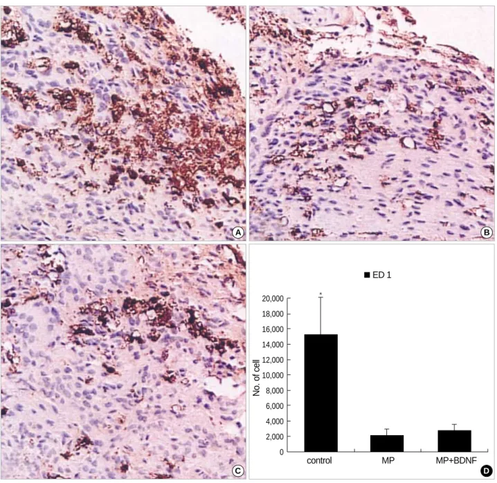
To assess infiltration of the contusion site by hematogenous cells (monocytes/macrophages), paraffin-embedded spinal cord tissue in the center of the contusion was immunostained with ED-1 antibody (15). During early stage (1, 3 & 7 days) after the injury, ED-1-positive cells increased substantially (Fig. 1A). Significantly fewer ED-1-positive cells were seen at the contusion site in the MP-only and MP+BDNF-infused animals (Fig. 1B, C). This difference was found to have sta- tistical significance after quantification of the injury area was performed using the computer-assisted image analysis system (Fig. 1D; p<0.05).
Axonal Survival and Regeneration
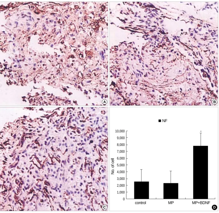
To evaluate axonal survival and regeneration, neurofilament (NF) and GAP-43 immunoreactivity were assessed at ten weeks of post injury. The significantly more NF-positive axons survived in the MP+BDNF-infused group (Fig. 2C) than in MP-only and control groups (Fig. 2A, B, D; p<0.05).
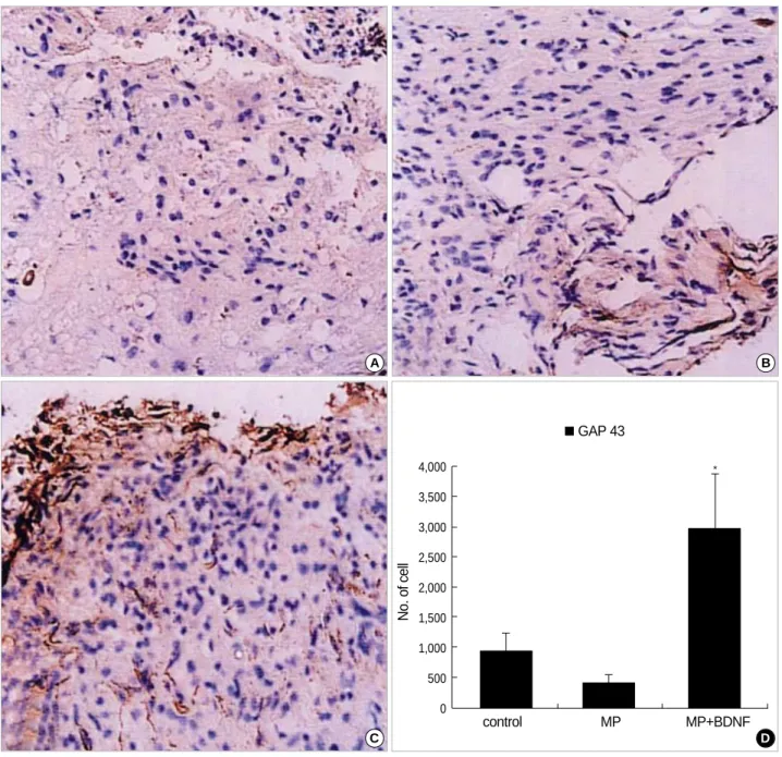
More GAP-43-positive neurites were also observed in the MP+BDNF-infused spinal cord (Fig. 3C) compared to the other two groups (Fig. 3A, B, D; p<0.05).
Motor Axonal Regeneration
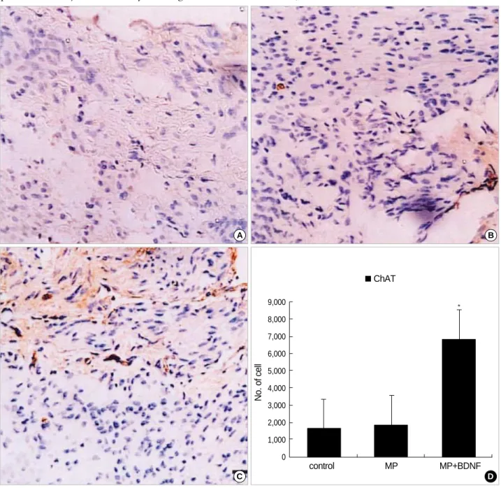
To identify the phenotype of regenerating fibers, ChAT immunoreactivity was assessed. As shown in Fig. 4, ChAT staining showed that motor axons predominated in axonal regeneration, and the MP+BDNF-infused group (Fig. 4C) regenerated many more motor axons than did the MP-only and control groups (Fig. 4A, B, D; p<0.05).
GAP-43 Gene Expression
Neuronal fibers and regenerated axons both express NF and GAP-43. To demonstrate whether axonal regeneration actually occurred, we compared early gene expression of GAP- 43, an endogenous indicator of axonal regeneration, in the different therapeutic groups. Injured spinal cord groups were infused with MP+BDNF, MP only, or were untreated, at the intervals of one, three, and seven days. The effects on GAP- 43 gene expression were examined at each interval using RT- PCR. The largest difference was observed during the first day.
RT-PCR revealed a 23.4±3% increase in GAP-43 gene expression in the MP+BDNF infused group, while no changes were detected in the MP-only and control groups (data not shown). There were no significant differences between the MP+BDNF and MP-only groups at days three or seven, although GAP-43 gene expression in both groups was still higher than it was in control group.
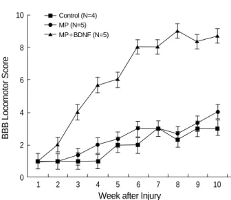
Functional Recovery
Unfortunately, some of the rats died of cystitis or other infec- tions, so the number surviving ten weeks after SCI was five in the BDNF+MP group, five in the MP-only group, and four in control group. Observers blinded to the treatments performed all functional assessments and analyses, using the BBB open field locomotion scale. The MP-only group and control groups uniformly exhibited either paralysis or occa- sional uncoordinated hindlimb spasms. Three weeks after SCI, initial paralysis was attenuated in animals infused with
MP+BDNF. Six weeks after injury, they demonstrated exten- sive joint movement in their hindlimbs, and their BBB score increased significantly relative to MP-only and control groups (Fig. 5, p<0.05). Rats in the MP-only group were not signif- icantly different from the control group.
DISCUSSION
Spinal cord contusion injury is a standard model for investi- gating neuron injury and regeneration. Authors evaluated the
Fig. 1.Immunohistochemical staining for ED-1 at the contusion injury site (×40). 3 days after the SCI, many ED-1-positive inflammatory cells (stained with brown color) are observed at the contusion site in the untreated spinal cord (A). In contrast, ED-1-positive inflammatory cells are markedly decreased in the MP-only (B) and MP+BDNF (C) infused animals. This difference, represented as means±S.D. in (D), is significant (p<0.05).
A B
C D
No. of cell
control MP MP+BDNF
ED 1
20,000 18,000 16,000 14,000 12,000 10,000 8,000 6,000 4,000 2,000 0
*
effects of a combination therapy of acute MP and continuous BDNF infusion on the initial anti-inflammatory reaction, axonal regeneration, and functional recovery. Immunostain- ing revealed a reduction in ED-1-positive inflammatory cells infiltrating the contusion site in MP and MP+BDNF groups.
Concomitantly, increased motor neuron regeneration and func- tional improvement were observed in the MP+BDNF group, compared to MP-only and control groups. Increased GAP-43 gene expression was seen in spinal cords from the rats in the MP+BDNF group one day after SCI. To our knowledge, this report is the first to demonstrate that a combination therapy of MP and BDNF can reduce the initial inflammatory reaction,
and boost axonal regeneration following SCI.
After the primary injury, the spinal cord lesion is enlarged by secondary inflammation, causing further injury. Since the activated macrophages and microglia release cytotoxic sub- stances that participate in secondary post-injury tissue destruc- tion, an important treatment issue is how to control initial inflammatory reactions following SCI. In this study, ED-1 was chosen as a marker to identify the presence of infiltrating cells (15). ED-1-positive cells in immunostained sections of injured spinal cords decreased dramatically and evenly in rats treated with MP alone or MP combined with BDNF infusion.
In contrast, more ED-1-positive inflammatory cells infiltrated
Fig. 2.Immunohistochemical staining for NF at the contusion injury site (×40). Ten weeks after SCI, fewer NF-positive neuronal fibers are observed at the lesion in the control (A) and MP-only groups (B). More NF-positive neuronal axons survive in the spinal cords of rats in the MP+BDNF group (C). This difference, represented as means±S.D. in (D), is significant (p<0.05).
A B
C D
No. of cell
control MP MP+BDNF
10,000 9,000 8,000 7,000 6,000 5,000 4,000 3,000 2,000 1,000 0
* NF
the injury site in untreated animals. MP with BDNF infusion keeps potent anti-inflammatory effects in the spinal cord con- tusion model, consistent with previous reports (16). Thus, adjuvant BDNF infusion did not affect the MP action on spinal cord, at least in terms of most important anti-inflam- matory effect in acute stage of SCI.
High-dose corticosteroid may exert several detrimental effects on neurological recovery (4, 17). By inhibiting immune cell activity and antigen processing by macrophages, corticos- teroid may interfere with neuronal regeneration. Glucocor- ticoids may also exacerbate postischemic neuronal necrosis and inhibit axonal sprouting. Some studies have shown that
MP depresses the production of growth-promoting factors such as BDNF and NT3 (9). Thus, MP treatment alone has not been sufficient, or may hinder, functional recovery follow- ing SCI. Therefore, we thought it reasonable to combine nerve- growth factor with MP after acute SCI. The more NF-posi- tive neuronal axons and the GAP-43-positive neurites were observed in spinal cords of MP+BDNF rats compared to MP- only and untreated rats, which suggests that neurons were rescued. GAP-43 immunoreactivity has been used to evalu- ate regenerating axons (18, 19). There is a close correlation between GAP-43 expression and successful regeneration of CNS neurons (20, 21). GAP-43 is concentrated at the axon-
Fig. 3.Immunohistochemical staining for GAP-43 at the contusion injury site (×40). Ten weeks after SCI, many GAP-43-positive neurites are found in the MP+BDNF group (C), while few are detected in the control (A) and MP-only treated (B) rats. This difference, represented as means±S.D. in (D), is significant (p<0.05).
A B
C D
No. of cell
control MP MP+BDNF
4,000 3,500 3,000 2,500 2,000 1,500 1,000 500 0
GAP 43
*
al growth cone, where it seems to play an important role in the transduction of growth cone guidance signals (18, 19). In this study, we hypothesized that significant numbers of GAP- 43 positive neurites in the MP+BDNF infused spinal cords could be interpreted as effective axonal regeneration. In addi- tion, most regenerating fibers were ChAT-positive motor neu- rons, and many more such fibers were observed in spinal cords infused with BDNF compared to other two groups. These results are consistent with previous reports (22).
We also investigated GAP-43 gene expression to determine whether axonal regeneration actually occurred. Quantitative RT-PCR revealed a 23.4±3% increase in GAP-43 gene ex- pression at the injured site one day following MP+BDNF
infusion, while no significant changes in GAP-43 gene expres- sion were detected in the MP-only treated and control animals.
This suggests that BDNF has the ability to enhance axonal regeneration in the early phase of acute SCI, consistent with recent reports (23).
In addition to immunohistochemical studies, we observed the effects of long-term infusion of BDNF on behavioral recov- ery, using the BBB open field locomotion scale. MP+BDNF treatment resulted in extensive hindlimb movements with weight support in almost all animals ten weeks after SCI.
Animals infused with MP only and untreated control animals did not exhibit extensive hindlimb movement. Three weeks after SCI, the BBB scores increased more in the MP+BDNF
Fig. 4.Immunohistochemical staining for ChAT at the contusion injury site (×40). More ChAT-positive axons appear in MP+BDNF rats (C) ten weeks after SCI than in the other two groups (A, B). This difference, represented as means±S.D. in (D), is significant (p<0.05).
A B
C D
No. of cell
control MP MP+BDNF
9,000 8,000 7,000 6,000 5,000 4,000 3,000 2,000 1,000 0
* ChAT
infused rats compared to MP-only treated and untreated rats.
Various forms of alternating hindlimb stepping or locomotor activity have been obtained in rats, cats, and humans following SCI. In the present study, animals treated with MP+BDNF exhibited active hindlimb movement, which is related to motor neuron regeneration stimulated by BDNF. It is thought that hindlimb activity is controlled within the lumbar spinal cord by interneuronal networks collectively referred to as cen- tral pattern generators (CPGs) (24). The effect of BDNF might be to reduce the activation threshold for existing CPGs, in addition to promoting axonal regeneration (25).
Contrary to several reports, animals treated with MP alone did not show good functional recovery in our study. Although scores of some of the MP-treated animals were statistically different at some points from control animals and it showed progressive recovery with time, this was not a consistent effect.
Furthermore, behavioral recovery curve generated for MP only treatment is virtually identical to that of control. This sug- gests that MP affects SCI during the acute phase only, and has little effect on long-term functional recovery. This may be related to the fact that MP reduces the growth factors and interfere neuronal regeneration and axonal sprouting (4, 9, 17), but it may also reflect the severity of the injury. Four ani- mals were lost during this study, and overall levels of function were not as high as reported by others. This may suggest that MP is less effective in severe SCI; it is not unusual in clinical situations for patients with severe SCI to fail to respond to mega-dose MP treatments, despite an appropriate therapeu- tic time window (5-8). It also suggests that continuous BDNF therapy may be more important than MP for long-term func- tional recovery.
The mechanisms of the recovery-enhancing effects of BDNF may be related to the fact that BDNF interacts with TrkB, a member of the trk receptor family, which transduces BDNF signaling in responsive neurons (26) . TrkB receptor is exp- ressed not only in developing motor neurons, but in mature motor neurons as well (27-29). Adult motor neurons have been shown to retrogradely transport BDNF in a receptor- mediated fashion (30) . The first evidence of this was the obser- vation that administration of exogenous BDNF markedly attenuated the decrease of ChAT immunoreactivity in adult facial motor neurons (31, 32), and in adult spinal motor neu- rons following distal axotomy (33). Subsequently, BDNF was shown to prevent cell death of adult motor neurons (34). How- ever, previous studies showed that this effect was transient because little effect was seen two to three weeks post injury.
Our study suggests that exogeneously administered BDNF attenuated the decrease of ChAT immunoreactivity, and pro- moted axonal outgrowth of injured adult motor neurons ten weeks following SCI. The neurotrophic effect of BDNF was shown to be long-lasting in adult motor neurons.
In spite of encouraging results, this study has several lim- itations. It was intended to test new therapeutic method, which is simulated clinical situation, it is necessary to iden- tify whether chronic BDNF infusion without initial MP treat- ment can evoke similar outcome compared to MP+BDNF infusion. Although MP+BDNF enhanced hindlimb step- ping and improved functional recovery following SCI, it was not enough recovery to walk. It might reflect an increase in reflexive movements that may not associated with motor axon regeneration because BDNF alone can cause reflexive air walk- ing in transection model (25). These will be elucidated with further experiments. Immunohistopathologic technique in this study is semi-quantitative, usually used in clinical pathol- ogy. Thus, further experiment using precise quantitative tech- nique is necessary to support motor axonal regeneration.
In summary, this study demonstrated that a combination therapy of MP+BDNF not only attenuated hematogenous inflammatory cell infiltration, but also enhanced motor axon regeneration following SCI. The present study offers a ratio- nal approach for the treatment of spinal cord injury in which both the attenuation of initial injury and the regeneration of motor axons are required. Further studies are needed to eval- uate the proper concentration and duration of BDNF infu- sion on a large number of animals that have sustained mod- erate SCI via other means.
ACKNOWLEDGEMENT
We gratefully acknowledge L.F. Eng and Y.L. Lee, Veterans Affair Palo Alto Health Care System, Department of Patholo- gy, Stanford University for their much-appreciated help and advice.
BBB Locomotor Score
10
8
6
4
2
0
Week after Injury
1 2 3 4 5 6 7 8 9 10
Control (N=4) MP (N=5) MP+BDNF (N=5)
Fig. 5.BBB (Basso Beattie Bresnahan) locomotor score (means
±S.D.). Rats that received MP+BDNF revealed active coordinat- ed hindlimb movement, six weeks after SCI. The MP-only and untreated rats exhibited either paralysis or occasional uncoordi- nated hindlimb spasms ten weeks after SCI. Differences each week were determined by post hoc means-corrected t-tests (p<0.05).
REFERENCES
1. Amar AP, Levy ML. Pathogenesis and pharmacological strategies for mitigating secondary damage in acute spinal cord injury. Neuro- surgery 1999; 44: 1027-39.
2. Coleman WP, Benzel D, Cahill DW, Ducker T, Geisler F, Green B, Gropper MR, Goffin J, Madsen PW 3rd, Maiman DJ, Ondra SL, Ros- ner M, Sasso RC, Trost GR, Zeidman S. A critical appraisal of the reporting of the National Acute Spinal Cord Injury Studies (II and III) of methylprednisolone in acute spinal cord injury. J Spinal Dis- ord 2000; 13: 185-99.
3. Hadley MN, Walters BC, Grabb PA, Oyesiku NM, Przybylski GJ, Resnick DK, Ryken TC, Mielke DH. Guidelines for the management of acute cervical spine and spinal cord injuries. Clin Neurosurg 2002;
49: 407-98.
4. Bracken MB. Pharmacological treatment of acute spinal cord injury:
current status and future projects. J Emerg Med 1993; 11 (Suppl 1):
43-8.
5. Galandiuk S, Raque G, Appel S, Polk HC Jr. The two-edged sword of large-dose steroids for spinal cord trauma. Ann Surg 1993; 218:
419-25.
6. George ER, Scholten DJ, Buechler CM, Jordan-Tibbs J, Mattice C, Albrecht RM. Failure of methylprednisolone to improve the outcome of spinal cord injuries. Am Surg 1995; 61: 659-63.
7. Gerndt SJ, Rodriguez JL, Pawlik JW, Taheri PA, Wahl WL, Micheals AJ, Papadopoulos SM. Consequences of high-dose steroid therapy for acute spinal cord injury. J Trauma 1997; 42: 279-84.
8. Gerhart KA, Johnson RL, Menconi J, Hoffman RE, Lammertse DP.
Utilization and effectiveness of methylprednisolone in a population- based sample of spinal cord injured persons. Paraplegia 1995; 33:
316-21.
9. Hayashi M, Ueyama T, Nemoto K, Tamaki T, Senba E. Sequential mRNA expression for immediate early genes, cytokines, and neuro- trophins in spinal cord injury. J Neurotrauma 2000; 17: 203-18.
10. Lindsay RM, Thoenen H, Barde YA. Placode and neural crest-derived sensory neurons are responsive at early developmental stages to brain- derived neurotrophic factor. Dev Biol 1985; 112: 319-28.
11. Alderson RF, Alterman AL, Barde YA, Lindsay RM. Brain-derived neurotrophic factor increases survival and differentiated functions of rat septal cholinergic neurons in culture. Neuron 1990; 5: 297-306.
12. Fawcett JP, Bamji SX, Causing CG, Aloyz R, Ase AR, Reader TA, McLean JH, Miller FD. Functional evidence that BDNF is an antero- grade neuronal trophic factor in the CNS. J Neurosci 1998; 18: 2808- 21.
13. Murphy GM, Jr., Jia XC, Yu AC, Lee YL, Tinklenberg JR, Eng LF.
Reverse transcription and polymerase chain reaction technique for quantification of mRNA in primary astrocyte cultures. J Neurosci Res 1993; 35: 643-51.
14. Basso DM, Beattie MS, Bresnahan JC. A sensitive and reliable loco- motor rating scale for open field testing in rats. J Neurotrauma 1995;
12: 1-21.
15. Dijkstra CD, Dopp EA, Joling P, Kraal G. The heterogeneity of mono- nuclear phagocytes in lymphoid organs: distinct macrophage subpop- ulations in the rat recognized by monoclonal antibodies ED1, ED2 and
ED3. Immunology 1985; 54: 589-99.
16. Xu J, Qu ZX, Hogan EL, Perot PL Jr. Protective effect of methylpred- nisolone on vascular injury in rat spinal cord injury. J Neurotrauma 1992; 9: 245-53.
17. Bracken MB, Shepard MJ, Collins WF Jr, Holford TR, Baskin DS, Eisenberg HM, Flamm E, Leo-Summers L, Maroon JC, Marshall LF.
Methylprednisolone or naloxone treatment after acute spinal cord injury: 1-year follow-up data. Results of the second National Acute Spinal Cord Injury Study. J Neurosurg 1992; 76: 23-31.
18. Brook GA, Plate D, Franzen R, Martin D, Moonen G, Schoenen J, Schmitt AB, Noth J, Nacimiento W. Spontaneous longitudinally ori- entated axonal regeneration is associated with the Schwann cell frame- work within the lesion site following spinal cord compression injury of the rat. J Neurosci Res 1998; 53: 51-65.
19. Tuszynski MH, Peterson DA, Ray J, Baird A, Nakahara Y, Gage FH.
Fibroblasts genetically modified to produce nerve growth factor induce robust neuritic ingrowth after grafting to the spinal cord. Exp Neurol 1994; 126: 1-14.
20. Kobayashi NR, Fan DP, Giehl KM, Bedard AM, Wiegand SJ, Tet- zlaff W. BDNF and NT-4/5 prevent atrophy of rat rubrospinal neu- rons after cervical axotomy, stimulate GAP-43 and alpha1-tubulin mRNA expression, and promote axonal regeneration. J Neurosci 1997;
17: 9583-95.
21. Ramon-Cueto A, Plant GW, Avila J, Bunge MB. Long-distance axonal regeneration in the transected adult rat spinal cord is promoted by olfactory ensheathing glia transplants. J Neurosci 1998; 18: 3803-15.
22. Kishino A, Ishige Y, Tatsuno T, Nakayama C, Noguchi H. BDNF prevents and reverses adult rat motor neuron degeneration and induces axonal outgrowth. Exp Neurol 1997; 144: 273-86.
23. Dougherty KD, Dreyfus CF, Black IB. Brain-derived neurotrophic factor in astrocytes, oligodendrocytes, and microglia/macrophages after spinal cord injury. Neurobiol Dis 2000; 7: 574-85.
24. Grillner S, Wallen P. Central pattern generators for locomotion, with special reference to vertebrates. Annu Rev Neurosci 1985; 8: 233-61.
25. Jakeman LB, Wei P, Guan Z, Stokes BT. Brain-derived neurotrophic factor stimulates hindlimb stepping and sprouting of cholinergic fibers after spinal cord injury. Exp Neurol 1998; 154: 170-84.
26. Squinto SP, Stitt TN, Aldrich TH, Davis S, Bianco SM, Radziejewski C, Glass DJ, Masiakowski P, Furth ME, Valenzuela DM, et al. trkB encodes a functional receptor for brain-derived neurotrophic factor and neurotrophin-3 but not nerve growth factor. Cell 1991; 65: 885-93.
27. Seeburger JL, Tarras S, Natter H, Springer JE. Spinal cord motoneu- rons express p75NGFR and p145trkB mRNA in amyotrophic lateral sclerosis. Brain Res 1993; 621: 111-5.
28. Frisen J, Verge VM, Cullheim S, Persson H, Fried K, Middlemas DS, Hunter T, Hokfelt T, Risling M. Increased levels of trkB mRNA and trkB protein-like immunoreactivity in the injured rat and cat spinal cord. Proc Natl Acad Sci USA 1992; 89: 11282-6.
29. Koliatsos VE, Clatterbuck RE, Winslow JW, Cayouette MH, Price DL. Evidence that brain-derived neurotrophic factor is a trophic factor for motor neurons in vivo. Neuron 1993; 10: 359-67.
30. DiStefano PS, Friedman B, Radziejewski C, Alexander C, Boland P, Schick CM, Lindsay RM, Wiegand SJ. The neurotrophins BDNF, NT-3, and NGF display distinct patterns of retrograde axonal trans-
port in peripheral and central neurons. Neuron 1992; 8: 983-93.
31. Yan Q, Elliott JL, Matheson C, Sun J, Zhang L, Mu X, Rex KL, Snider WD. Influences of neurotrophins on mammalian motoneurons in vivo.
J Neurobiol 1993; 24: 1555-77.
32. Yan Q, Matheson C, Lopez OT, Miller JA. The biological responses of axotomized adult motoneurons to brain-derived neurotrophic factor.
J Neurosci 1994; 14: 5281-91.
33. Friedman B, Kleinfeld D, Ip NY, Verge VM, Moulton R, Boland P,
Zlotchenko E, Lindsay RM, Liu L. BDNF and NT-4/5 exert neuro- trophic influences on injured adult spinal motor neurons. J Neurosci 1995; 15: 1044-56.
34. Novikov L, Novikova L, Kellerth JO. Brain-derived neurotrophic factor promotes survival and blocks nitric oxide synthase expression in adult rat spinal motoneurons after ventral root avulsion. Neu- rosci Lett 1995; 200: 45-8.